
Neck — MCQs

On this page
How many cartilages are present in the larynx?
The fourth thyroid vein (Kocher's vein) is occasionally found between which two thyroid veins?
The atlanto-axial joint is classified as which type of joint?
What is the arterial supply of the trachea?
The posterior belly of the digastric muscle is attached to which bony landmark?
The inferior laryngeal artery is a branch of which of the following arteries?
What is the narrowest part of an infant's larynx?
The recurrent laryngeal nerve runs along which border of the pharyngeal constrictor muscles?
All statements regarding the cervical part of the esophagus are true except?
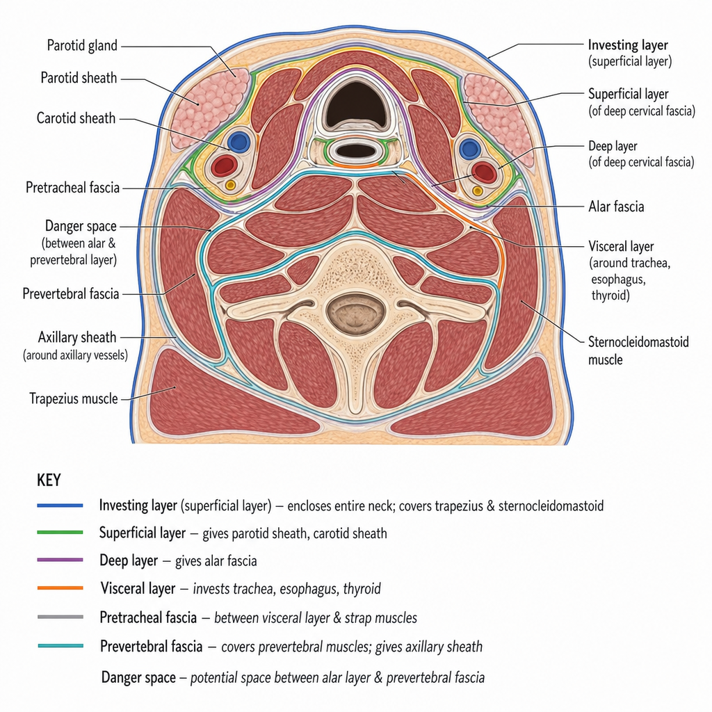
In the given cross-section of the deep cervical fascia, which of the following statements is NOT true?
Practice by Chapter
Cervical Fascia
Practice Questions
Triangles of the Neck
Practice Questions
Deep Structures of the Neck
Practice Questions
Thyroid and Parathyroid Glands
Practice Questions
Vasculature of the Neck
Practice Questions
Lymphatic Drainage
Practice Questions
Cervical Plexus
Practice Questions
Root of the Neck
Practice Questions
Applied Anatomy and Clinical Correlations
Practice Questions
Surface Anatomy of the Neck
Practice Questions
Want unlimited practice?
Get full access to all questions, explanations, and performance tracking.
Scan to download app