
Neck — MCQs

On this page
Which of the following muscle is not innervated by the nerve shown in the image?
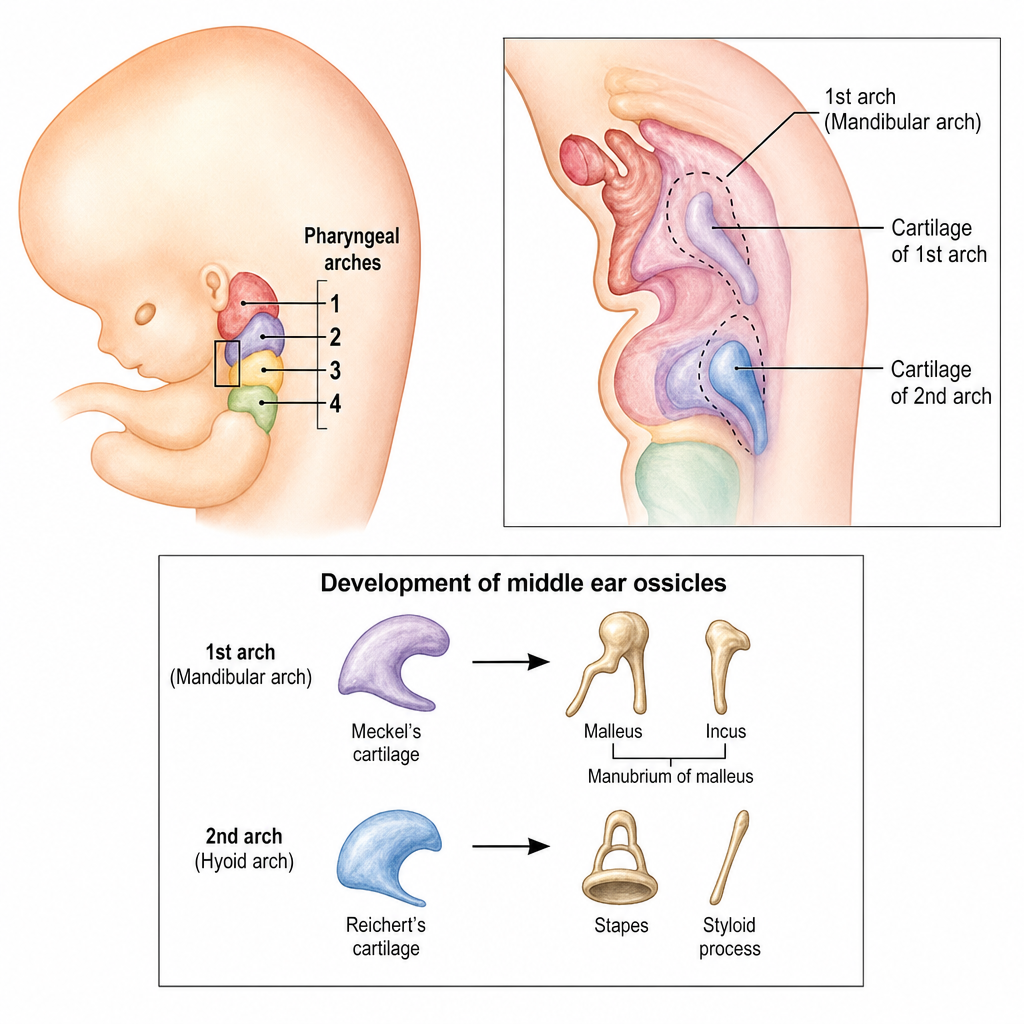
A neonate is noted at birth to have a small jaw, cleft palate, and glossoptosis causing airway obstruction. A clinical geneticist suspects a defect in neural crest cell migration into a specific pharyngeal arch during the fourth week of embryonic development. The cartilaginous element of the affected arch that normally forms two specific middle ear ossicles is which of the following?
Contents of the suboccipital triangle are formed by all of the following structures, EXCEPT?
Level III neck nodes are located in which region?
A 10-year-old child suddenly choked while consuming food. After a failed Valsalva's maneuver, a needle cricothyrotomy was performed. Into which of the following regions would this procedure open?
Practice by Chapter
Cervical Fascia
Practice Questions
Triangles of the Neck
Practice Questions
Deep Structures of the Neck
Practice Questions
Thyroid and Parathyroid Glands
Practice Questions
Vasculature of the Neck
Practice Questions
Lymphatic Drainage
Practice Questions
Cervical Plexus
Practice Questions
Root of the Neck
Practice Questions
Applied Anatomy and Clinical Correlations
Practice Questions
Surface Anatomy of the Neck
Practice Questions
Want unlimited practice?
Get full access to all questions, explanations, and performance tracking.
Scan to download app