An electron microscopy of muscle biopsy shows 'parking lot' appearance. Which additional finding would confirm myotonic dystrophy?
Not a monomeric intermediate filament:
Colorado stain is related to:
Fluorescein dye for ophthalmological diagnosis is injected into:
During autopsy for virology study which agent is used for storing tissue:
Patient presenting with cutaneous vasculitis, glomerulonephritis, peripheral neuropathy, Which investigation is to be performed next that will help you diagnose the condition?
A patient presents with pulmonary hemorrhage and is P-ANCA positive. What is the most likely diagnosis?
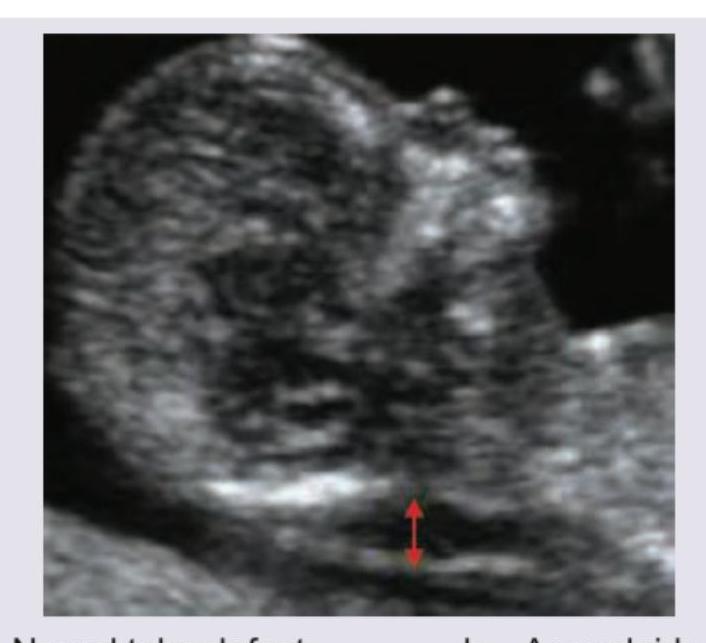
The following USG scan should prompt you to screen for which of the following disorders?

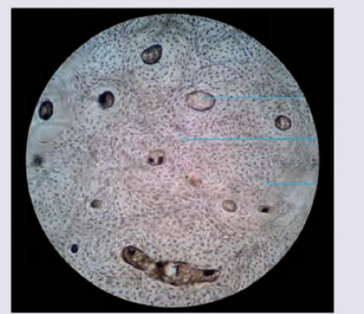
The image shows presence of:

Which cells are present in the collecting duct?
Explanation: ***Ring fibers*** - **Ring fibers** are a classic histopathological feature seen in **myotonic dystrophy**, characterized by peripheral myofibrils arranged circularly around a central core [1]. - The "parking lot" appearance on electron microscopy refers to collections of **sarcoplasmic reticulum** and **T-tubules**, which can be seen in various myopathies but are often prominent in myotonic dystrophy, complementing the presence of ring fibers [1]. *Ragged red fibers* - **Ragged red fibers** are characteristic of **mitochondrial myopathies**, indicating abnormal proliferation of mitochondria beneath the sarcolemma. - They are typically identified with **Gomori trichrome stain** and are not a feature of myotonic dystrophy. *Central cores* - **Central cores** are a hallmark of **central core disease**, a congenital myopathy, and are regions within muscle fibers where oxidative enzyme activity is absent. - These are not typically associated with myotonic dystrophy; rather, they suggest a different underlying genetic defect affecting muscle structure. *Nemaline rods* - **Nemaline rods** are rod-like inclusions observed in muscle fibers in **nemaline myopathy**, an inherited disorder often associated with mutations in genes encoding components of the thin filament. - They are distinct from the pathological findings in myotonic dystrophy and point to a specific type of congenital myopathy. **References:** [1] Cross SS. Underwood's Pathology: A Clinical Approach. 6th ed. Common Clinical Manifestations Of Central And Peripheral Nervous System Disease, pp. 732-733.
Explanation: ***Tubulin*** - **Tubulin** is the monomeric building block of **microtubules**, which are distinct from intermediate filaments. - Microtubules are involved in cell motility, intracellular transport, and maintaining cell shape, but they do not belong to the intermediate filament family. *Desmin* - **Desmin** is a type III **intermediate filament** that is primarily expressed in muscle cells. - It plays a crucial role in organizing the **sarcomeres** and providing structural integrity to muscle fibers. *Keratin* - **Keratin** is the most diverse family of **intermediate filaments** and is primarily found in epithelial cells. - It provides **mechanical strength** to cells and tissues, forming structures like hair, nails, and the outer layer of skin. *Vimentin* - **Vimentin** is a type III **intermediate filament** that is widely expressed in cells of **mesenchymal origin**, such as fibroblasts, endothelial cells, and leukocytes. - It contributes to cell shape, motility, and the integrity of the **cytoskeleton**.
Explanation: ***Dental fluorosis*** - The term "Colorado stain" is an older, historical term used to describe the **characteristic brown discoloration and mottling of teeth** caused by **dental fluorosis**. - This condition occurs due to **excessive fluoride intake** during tooth development, particularly in areas with high natural fluoride levels in drinking water. *Staining from medications* - While certain medications can cause tooth discoloration (e.g., **tetracycline staining**), this is a general category and not specifically referred to as "Colorado stain." - **Tetracycline staining** typically presents as grey-brown or yellow-brown intrinsic stains, distinct from the mottled appearance of fluorosis. *Staining from food or tobacco* - Staining from food and tobacco is typically **extrinsic**, meaning it occurs on the surface of the tooth. - This type of staining usually presents as yellow, brown, or black stains that can often be removed by professional cleaning, unlike the intrinsic discoloration of fluorosis. *Cavities not visually apparent* - Dental caries (cavities) are **structural defects** in the tooth and, while sometimes discolored, are not referred to as "Colorado stain." - Early caries might be radiographically visible before clinical detection but are not a type of staining.
Explanation: ***Antecubital vein*** - Fluorescein angiography requires rapid delivery of the dye into the **systemic circulation** for quick visualization of retinal and choroidal vasculature. - The **antecubital vein** is a readily accessible, large superficial vein that allows for quick and efficient intravenous dye injection. *Popliteal vein* - The popliteal vein is located behind the **knee** and is not a standard or practical site for routine intravenous injections, especially when rapid delivery to the eye is needed. - Its location makes it less accessible and potentially more uncomfortable for the patient compared to an arm vein. *Femoral vein* - The femoral vein is a large, deep vein in the **groin**, typically reserved for central venous access or specific procedures due to the increased risk of complications like infection or hematoma. - It is not routinely used for peripheral intravenous injections such as fluorescein, where a more superficial and accessible vein is preferred. *Subclavian vein* - The subclavian vein is a **central vein** located under the clavicle, accessed via a more invasive procedure, usually for central venous catheters or hemodialysis access. - It carries higher risks compared to peripheral venipuncture and is not chosen for simple diagnostic dye injections like fluorescein.
Explanation: ***50% glycerine*** - **Glycerine** is commonly used for preserving tissues for virological studies because it helps to maintain viral viability by preventing **desiccation** and **denaturation** of viral particles. - It acts as a **cryoprotectant**, stabilizing cell membranes and protein structures, which is crucial for subsequent **viral isolation** and detection. *Alcohol* - **Alcohol** acts as a **fixative** by denaturing proteins and dehydrating tissues, which would inactivate most viruses and make them unsuitable for viability studies. - While useful for histopathology, it is not appropriate for preserving viral infectivity or integrity. *Rectified spirit* - **Rectified spirit** is a highly concentrated form of alcohol (typically 95% ethanol), and like alcohol, it causes **protein denaturation** and **dehydration**. - This property makes it unsuitable for preserving viral viability for subsequent virological studies. *Sodium chloride* - **Sodium chloride** alone in isotonic solutions can maintain tissue hydration but does not provide adequate **viral stabilization** or protection against degradation. - It would not prevent **enzymatic degradation** or maintain viral infectivity over time, especially at room temperature.
Explanation: ### ANCA - The combination of **cutaneous vasculitis**, **glomerulonephritis**, and **peripheral neuropathy** points towards a small-vessel vasculitis, for which **ANCA (anti-neutrophil cytoplasmic antibodies)** testing is crucial [1]. - ANCA is highly specific for conditions like **Granulomatosis with Polyangiitis (GPA)** and **Microscopic Polyangiitis (MPA)** [1]. ### RA factor - **Rheumatoid factor (RF)** is primarily associated with **rheumatoid arthritis**, which typically presents with symmetrical polyarthritis, not the constellation of symptoms described. - While RF can be positive in some vasculitides, it is not the most specific initial test for the given clinical presentation. ### Hbsag - **Hepatitis B surface antigen (HbsAg)** typically screens for **Hepatitis B infection**, which can cause **polyarteritis nodosa (PAN)**, a medium-vessel vasculitis. - However, the patient's symptoms (cutaneous vasculitis, glomerulonephritis) are more characteristic of **small-vessel vasculitis**, making ANCA a more direct investigation [1]. ### MIF - **MIF (Macrophage Migration Inhibitory Factor)** is a cytokine involved in inflammation, but it is not a routine diagnostic marker for vasculitis. - It is not used as a primary investigation to diagnose specific autoimmune or inflammatory conditions like vasculitis.
Explanation: ***Microscopic polyangiitis*** - This condition is characterized by **pulmonary hemorrhage** (often manifesting as diffuse alveolar hemorrhage) and **P-ANCA positivity**, which is typically associated with antibodies against **myeloperoxidase (MPO)**. [1] - It is a **small-vessel vasculitis** that frequently affects the kidneys (glomerulonephritis) and lungs without granuloma formation. *Churg-Strauss syndrome* - While Churg-Strauss syndrome (now known as **Eosinophilic Granulomatosis with Polyangiitis**, EGPA) can be P-ANCA positive, it is typically associated with a history of **asthma**, **allergic rhinitis**, and **eosinophilia**. [1] - Pulmonary involvement often includes **infiltrates** and nodules, but diffuse alveolar hemorrhage with severe pulmonary hemorrhage is less common as the primary presentation compared to MPA. *Wegener granulomatosis* - Wegener granulomatosis (now known as **Granulomatosis with Polyangiitis**, GPA) primarily presents with **upper and lower respiratory tract granulomatous inflammation** and **glomerulonephritis**. - It is typically associated with **C-ANCA positivity** (antibodies against proteinase 3, PR3), not P-ANCA. *Polyarteritis nodosa (PAN)* - Polyarteritis nodosa is a **medium-vessel vasculitis** that typically affects the **kidneys, gastrointestinal tract, skin, and nervous system**. [1] - It is classically **ANCA-negative** and does not typically cause pulmonary hemorrhage or diffuse alveolar hemorrhage.
Explanation: ***Aneuploidy*** - The ultrasound image shows increased nuchal translucency (indicated by the red arrow), which is a key marker for **chromosomal abnormalities** like Down syndrome (Trisomy 21). - Increased nuchal translucency combined with other features like **absent nasal bone** (not clearly visible in this image but often associated) warrants further screening for aneuploidy. *Neural tube defect* - Neural tube defects are characterized by abnormalities of the brain and spine, such as **anencephaly** or **spina bifida**, which are not directly indicated by increased nuchal translucency. - While some chromosomal abnormalities can be associated with neural tube defects, nuchal translucency specifically points more strongly to aneuploidy. *Achondroplasia* - Achondroplasia is a form of **dwarfism** recognized by disproportionately short limbs and macrocephaly, which are typically identified later in pregnancy during detailed anatomical surveys. - Increased nuchal translucency is not a primary screening marker for achondroplasia. *Artifact on routine scans* - While artifacts can occur, increased nuchal translucency is a well-established and **clinically significant finding** that requires specific measurements and interpretation in screening for fetal abnormalities. - This measurement is a standard part of the **first-trimester screening** for chromosomal disorders.
Explanation: ***Fibrocartilage*** - The image displays characteristic features of fibrocartilage, including visible bundles of **collagen fibers** (appearing wavy and somewhat disorganized) interspersed with **chondrocytes** residing in lacunae. - The arrangement and presence of abundant collagen make it suitable for locations requiring robust **tensile strength** and **shock absorption**, such as intervertebral discs and menisci. *Compact bone* - Compact bone would typically show **Haversian systems (osteons)** with concentric lamellae surrounding a central canal, which are not evident in this image. - The cellular components, **osteocytes**, would be found within lacunae, but the overall matrix and organization are distinct from cartilage. *Hyaline cartilage* - Hyaline cartilage has a **smooth, glassy matrix** with very fine collagen fibers that are not usually visible under light microscopy, unlike the distinct bundles seen here. - Chondrocytes in hyaline cartilage often appear in **isogenous groups** (clusters of cells), which are not prominently featured in this image. *Myositis ossificans* - **Myositis ossificans** is a condition where bone forms within muscle tissue, typically following trauma, and would show mature or immature bone tissue, not cartilage. - Histologically, it would present with **osteoid formation** and calcification within muscle, along with inflammatory cells, which are absent in this image.
Explanation: The collecting duct is the final segment of the renal tubule system, playing a critical role in fluid balance and acid-base homeostasis. ### **Explanation of the Correct Answer** The collecting duct is lined by a simple cuboidal epithelium consisting of two distinct cell types: 1. **Principal Cells (P cells):** These are the predominant cells. They possess receptors for **ADH (Vasopressin)**, which regulates water reabsorption via Aquaporin-2 channels [3], and **Aldosterone**, which mediates sodium reabsorption and potassium secretion. 2. **Intercalated Cells (I cells):** These are fewer in number and are primarily involved in acid-base balance [2]. **Type A** cells secrete $H^+$ (acidosis compensation), while **Type B** cells secrete $HCO_3^-$ (alkalosis compensation). ### **Analysis of Incorrect Options** * **B. Parietal and Oxyntic cells:** These are synonyms for the same cell type found in the **stomach lining** (gastric glands) responsible for secreting Hydrochloric acid (HCl) and Intrinsic Factor. * **C. Lacis cells:** Also known as extraglomerular mesangial cells, these are part of the **Juxtaglomerular Apparatus (JGA)** located between the afferent and efferent arterioles. * **D. Podocytes:** These are highly specialized visceral epithelial cells of the **Bowman’s capsule** [1] that wrap around glomerular capillaries to form the filtration slits. ### **High-Yield NEET-PG Pearls** * **Histology Tip:** Principal cells have short microvilli and a single primary cilium, whereas Intercalated cells have prominent microplicae (surface folds). * **Pharmacology Link:** Potassium-sparing diuretics (like Spironolactone and Amiloride) act specifically on the **Principal cells**. * **Embryology:** Unlike the rest of the nephron (derived from Metanephric blastema), the collecting duct develops from the **Ureteric bud**.
Get full access to all questions, explanations, and performance tracking.
Start For Free