In a preterm baby with respiratory distress syndrome, which of the following lipids would be deficient?
The activation of muscarinic receptors in bronchiolar smooth muscle is associated with:
All of the following are features of Lymph node histology except:
Bifurcation of the trachea is at which level?
Which of the following is a stain for heart failure cells?
Investigation of choice for Posterior urethral valves?
Granuloma is a pathological feature of all, except which of the following?
Which of the following diseases is NOT associated with Anti-Neutrophil Cytoplasmic Antibodies (ANCA)?
The nasopharynx is primarily lined by which type of epithelium?
All cartilage is covered by perichondrium, except
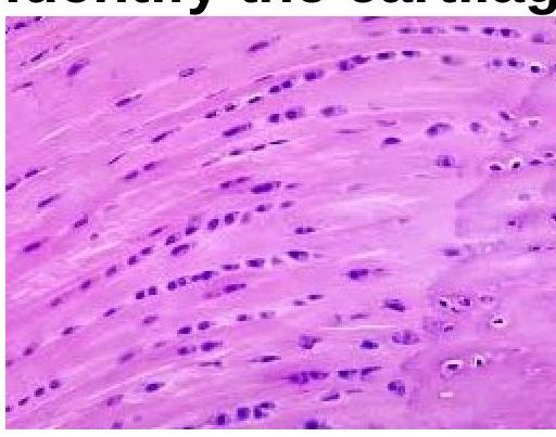
Explanation: ***Lecithin*** - **Lecithin** (also known as **phosphatidylcholine**) is the primary component of **surfactant** in the lungs, which reduces surface tension and prevents alveolar collapse. - In **preterm babies**, insufficient production of lecithin due to immature lung development leads to **respiratory distress syndrome (RDS)**. *Cardiolipin* - **Cardiolipin** is a major phospholipid found in the **inner mitochondrial membrane**, crucial for oxidative phosphorylation. - Deficiency is associated with mitochondrial disorders like **Barth syndrome**, not primary respiratory distress. *Sphingomyelin* - **Sphingomyelin** is a significant component of **cell membranes** and **myelin sheaths**, important for nerve insulation. - While present in the lungs, its primary role is not in surface tension reduction, and its deficiency is not directly linked to RDS. *Phosphatidylinositol* - **Phosphatidylinositol** is a precursor for various **signaling molecules** and plays a role in cell membrane structure. - While involved in cellular processes, it is not the critical surfactant component whose deficiency causes RDS.
Explanation: ***Increase in IP3 and DAG*** - **Muscarinic receptors** on bronchial smooth muscle (M3 receptors) are **Gq protein-coupled receptors** [1]. - Activation of **Gq proteins** leads to the activation of **phospholipase C**, which hydrolyzes **PIP2** into **IP3** and **DAG** [1, 3]. *Inhibition of protein kinase C* - **DAG** (diacylglycerol), produced from the breakdown of PIP2, **activates protein kinase C (PKC)**, rather than inhibiting it [2]. - This activation of PKC contributes to downstream cellular responses, including smooth muscle contraction [1]. *Activation of adenylyl cyclase* - **Adenylyl cyclase** is typically activated by **Gs protein-coupled receptors**, leading to an increase in **cAMP**. - **Muscarinic (M3) receptors** are **Gq-coupled**, so they do not activate adenylyl cyclase; instead, they operate through the phospholipase C pathway [1, 3]. *Opening of Na+/K+ cation channels* - While some neurotransmitter receptors are **ligand-gated ion channels** (e.g., nicotinic receptors), muscarinic receptors are **G protein-coupled receptors** [1]. - Their activation does not directly lead to the opening of **Na+/K+ cation channels**; rather, they initiate intracellular signaling cascades.
Explanation: ***Red pulp and White pulp are present*** - **Red pulp** and **white pulp** are characteristic histological features of the **spleen**, not lymph nodes [1]. - The white pulp contains lymphoid follicles (PALS - periarteriolar lymphoid sheaths), while the red pulp is involved in filtering blood and destroying old red blood cells [1]. - This is the feature that does NOT belong to lymph node histology. *Both Efferent and Afferent are present* - Lymph nodes have multiple **afferent lymphatic vessels** that bring lymph into the node and usually one or two **efferent lymphatic vessels** that carry lymph away [2]. - This arrangement allows for efficient filtering of lymph and immune surveillance [2]. - This IS a feature of lymph nodes. *Subcapsular sinus present* - The **subcapsular sinus** is a space located directly beneath the capsule of the lymph node, which receives lymph from the afferent lymphatic vessels. - It contains a network of reticular fibers and macrophages, acting as the initial filtering area. - This IS a feature of lymph nodes. *Cortex and Medulla are present* - Lymph nodes are histologically divided into an outer **cortex** and an inner **medulla**. - The cortex contains lymphoid follicles (B-cell areas) and paracortical areas (T-cell areas), while the medulla consists of medullary cords and sinuses. - This IS a feature of lymph nodes.
Explanation: ***Opposite the disc between the T4-T5 vertebrae*** - The **trachea** bifurcates into the right and left main bronchi at the level of the **carina**. - In adults, this anatomical landmark consistently corresponds to the intervertebral disc between the **fourth and fifth thoracic vertebrae (T4-T5)**. *Opposite the disc between the T3-T4 vertebrae* - This level is generally **above the tracheal bifurcation** in most individuals. - The superior margin of the **manubrium sterni** is typically at the level of the T3 vertebral body, which is too high for the tracheal carina. *Opposite the disc between the T5-T6 vertebrae* - This level is generally **below the tracheal bifurcation**. - The **inferior mediastinum** begins roughly at the T5 level, which is after the carina. *Opposite the disc between the T7-T8 vertebrae* - This level is significantly **below the carina** and corresponds to the approximate level of the inferior aspect of the **heart** or the **diaphragmatic domes**. - No major tracheal branching occurs at this lower thoracic vertebral level.
Explanation: ***Prussian blue*** - Heart failure cells are **alveolar macrophages** that have phagocytosed **hemosiderin** (iron-laden pigment) from extravasated red blood cells due to **pulmonary congestion** in heart failure. - The **Prussian blue stain** specifically detects the ferric iron (Fe3+) within hemosiderin, making it the appropriate stain for identifying heart failure cells. *PAS* - **Periodic Acid-Schiff (PAS)** stain detects **carbohydrates** such as glycogen, mucins, and glycoproteins, and is used for conditions like **glycogen storage diseases** or certain fungal infections. - It does not specifically identify iron or hemosiderin, hence it is not used for heart failure cells. *Sudan black* - **Sudan black** is a **lipid stain** used to identify intracellular **lipids** and distinguish between different types of leukemia based on the presence of myeloperoxidase. - It is not designed to detect iron or hemosiderin and thus is not used for heart failure cells. *Oil red O* - **Oil Red O** is another common stain for demonstrating neutral **lipids** and triglycerides in frozen tissue sections. - Like Sudan black, it is a lipid stain and therefore would not be helpful in identifying the iron-laden hemosiderin characteristic of heart failure cells.
Explanation: ***Micturating Cystourethrography (MCU)*** - The **Micturating Cystourethrography (MCU)** is the gold standard for diagnosing posterior urethral valves (PUV) as it directly visualizes the posterior urethra during voiding. - It classically shows a **dilated posterior urethra** with a narrow opening at the level of the valves, often accompanied by **vesicoureteral reflux** and bladder wall abnormalities. *Ultrasound* - **Antenatal ultrasound** can suggest PUV by showing bilateral **hydronephrosis**, a dilated bladder, and thick-walled bladder with a "keyhole sign" (dilated posterior urethra). - However, ultrasound alone **cannot definitively diagnose** the valves or rule out other causes of obstruction. *Retrograde urethrography* - **Retrograde urethrography (RGU)** involves injecting contrast against the flow of urine, which can mask the presence of posterior urethral valves, as they are typically obstructive to antegrade flow. - While RGU can highlight urethral strictures and other anterior urethral pathologies, it is **not ideal** for visualizing posterior urethral valves. *Intravenous Pyelography* - **Intravenous Pyelography (IVP)** assesses kidney function and the collecting system, but it provides **limited detailed visualization** of the urethra itself. - While it might show features of obstructive uropathy like **hydronephrosis** or delayed excretion, it cannot directly confirm the presence or location of posterior urethral valves.
Explanation: ***Microscopic polyangitis*** - This condition is associated with **necrotizing vasculitis** without significant **granulomatous inflammation** [1]. - Primarily affects small vessels and typically features **pauci-immune** glomerulonephritis [1]. *Giant cell arteritis* - Characterized by **granulomatous inflammation** in the temporal arteries, leading to headaches and vision loss [2]. - It often shows **multinucleated giant cells** in biopsy specimens, confirming the diagnosis [2]. *Churg strauss disease* - Also known as **Eosinophilic Granulomatosis with Polyangiitis**, it features **granulomas** and affects small to medium vessels. - Typically presents with asthma, nasal polyps, and significant **eosinophilia**. *Wegner's granulomatosis* - Now referred to as **Granulomatosis with Polyangiitis**, it prominently features **necrotizing granulomas** in the respiratory tract and kidneys [3]. - Associated with **c-ANCA** (anti-neutrophil cytoplasmic antibodies), confirming its granulomatous nature [3]. **References:** [1] Kumar V, Abbas AK, et al.. Robbins and Cotran Pathologic Basis of Disease. 9th ed. Diseases of Infancy and Childhood, pp. 518-519. [2] Kumar V, Abbas AK, et al.. Robbins and Cotran Pathologic Basis of Disease. 9th ed. Diseases of Infancy and Childhood, pp. 516-517. [3] Kumar V, Abbas AK, et al.. Robbins and Cotran Pathologic Basis of Disease. 9th ed. Diseases of Infancy and Childhood, pp. 519-520.
Explanation: ***Henoch schonlein purpura*** - **Henoch-Schönlein purpura (HSP)** is not associated with **ANCA**; it primarily involves IgA deposition [1]. - Commonly presents with **purpura**, **abdominal pain**, and **glomerulonephritis**, differentiating it from ANCA-associated vasculitides [1]. *Wegener's granulomatosis* - **Wegener's granulomatosis**, now known as **Granulomatosis with polyangiitis**, is strongly associated with **c-ANCA** and anti-PR3 antibodies. - It typically presents with **respiratory** and **renal symptoms** due to vasculitis [2]. *Microscopic PAN* - **Microscopic polyangiitis (PAN)** is associated with **p-ANCA** and myeloperoxidase (MPO) antibodies. - It leads to **glomerulonephritis** and **pulmonary hemorrhage**, indicating its vasculitic nature. *Churg Strauss syndrome* - **Churg-Strauss syndrome**, or **Eosinophilic Granulomatosis with Polyangiitis**, is associated with **p-ANCA** and perinuclear staining [1]. - Often presents with **asthma**, **eosinophilia**, and systemic vasculitis affecting multiple organs [1].
Explanation: ***Ciliated columnar*** - The **nasopharynx** is lined by **pseudostratified ciliated columnar epithelium** with goblet cells, also known as respiratory epithelium [2]. - This specialized epithelium is crucial for **warming**, **humidifying**, and **filtering** inhaled air before it reaches the lungs [1]. *Stratified squamous nonkeratinized* - This type of epithelium is found in areas subject to **abrasion** and needing protection, such as the **oral cavity**, pharynx (oropharynx and laryngopharynx), and esophagus. - It is not primary in the nasopharynx, which requires ciliary action for particle removal. *Stratified squamous keratinized* - This robust epithelium is characteristic of areas that require significant **protection against friction** and **drying**, such as the **epidermis of the skin**. - It is not found in the nasopharynx due to its lack of flexibility and ciliary function. *Cuboidal* - **Cuboidal epithelium** is typically found in glands and kidney tubules, where its function includes **secretion** and **absorption**. - It lacks the specialized cilia and goblet cells necessary for the respiratory function of the nasopharynx [2].
Explanation: ***Fibrocartilage*** - **Fibrocartilage** completely lacks a perichondrium as a defining characteristic of this cartilage type. - This type of cartilage is found in structures like **intervertebral discs**, **pubic symphysis**, and **menisci**, where it provides strong support and shock absorption. - It merges imperceptibly with surrounding dense connective tissue without a distinct perichondrial covering. - Among all cartilage types, **fibrocartilage is the only type that NEVER has perichondrium**. *Hyaline* - Most **hyaline cartilage** (such as in the **trachea, bronchi, nose, larynx, and ribs**) is covered by a **perichondrium** that provides growth and nutrition. - **Important exception:** **Articular cartilage** (covering joint surfaces) lacks perichondrium because it receives nutrition from synovial fluid, but this is a specific exception within the hyaline category [1]. - Since hyaline cartilage CAN have perichondrium in most locations, it is not the best answer to this "except" question. *Elastic* - **Elastic cartilage**, found in structures such as the **external ear (auricle)** and **epiglottis**, is always surrounded by a **perichondrium**. - The perichondrium supports growth, provides nutrition, and aids in repair after injury. *All types of cartilage are covered by perichondrium* - This statement is **incorrect** because fibrocartilage never has a perichondrium. - Additionally, articular hyaline cartilage also lacks perichondrium, making this statement doubly false [1].
Get full access to all questions, explanations, and performance tracking.
Start For Free