
Histology — MCQs

On this page
What is the lining epithelium of the fallopian tube?
Which is the innermost layer of the corneal epithelium?
The epithelium lining the cervix mucosa is:
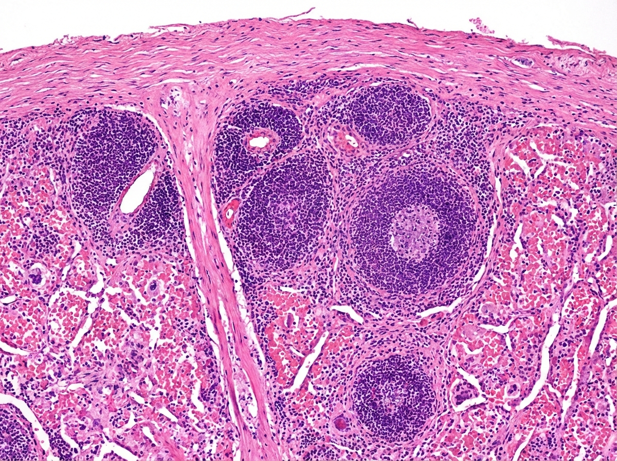
The given histological specimen is of:
Lacis cells are located at?
Which of the following is true about iron deficiency anemia?
Nissl's substance is mainly composed of which of the following?
Microscopic examination of the articular surface of a synovial joint demonstrates?
Which of the following statements concerning Hassall corpuscles is true?
Elastic cartilage is found in which of the following locations?
Practice by Chapter
Basic Tissue Types
Practice Questions
Cell Biology and Organelles
Practice Questions
Epithelial Tissue
Practice Questions
Connective Tissue
Practice Questions
Muscular Tissue
Practice Questions
Nervous Tissue
Practice Questions
Cardiovascular System Histology
Practice Questions
Lymphoid Organs and Immune System
Practice Questions
Endocrine System Histology
Practice Questions
Respiratory System Histology
Practice Questions
Digestive System Histology
Practice Questions
Urinary and Reproductive System Histology
Practice Questions
Want unlimited practice?
Get full access to all questions, explanations, and performance tracking.
Scan to download app