Oil Red O staining is used for which type of specimen preparation?
Ito cells contain which of the following?
What is thrombospondin?
Which one of the following sensory receptors is found in the epidermis?
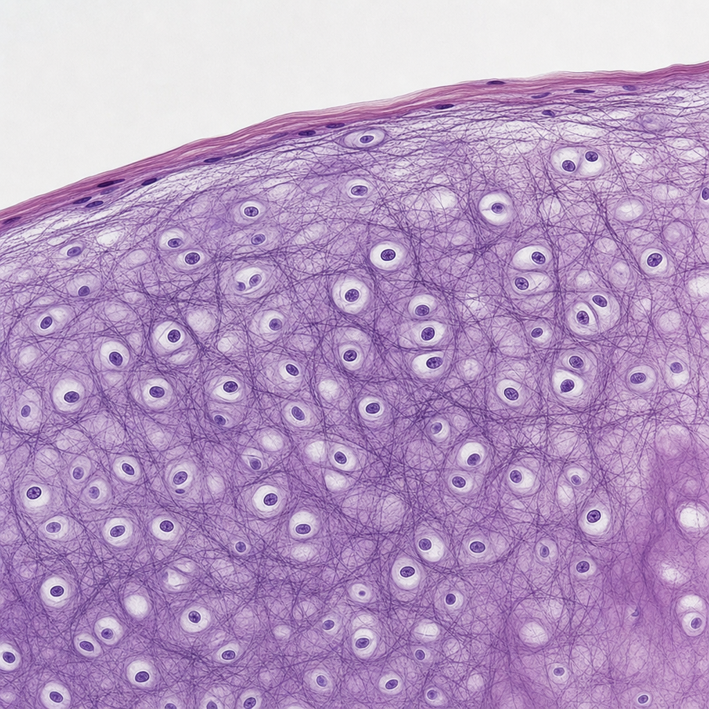
What percentage of testicular cancer is associated with cryptorchidism?
The epithelium lining the cervix mucosa is:
The given histological specimen is of:

Pseudostratified columnar epithelium is present in which of the following structures?
Presence of nephroblastomatosis in a biopsy specimen from Wilm's tumor of the left kidney indicates a high possibility of?
Which of the following is true about iron deficiency anemia?
Explanation: **Explanation:** **1. Why Frozen Section is Correct:** Oil Red O is a lysochrome (fat-soluble) dye used to demonstrate **neutral lipids and triglycerides**. The fundamental principle of lipid staining is that the dye must be more soluble in the lipid than in its solvent. In routine histological processing (paraffin embedding), specimens undergo dehydration with **alcohol** and clearing with **xylene**. These organic solvents dissolve lipids, leaving behind empty vacuoles (as seen in adipocytes). To preserve lipids within the tissue, the specimen must bypass these solvents. **Frozen sections** (using a cryostat) are the gold standard because they maintain the chemical integrity of lipids, allowing the Oil Red O to physically dissolve into the fat droplets, staining them bright red. **2. Why Other Options are Incorrect:** * **Alcohol & Formalin fixed specimens (C & D):** While formalin itself does not dissolve lipids, the subsequent processing steps required for paraffin embedding (dehydration via graded alcohols and clearing agents) remove the lipids entirely. * **Glutaraldehyde fixed specimen (B):** This is primarily used for Electron Microscopy to provide superior protein cross-linking but does not prevent lipid loss during the routine dehydration required for light microscopy. **High-Yield Clinical Pearls for NEET-PG:** * **Sudan Black B:** Another common lipid stain; it is more sensitive for phospholipids and myelin. * **Clinical Application:** Oil Red O is used to diagnose **Fat Embolism Syndrome** (in lung/kidney biopsies) and to identify lipid-rich tumors like liposarcomas or xanthomas. * **Osmium Tetroxide:** The only fixative that also acts as a stain for lipids, turning them black (used in EM). * **Mounting:** Always use **aqueous mounting media** (e.g., glycerin jelly) for lipid stains, as organic mounting media will dissolve the stain.
Explanation: ### Explanation **Ito cells**, also known as **Hepatic Stellate Cells (HSCs)**, are specialized perisinusoidal cells located in the **Space of Disse** (the area between the hepatocytes and the sinusoidal endothelium) in the liver [1]. **Why Vitamin A is correct:** The primary physiological function of Ito cells in a healthy liver is the **storage of Vitamin A (Retinoids)**. They contain characteristic lipid droplets in their cytoplasm where approximately 80% of the body's total Vitamin A is stored in the form of retinyl esters. **Why other options are incorrect:** * **Vitamin C:** This is a water-soluble vitamin primarily absorbed in the distal ileum and utilized as a cofactor for collagen synthesis; it is not stored in specialized hepatic cells. * **Vitamin D:** While the liver performs the first hydroxylation of Vitamin D (converting it to 25-hydroxyvitamin D), the vitamin itself is primarily stored in adipose tissue and skeletal muscle, not specifically within Ito cells. **High-Yield Clinical Pearls for NEET-PG:** * **Fibrosis:** In response to chronic liver injury (e.g., alcohol, viral hepatitis), Ito cells lose their Vitamin A droplets and undergo "activation." They transform into **myofibroblasts**, which secrete excessive Type I and Type III collagen, leading to **liver cirrhosis**. * **Markers:** Activated Ito cells express **alpha-smooth muscle actin (α-SMA)** and Desmin. * **Location:** Always remember they are found in the **Space of Disse** [1]. * **Function:** They also play a role in regulating sinusoidal blood flow and secreting growth factors for hepatocyte regeneration.
Explanation: ### Explanation **Thrombospondin-1 (TSP-1)** is a multi-domain glycoprotein found in the extracellular matrix and stored within the alpha-granules of platelets. In the context of histology and tumor biology, it is primarily recognized as a potent **endogenous inhibitor of angiogenesis**. **1. Why Option D is Correct:** Thrombospondin-1 inhibits angiogenesis through a dual mechanism: * **Direct Effect:** It binds to the **CD36 receptor** on endothelial cells, triggering apoptosis (programmed cell death). * **Indirect Effect:** It binds to and sequesters Vascular Endothelial Growth Factor (**VEGF**), preventing it from interacting with its receptors, thereby halting the formation of new blood vessels. This makes it a key player in the "angiogenic switch" during tumor progression. **2. Why Other Options are Incorrect:** * **Option A & B:** While the name "thrombospondin" suggests a role in thrombosis, it is not a primary coagulation factor (like Fibrinogen or Prothrombin). While it is released during platelet activation and aids in platelet aggregation, its defining physiological role in competitive exams is its anti-angiogenic property. * **Option C:** Contractile proteins refer to Actin and Myosin found in muscle fibers. Thrombospondin is an adhesive glycoprotein of the extracellular matrix, not a contractile element. **3. High-Yield Clinical Pearls for NEET-PG:** * **Tumor Suppressor Link:** The expression of Thrombospondin-1 is positively regulated by the **p53 tumor suppressor gene**. Loss of p53 leads to decreased TSP-1 levels, favoring tumor angiogenesis. * **Alpha-Granules:** Remember that TSP-1 is stored in **alpha-granules** of platelets (along with PDGF, TGF-β, and PF4), not dense granules. * **Wound Healing:** Beyond angiogenesis, it plays a role in cell-to-cell and cell-to-matrix interactions during tissue repair. ### Available References (numbered 1 to 5)
Explanation: ### Explanation The skin is divided into the **epidermis** (outer epithelial layer) and the **dermis** (inner connective tissue layer). Most specialized sensory receptors are located within the dermis; however, the **Merkel disc** is a notable exception. **1. Why Merkel Disc is Correct:** Merkel discs (or Merkel cell-neurite complexes) are located in the **basal layer (stratum basale) of the epidermis** [1]. They consist of a specialized epithelial cell (Merkel cell) in close contact with an expanded nerve terminal. They are **slowly adapting Type I mechanoreceptors** responsible for sensing fine touch, pressure, and texture (e.g., reading Braille). **2. Analysis of Incorrect Options:** * **Meissner’s corpuscles:** These are rapidly adapting mechanoreceptors for fine touch and low-frequency vibration. They are located in the **dermal papillae** (just beneath the epidermis), not within the epidermis itself. * **Ruffini endings:** These are slowly adapting receptors that detect skin stretch and torque. They are located deep within the **dermis**. * **Pacinian corpuscles:** These are large, onion-like structures that detect deep pressure and high-frequency vibration. They are found in the **deep dermis and hypodermis** (subcutaneous tissue). **Clinical Pearls & High-Yield Facts:** * **Free Nerve Endings:** Along with Merkel discs, these are the only other receptors that penetrate the epidermis. * **Merkel Cell Carcinoma:** A rare but highly aggressive neuroendocrine skin cancer derived from these cells. * **Location Density:** Meissner’s and Merkel receptors are most numerous in hairless (glabrous) skin, such as fingertips and lips. * **Mnemonics:** Remember "**M**erkel and **M**eissner are for **M**apping touch," but only **M**erkel stays in the **M**alpighian (basal) layer of the epidermis.
Explanation: Explanation: **Cryptorchidism** (undescended testis) is the most significant risk factor for the development of testicular germ cell tumors [1]. Approximately **10%** of all cases of testicular cancer occur in men with a history of cryptorchidism. 1. **Why 10% is correct:** While the relative risk of developing cancer in an undescended testis is 3 to 4 times higher than in the general population, the absolute prevalence of the condition is low enough that it accounts for only about 1 in 10 (10%) of total testicular cancer cases. The risk is highest for intra-abdominal testes and is significantly reduced if orchiopexy is performed before puberty [1]. 2. **Why other options are incorrect:** * **30%, 50%, and 70%:** These values are overestimates. While cryptorchidism is a major risk factor, the vast majority (90%) of testicular cancers occur in men with normally descended testes. High percentages like 50-70% would imply that cryptorchidism is a prerequisite for the disease, which is clinically inaccurate. **High-Yield Clinical Pearls for NEET-PG:** * **Most Common Tumor:** The most common histological type associated with cryptorchidism is **Seminoma**. * **Contralateral Risk:** In patients with unilateral cryptorchidism, there is also an increased risk of cancer in the **contralateral, normally descended testis** (suggesting a possible underlying dysgenesis). * **Orchiopexy:** Surgical correction (orchiopexy) does not completely eliminate the cancer risk but makes the testis accessible for clinical examination and self-monitoring [1]. * **Location:** The higher the testis is located (e.g., abdominal vs. inguinal), the higher the risk of malignancy.
Explanation: The cervix is divided into two distinct anatomical and histological regions: the **endocervix** and the **ectocervix**. **1. Why Simple Columnar Epithelium is Correct:** The endocervical canal (the inner part of the cervix) is lined by a **simple columnar epithelium**. These cells are tall, mucus-secreting, and form deep invaginations known as cervical glands (crypts). This epithelium is responsible for producing cervical mucus, which changes in consistency during the menstrual cycle under hormonal influence. **2. Analysis of Incorrect Options:** * **Simple Squamous Epithelium (A):** This is found in areas requiring passive diffusion (e.g., alveoli, endothelium) and is not present in the female reproductive tract. * **Stratified Squamous Epithelium (C):** This lines the **ectocervix** (the portion projecting into the vagina) and the vagina itself [1]. It is non-keratinized and provides protection against mechanical stress. * **Ciliated Columnar Epithelium (D):** This is the characteristic lining of the **Fallopian tubes**, where cilia help transport the ovum. While some ciliated cells may be found in the endocervix, the predominant type is secretory simple columnar. **3. High-Yield Clinical Pearls for NEET-PG:** * **Squamocolumnar Junction (SCJ):** The point where the simple columnar epithelium (endocervix) meets the stratified squamous epithelium (ectocervix). * **Transformation Zone:** The area where columnar epithelium undergoes metaplasia into squamous epithelium [2]. This is the **most common site for Cervical Cancer (Squamous Cell Carcinoma)** and is the area sampled during a Pap smear [2]. * **Nabothian Cysts:** Formed when the squamous epithelium overgrows and blocks the orifices of the endocervical columnar glands.
Explanation: ***Spleen*** - The histological specimen shows characteristic **white pulp** containing **Malpighian corpuscles** (lymphoid follicles) and **periarteriolar lymphoid sheaths (PALS)** surrounding central arterioles. - The **red pulp** consists of **cords of Billroth** and **venous sinusoids**, along with a **fibrous capsule** with extending **trabeculae**. *Thymus* - Contains **Hassall's corpuscles** (characteristic concentric epithelial structures) which are absent in this specimen. - Shows distinct **cortical and medullary regions** organized into **lobules** separated by connective tissue septa. *Lymph node* - Features a distinct **subcapsular sinus** and **afferent lymphatics** which are not present in this specimen. - Shows clear **cortex, paracortex, and medulla** organization with **germinal centers** in follicles. *Seminal vesicles* - Lined by **pseudostratified columnar epithelium** with **irregular mucosal folds** creating a honeycomb appearance. - Contains a thick **smooth muscle wall** and secretory epithelium, completely different from lymphoid tissue architecture.
Explanation: **Explanation:** The correct answer is **Vas deferens**. **1. Why Vas deferens is correct:** Pseudostratified columnar epithelium consists of a single layer of cells that appear stratified because their nuclei are located at different levels [1]. All cells touch the basement membrane, but not all reach the luminal surface. In the **Vas deferens** and the **Epididymis**, this epithelium is specifically characterized by the presence of **stereocilia** (long, non-motile microvilli) which aid in the absorption of fluid and the maturation of sperm. **2. Why the other options are incorrect:** * **Esophagus:** Lined by **Non-keratinized stratified squamous epithelium**, which is designed to withstand the mechanical stress and friction of swallowing food. * **Cornea:** The anterior surface of the cornea is lined by **Non-keratinized stratified squamous epithelium**, while the posterior surface (endothelium) is simple squamous. * **Thyroid:** The thyroid follicles are lined by **Simple cuboidal epithelium**. These cells can become columnar when highly active or squamous when inactive. **3. High-Yield Clinical Pearls for NEET-PG:** * **Respiratory Type:** Pseudostratified ciliated columnar epithelium with goblet cells is the hallmark of the **Trachea** and larger bronchi (Respiratory Epithelium) [2]. * **Non-ciliated Type:** Found in the **male urethra** and large ducts of parotid glands. * **Stereociliated Type:** Found exclusively in the **Epididymis** and **Vas deferens**. * **Exam Tip:** If a question mentions "Pseudostratified" and "Male Reproductive System," always look for the Epididymis or Vas deferens. If it mentions "Respiratory System," look for the Trachea.
Explanation: Explanation: Nephroblastomatosis refers to the presence of multiple or diffuse nephrogenic rests (persistent foci of embryonal cells) in the kidney. These rests are considered the precursor lesions of Wilms tumor (nephroblastoma). 1. Why Option C is correct: Nephrogenic rests are found in approximately 35% of unilateral Wilms tumors but are present in nearly 100% of bilateral cases [1]. If a biopsy of a unilateral tumor (e.g., the left kidney) shows nephroblastomatosis, it indicates that the "field" of renal tissue is predisposed to malignant transformation. This significantly increases the risk of developing a metachronous Wilms tumor in the contralateral (right) kidney [1]. 2. Why other options are incorrect: * Option A: While Denys-Drash syndrome is associated with Wilms tumor (due to WT1 mutation), it is specifically characterized by gonadal dysgenesis and early-onset nephropathy (diffuse mesangial sclerosis). Nephroblastomatosis is a histological precursor, not a pathognomonic marker for this specific syndrome. * Option B: Mutations or "imprinting" defects in IGF-2 (specifically at the WT2 locus on chromosome 11p15.5) are associated with Beckwith-Wiedemann Syndrome [1]. While these patients have nephrogenic rests, the presence of the rests themselves is a clinical indicator of bilateral risk rather than a specific test for IGF-2 mutation. * Option D: Nephroblastomatosis is a precursor stage, not a sign of malignancy or spread. Lymph node metastasis is determined by the histological grade (anaplasia) and surgical staging, not by the presence of nephrogenic rests [2]. High-Yield Clinical Pearls for NEET-PG: * Nephrogenic Rests: Two types exist—Perilobar (associated with Beckwith-Wiedemann) and Intralobar (associated with WAGR and Denys-Drash). * WAGR Syndrome: Wilms tumor, Aniridia, Genitourinary anomalies, and intellectual disability (formerly Retardation). * Triphasic Histology: Classic Wilms tumor shows Blastemal, Stromal, and Epithelial components.
Explanation: **Explanation:** Iron Deficiency Anemia (IDA) is the most common cause of microcytic hypochromic anemia worldwide. It occurs when iron stores are depleted, leading to impaired hemoglobin synthesis. **Why Option B is Correct:** **Total Iron Binding Capacity (TIBC)** is a functional measurement of the amount of **Transferrin** (the transport protein for iron) available in the blood. In IDA, the liver increases the production of Transferrin in an attempt to capture more iron for the body. Consequently, as iron levels drop, the "empty" binding sites increase, leading to an **increased TIBC** [1]. **Analysis of Incorrect Options:** * **A. Increased serum ferritin:** Ferritin is the storage form of iron. In IDA, these stores are the first to be depleted; therefore, **decreased serum ferritin** is the most sensitive early marker of IDA [1]. * **C. Increased transferrin saturation:** Transferrin saturation is the ratio of serum iron to TIBC. Since serum iron is low and TIBC is high in IDA, the **saturation decreases** (typically <15%) [1]. * **D. Macrocytic hypochromic anemia:** IDA characteristically produces **Microcytic (low MCV) and Hypochromic (low MCHC)** RBCs [1]. Macrocytic anemia is seen in Vitamin B12 or Folate deficiency. **NEET-PG High-Yield Pearls:** * **Gold Standard Diagnosis:** Bone marrow aspiration (Prussian blue staining) showing absent haemosiderin in erythroblasts. * **Earliest Lab Finding:** Decreased serum ferritin. * **Blood Film:** Microcytic hypochromic cells, anisocytosis (increased RDW), and characteristic **"pencil cells"** (elliptocytes). * **Mentzer Index:** (MCV/RBC count) >13 suggests IDA, while <13 suggests Thalassemia trait.
Basic Tissue Types
Practice Questions
Cell Biology and Organelles
Practice Questions
Epithelial Tissue
Practice Questions
Connective Tissue
Practice Questions
Muscular Tissue
Practice Questions
Nervous Tissue
Practice Questions
Cardiovascular System Histology
Practice Questions
Lymphoid Organs and Immune System
Practice Questions
Endocrine System Histology
Practice Questions
Respiratory System Histology
Practice Questions
Digestive System Histology
Practice Questions
Urinary and Reproductive System Histology
Practice Questions
Get full access to all questions, explanations, and performance tracking.
Start For Free