
Histology — MCQs

On this page
Which type of collagen is maximum in skin?
All of the following are features of Lymph node histology except:
Type I collagen is present in all EXCEPT:
Ligamentum flavum consists of which fibres:
Which cell type is responsible for maintaining bone tissue?
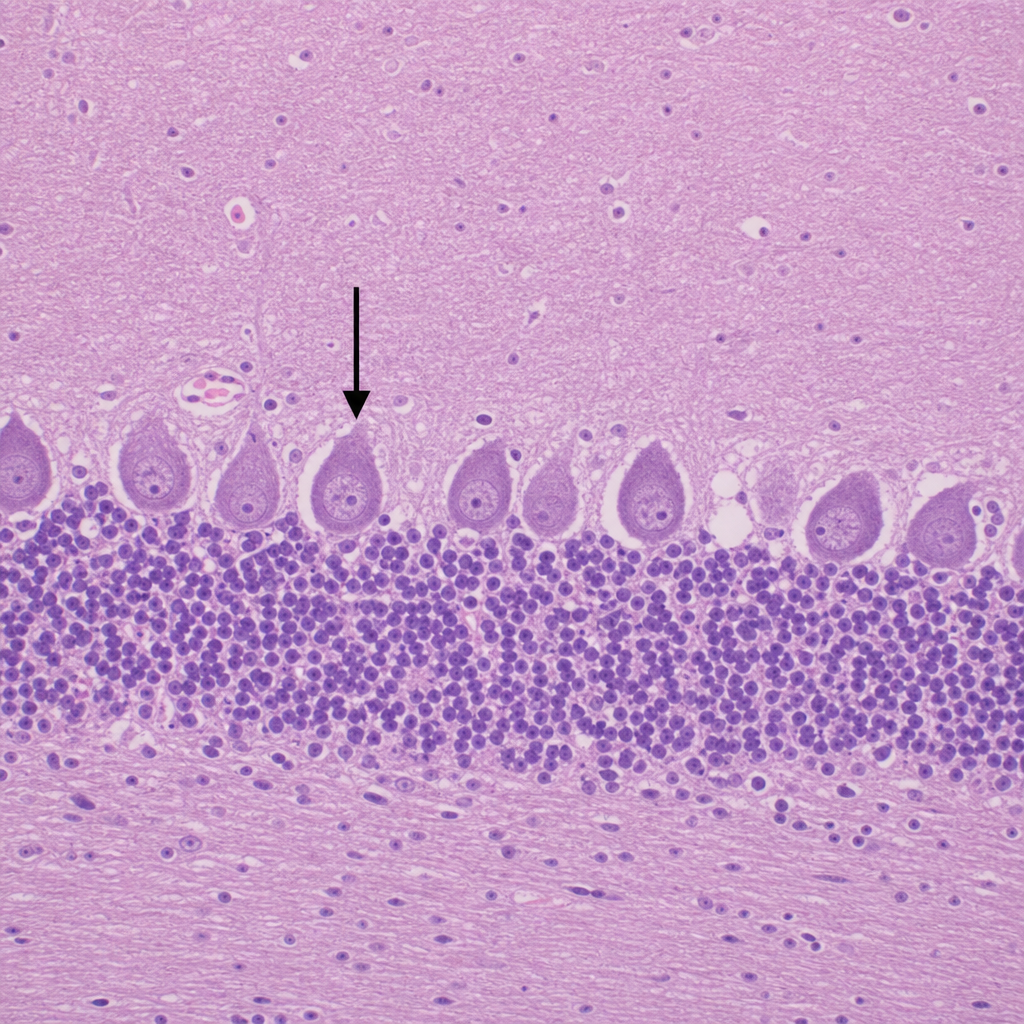
Identify the cell type marked in the cerebellum.
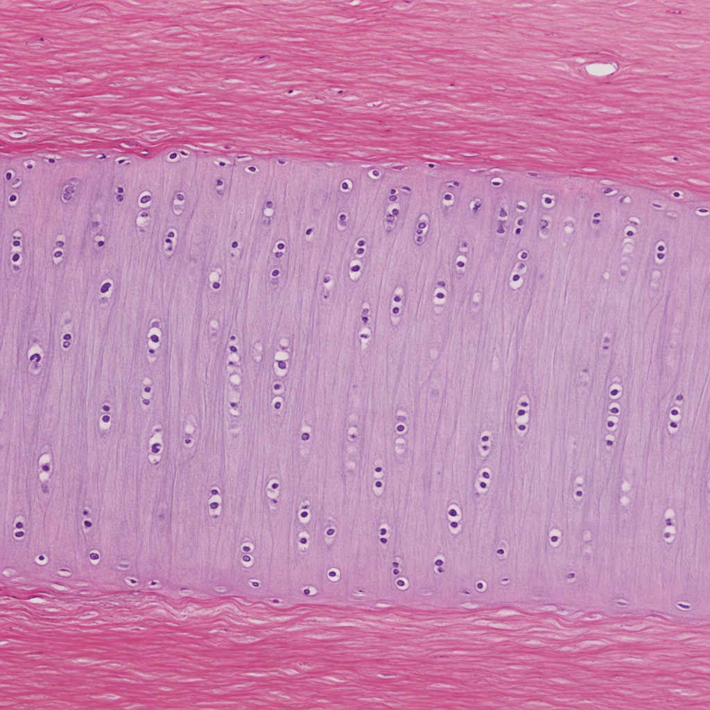
Identify the type of cartilage shown in the image.
Hard palate contains:
What constitutes the Malpighian layer of skin?
Which protein is commonly found in the structure of skin and hair?
Practice by Chapter
Basic Tissue Types
Practice Questions
Cell Biology and Organelles
Practice Questions
Epithelial Tissue
Practice Questions
Connective Tissue
Practice Questions
Muscular Tissue
Practice Questions
Nervous Tissue
Practice Questions
Cardiovascular System Histology
Practice Questions
Lymphoid Organs and Immune System
Practice Questions
Endocrine System Histology
Practice Questions
Respiratory System Histology
Practice Questions
Digestive System Histology
Practice Questions
Urinary and Reproductive System Histology
Practice Questions
Want unlimited practice?
Get full access to all questions, explanations, and performance tracking.
Scan to download app