All of the following are true about type-II pneumocytes, EXCEPT:
Fat is present in relation to which anatomical structure?
Elastic cartilage is found in which of the following locations?
What is the primary treatment modality for extragonadal germ cell tumors?
Myelin is stained by which of the following techniques?
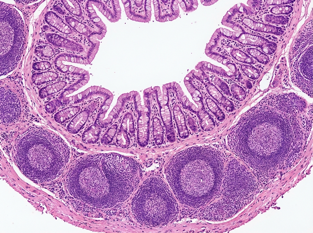
The given histological specimen is:

A peripheral smear demonstrates leukemia composed of small mature lymphocytes without blast forms. What is the most likely age of this patient?
Which of the following types of dendritic cells are characteristically found in the thymus?
Articular cartilage is primarily composed of which type of collagen?
Which cells of the liver play a role in the absorption of Vitamin A and Vitamin D?
Explanation: **Explanation:** The correct answer is **C (No secretory function)** because it is a false statement. Type II pneumocytes are highly active metabolic cells whose primary function is the **secretion of pulmonary surfactant** [1]. **1. Why Option C is the correct answer (False statement):** Type II pneumocytes contain characteristic membrane-bound organelles called **lamellar bodies** [1]. These bodies store and secrete surfactant (a mixture of phospholipids like DPPC and proteins), which reduces surface tension in the alveoli, preventing lung collapse during expiration [2]. **2. Analysis of Incorrect Options (True statements):** * **Option A (Progenitor cells):** Type II pneumocytes act as the "stem cells" of the alveolar epithelium [1]. When Type I cells are damaged (e.g., by toxic gases or inflammation), Type II cells undergo mitosis to replace themselves and differentiate into Type I cells. * **Option B (Septal cells):** They are also known as septal cells or Great Alveolar cells. They are typically found at the alveolar angles (junctions) and occupy about 5% of the alveolar surface area, despite being more numerous than Type I cells. * **Option D (Lamellar bodies):** These are the histological hallmark of Type II pneumocytes [1]. On electron microscopy, they appear as concentric layers of lipid membranes. **High-Yield Clinical Pearls for NEET-PG:** * **Development:** Surfactant production begins around **24–26 weeks** of gestation, but reaches adequate levels only after **35 weeks**. * **Clinical Correlation:** Deficiency of surfactant in premature neonates leads to **Infant Respiratory Distress Syndrome (IRDS)** or Hyaline Membrane Disease [2]. * **Morphology:** Type I cells are squamous (flat) for gas exchange, while Type II cells are **cuboidal** with apical microvilli.
Explanation: The skin is histologically divided into three primary layers: the epidermis, the dermis, and the hypodermis (subcutaneous tissue) [1]. **1. Why Sub-cutaneous tissue is correct:** The **subcutaneous tissue (hypodermis)** is the primary site for fat storage in the integumentary system. It consists of loose connective tissue and **adipose tissue** (organized into lobules by fibrous septa) [2]. Its physiological roles include thermal insulation, mechanical cushioning, and acting as an energy reservoir [2]. **2. Why the other options are incorrect:** * **Epidermis:** This is the outermost, avascular layer composed of stratified squamous keratinized epithelium [1]. It contains keratinocytes, melanocytes, Langerhans cells, and Merkel cells [1], but **no fat**. * **Dermis:** This layer consists of dense irregular connective tissue containing collagen, elastic fibers, blood vessels, nerves, and hair follicles [1]. While it supports the skin, it does not contain adipose tissue layers. * **Nail bed:** This is the specialized epithelial surface (sterile matrix) upon which the nail plate rests. It is highly vascular but lacks a subcutaneous fat layer to ensure the nail plate remains firmly attached to the underlying distal phalanx. **Clinical Pearls & High-Yield Facts:** * **Panniculus Adiposus:** The technical term for the fatty layer of the subcutaneous tissue. * **Injection Site:** The subcutaneous layer is the target for insulin and heparin injections due to its vascularity and predictable absorption rates. * **Skin Ligaments (Retinacula cutis):** These fibrous bands extend from the dermis through the subcutaneous fat to the deep fascia; their length and density determine skin mobility. * **Brown Fat:** In neonates, specialized brown adipose tissue is found in the subcutaneous layer (interscapular region) for non-shivering thermogenesis [2].
Explanation: The fundamental concept in cartilage histology is the composition of the extracellular matrix. **Elastic cartilage** is characterized by a dense network of branching elastic fibers in addition to Type II collagen. This provides the tissue with significant flexibility and the ability to recoil after deformation. The classic locations for elastic cartilage follow the **"3 E’s" rule**: **E**piglottis, **E**xternal ear (pinna/auditory canal), and **E**ustachian tube (also the corniculate and cuneiform cartilages of the larynx). **Analysis of Incorrect Options:** * **A. Articular cartilage:** This is **Hyaline cartilage** [1]. It lacks elastic fibers and is designed to provide a smooth, low-friction surface for joints. It is the most common type of cartilage in the body. * **B. Costal cartilage:** This is also **Hyaline cartilage**. It connects the ribs to the sternum, providing structural support with limited flexibility. * **D. Intervertebral disc:** This is **Fibrocartilage**. It contains thick bundles of **Type I collagen**, making it the strongest type of cartilage, designed to withstand heavy pressure and shearing forces. It is also found in the pubic symphysis and knee menisci. **High-Yield NEET-PG Pearls:** * **Staining:** Elastic cartilage is best visualized using special stains like **Orcein** or **Verhoeff-Van Gieson (VVG)**, which turn the elastic fibers black/purple. * **Perichondrium:** Both Hyaline (except articular) and Elastic cartilage possess a perichondrium. **Fibrocartilage lacks a perichondrium.** * **Calcification:** Unlike hyaline cartilage, elastic cartilage **does not calcify** with age.
Explanation: **Explanation:** Extragonadal Germ Cell Tumors (EGGCTs) are rare neoplasms that arise from primordial germ cells that failed to migrate to the gonadal ridges during embryogenesis. They are typically found in midline structures such as the mediastinum, retroperitoneum, and pineal gland. **Why Chemotherapy is Correct:** The primary treatment modality for EGGCTs is **platinum-based chemotherapy** (typically the BEP regimen: Bleomycin, Etoposide, and Cisplatin) [1]. Germ cell tumors are highly chemosensitive. Because EGGCTs are often bulky, located in surgically challenging midline areas, and have a high propensity for systemic micrometastasis, systemic chemotherapy is required as the first-line treatment to downstage the tumor and address potential spread [1]. **Why Other Options are Incorrect:** * **Radiotherapy:** While some dysgerminomas/seminomas are radiosensitive, radiation is generally reserved for specific sites (like intracranial germinomas) or palliative care [1]. It is not the primary modality for systemic or bulky EGGCTs. * **Surgery:** In EGGCTs, surgery is rarely the primary treatment. It is usually performed **post-chemotherapy** to resect residual masses (especially in non-seminomatous types) to check for viable tumor or teratoma [1]. * **Immunotherapy:** This is currently not a standard first-line treatment for germ cell tumors, though it is being investigated for refractory cases. **High-Yield Clinical Pearls for NEET-PG:** * **Most common site:** The mediastinum is the most common extragonadal site in adults [1]. * **Tumor Markers:** Always check **AFP** (elevated in Yolk Sac Tumors) and **β-hCG** (elevated in Choriocarcinomas). * **Association:** Mediastinal non-seminomatous GCTs are associated with hematologic malignancies (e.g., Acute Myeloid Leukemia). * **Klinefelter Syndrome (47, XXY):** There is a strong clinical association between Klinefelter syndrome and the development of mediastinal EGGCTs.
Explanation: **Explanation:** **1. Why Bielschowsky stain is correct:** The **Bielschowsky stain** is a silver impregnation technique used primarily to visualize nerve fibers, neurofibrils, and **myelin** in the central nervous system [3]. It utilizes silver nitrate to highlight axons and is particularly useful in identifying pathological changes like senile plaques and neurofibrillary tangles in Alzheimer’s disease. While Luxol Fast Blue is the most specific stain for myelin [2], silver stains like Bielschowsky and Bodian are high-yield alternatives frequently tested in histology. **2. Analysis of Incorrect Options:** * **A. Warthin-Starry stain:** This is a silver nitrate-based stain used specifically for detecting **Spirochetes** (e.g., *Treponema pallidum*), *H. pylori*, and *Bartonella henselae*. * **B. Von Kossa stain:** This is the gold standard for demonstrating **Calcium** deposits (mineralization) in tissue sections. It works by transforming calcium salts into silver salts. * **C. Romanowsky stain:** This is a group of stains (including Leishman, Giemsa, and Wright stains) used primarily for **Peripheral Blood Smears** and bone marrow aspirates to differentiate blood cells. **3. Clinical Pearls & High-Yield Facts for NEET-PG:** * **Specific Myelin Stain:** **Luxol Fast Blue (LFB)** is the most specific stain for myelin [2]. * **Peripheral Nerve Myelin:** Formed by Schwann cells; **CNS Myelin:** Formed by Oligodendrocytes [1]. * **Demyelinating Disease:** Multiple Sclerosis (CNS) and Guillain-Barré Syndrome (PNS) [4]. * **Other Silver Stains:** * *Gomori’s Methenamine Silver (GMS):* Fungi and *Pneumocystis jirovecii*. * *Masson’s Fontana:* Melanin and Argentaffin cells.
Explanation: ***Vermiform appendix*** - Contains distinctive **lymphoid follicles** in the submucosa and **simple columnar epithelium** with **crypts of Lieberkühn**. - Shows a **narrow lumen** surrounded by abundant **lymphatic tissue**, which is characteristic of the appendix. *Urethra* - Lined with **transitional epithelium** (urothelium) that changes from stratified to pseudostratified columnar. - Lacks the prominent **lymphoid follicles** and **crypts** seen in appendicular tissue. *Vas deferens* - Features a thick **three-layered smooth muscle wall** and **pseudostratified columnar epithelium** with **stereocilia**. - Has a **star-shaped lumen** and lacks the extensive **lymphoid tissue** of the appendix. *Stenson's duct* - Shows **stratified columnar epithelium** and is associated with **parotid salivary acini**. - Lacks **lymphoid follicles** and **intestinal-type crypts** characteristic of the appendix.
Explanation: ### Explanation The peripheral smear description of **small mature lymphocytes without blast forms** is the classic hematological hallmark of **Chronic Lymphocytic Leukemia (CLL)**. **1. Why 65 years is correct:** CLL is primarily a disease of the elderly, with a median age at diagnosis of approximately 70 years. It is characterized by the clonal proliferation of morphologically mature but immunologically incompetent B-lymphocytes. On a peripheral smear, these cells appear as small, round lymphocytes with scant cytoplasm and "block-like" chromatin. A key diagnostic feature often seen alongside these cells is **Smudge cells** (Gumprecht shadows), which are fragile lymphocytes ruptured during slide preparation. **2. Why other options are incorrect:** * **1 year (Option A):** This age group is most commonly associated with **Acute Lymphoblastic Leukemia (ALL)**. ALL presents with "blasts" (large cells with high N:C ratio and nucleoli), not mature lymphocytes. * **20 years (Option B):** While lymphomas can occur in young adults, a leukemia of mature lymphocytes is extremely rare in this age group. * **45 years (Option C):** This age is more characteristic of **Chronic Myeloid Leukemia (CML)**, which would show a full spectrum of myeloid cells (myelocytes, metamyelocytes) rather than mature lymphocytes. **3. High-Yield Clinical Pearls for NEET-PG:** * **Most common leukemia:** CLL is the most common leukemia in adults in Western countries. * **Immunophenotype:** CLL cells characteristically express **CD5** (a T-cell marker) along with B-cell markers **CD19, CD20, and CD23**. * **Richter Transformation:** In about 5-10% of cases, CLL can transform into a high-grade Diffuse Large B-cell Lymphoma (DLBCL). * **Pathognomonic sign:** Look for the mention of "Smudge cells" or "Basket cells" in the question stem to confirm CLL.
Explanation: The **interdigitating dendritic cell (IDC)** is the characteristic dendritic cell of the **thymus**, primarily located in the **medulla** and at the corticomedullary junction. These cells are highly specialized Antigen Presenting Cells (APCs) that express high levels of MHC Class I and II molecules. Their primary role in the thymus is **Negative Selection**: they present self-antigens to developing T-cells; those T-cells that bind too strongly to self-antigens undergo apoptosis, ensuring central self-tolerance and preventing autoimmunity. **Analysis of Incorrect Options:** * **A. Interstitial dendritic cells:** These are found in the connective tissue of most organs (e.g., heart, lungs, liver) but are not the characteristic functional cells of the thymic parenchyma. * **C. Circulating dendritic cells:** These are immature dendritic cells (veiled cells) found in the blood or lymph, currently in transit to peripheral tissues or secondary lymphoid organs. * **D. Follicular dendritic cells (FDCs):** These are found in the **germinal centers of B-cell follicles** in the lymph nodes and spleen. They trap antigen-antibody complexes to present to B-cells. Since the thymus is a primary lymphoid organ for T-cell maturation and lacks B-cell follicles, FDCs are not typically found there. **High-Yield Facts for NEET-PG:** * **Hassall’s Corpuscles:** These are characteristic acidophilic structures in the thymic medulla formed by Type VI epithelioreticular cells. * **Blood-Thymus Barrier:** Found only in the **cortex**, preventing premature exposure of developing T-cells to blood-borne antigens. * **DiGeorge Syndrome:** Failure of the 3rd and 4th pharyngeal pouches to develop, leading to thymic aplasia and T-cell deficiency [1].
Explanation: Articular cartilage is a specialized form of **hyaline cartilage** that covers the weight-bearing surfaces of joints [1]. Its primary structural framework consists of **Type II collagen** fibers (comprising about 90-95% of the total collagen content) [1]. These fibers form a dense network that provides tensile strength and anchors the proteoglycan matrix, allowing the cartilage to withstand shear forces and pressure during joint movement [1]. **Analysis of Options:** * **Type I Collagen (Option A):** Found in "tough" tissues like **bone, skin, tendons, and fibrocartilage** (e.g., intervertebral discs, pubic symphysis). It is designed to resist tension rather than compression. * **Type III Collagen (Option B):** Also known as **reticular fibers**. It forms a supportive meshwork in soft organs like the liver, spleen, and lymph nodes, and is prominent during the early stages of wound healing (granulation tissue). * **Type IV Collagen (Option D):** This type does not form fibrils; instead, it forms a two-dimensional mesh that is a critical structural component of the **basal lamina** (basement membrane). **High-Yield Clinical Pearls for NEET-PG:** 1. **Mnemonic for Collagen:** * Type **I**: **B**one (and Skin/Tendon) * Type **II**: **C**artilage (Hyaline and Elastic) * Type **III**: **R**eticular fibers (Blood vessels/Spleen) * Type **IV**: Under the **F**loor (Basement membrane) 2. **Osteoarthritis:** Characterized by the progressive degradation of Type II collagen and proteoglycans in articular cartilage. 3. **Cartilage Types:** While hyaline and elastic cartilage contain Type II, **fibrocartilage** is unique because it contains a significant amount of **Type I collagen** to handle heavy tensile loads.
Explanation: **Explanation:** The correct answer is **Stellate cells of Ito** (also known as Hepatic Stellate Cells or Lipocytes) [1]. **1. Why Stellate cells of Ito is correct:** These cells are located in the **Space of Disse** (the perisinusoidal space between hepatocytes and sinusoidal endothelium) [1]. Their primary physiological function is the **storage of exogenous Vitamin A** (retinoids) and other fat-soluble vitamins like **Vitamin D** in the form of cytoplasmic lipid droplets. In a healthy liver, they store approximately 80% of the body's total Vitamin A. **2. Why other options are incorrect:** * **Kupffer cells:** These are specialized **macrophages** located within the liver sinusoids [1]. Their primary role is phagocytosis of pathogens, cell debris, and old red blood cells; they do not store vitamins. * **Stem cells:** In the liver, these are often referred to as "Oval cells" (located in the Canals of Hering). They function in regeneration and differentiation into hepatocytes or cholangiocytes during severe injury, not in vitamin metabolism. * **Sinusoids:** These are low-pressure vascular channels (capillaries) that allow for the exchange of substances between blood and hepatocytes [2]. They are a structural space, not a cell type responsible for vitamin storage. **3. Clinical Pearls for NEET-PG:** * **Fibrosis:** In chronic liver injury (e.g., cirrhosis), Ito cells lose their vitamin-storing capacity and transform into **myofibroblasts**, which secrete excess **Type I collagen**, leading to liver fibrosis [1]. * **Marker:** Desmin is a common histological marker for these cells. * **Space of Disse:** It is the site where nutrient exchange occurs and where lymph formation begins [2].
Basic Tissue Types
Practice Questions
Cell Biology and Organelles
Practice Questions
Epithelial Tissue
Practice Questions
Connective Tissue
Practice Questions
Muscular Tissue
Practice Questions
Nervous Tissue
Practice Questions
Cardiovascular System Histology
Practice Questions
Lymphoid Organs and Immune System
Practice Questions
Endocrine System Histology
Practice Questions
Respiratory System Histology
Practice Questions
Digestive System Histology
Practice Questions
Urinary and Reproductive System Histology
Practice Questions
Get full access to all questions, explanations, and performance tracking.
Start For Free