
Histology — MCQs

On this page
Barr body was first detected in:
Cells as they approach towards stratum corneum show the following features
Which of the following site doesn't contain brown adipose tissues?
Which of the following statements is TRUE about osteoblasts and chondroblasts?
Space of Disse is seen in
Stem cells in skin are found in all, EXCEPT:
In articular cartilage, most active chondrocytes are seen in ?
At which of the following sites do osteoclasts remove bone?
Desmosomes are helpful in connecting -
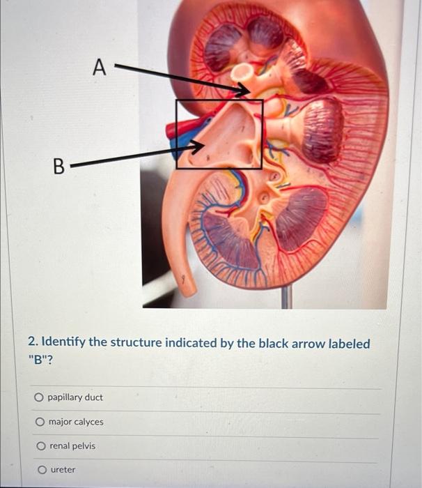
Name the structure marked with an arrow
Practice by Chapter
Basic Tissue Types
Practice Questions
Cell Biology and Organelles
Practice Questions
Epithelial Tissue
Practice Questions
Connective Tissue
Practice Questions
Muscular Tissue
Practice Questions
Nervous Tissue
Practice Questions
Cardiovascular System Histology
Practice Questions
Lymphoid Organs and Immune System
Practice Questions
Endocrine System Histology
Practice Questions
Respiratory System Histology
Practice Questions
Digestive System Histology
Practice Questions
Urinary and Reproductive System Histology
Practice Questions
Want unlimited practice?
Get full access to all questions, explanations, and performance tracking.
Scan to download app