
Histology — MCQs

On this page
Merkel cells are found in which of the following tissues?
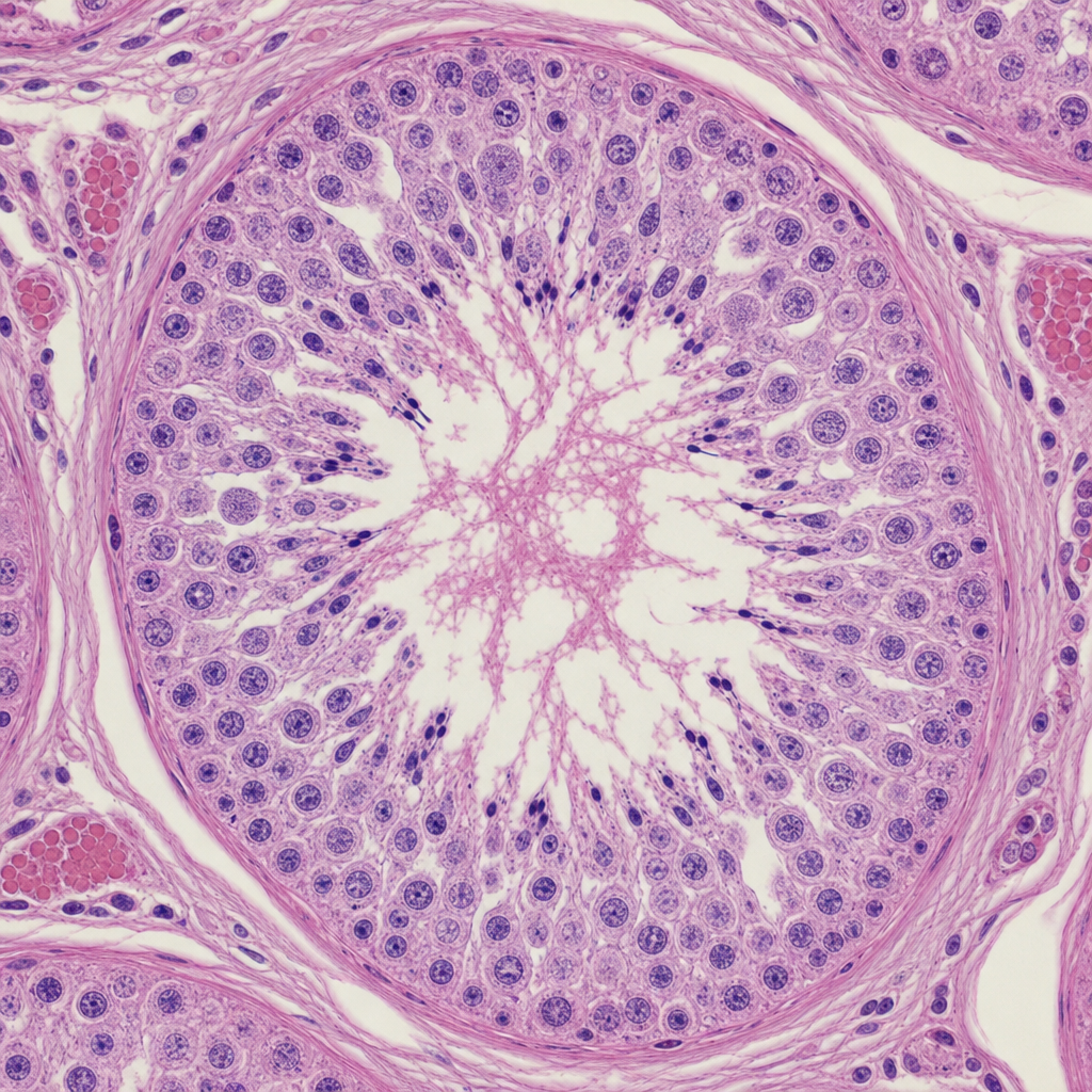
The slide of the testis is shown below. The supporting cells of the testis are:
The main type of collagen in anchoring fibrils (component of dermoepidermal junction) is:
Periosteal matrix acts on bone tissue to bring about:
Langerhans cells in skin fall under which category?
In which of the following structures is regularly arranged parallel collagen present?
Submucosal plexus is -
Inorganic component of bone is:
The bone matrix has the following crystals -
The Eustachian tube is made up of
Practice by Chapter
Basic Tissue Types
Practice Questions
Cell Biology and Organelles
Practice Questions
Epithelial Tissue
Practice Questions
Connective Tissue
Practice Questions
Muscular Tissue
Practice Questions
Nervous Tissue
Practice Questions
Cardiovascular System Histology
Practice Questions
Lymphoid Organs and Immune System
Practice Questions
Endocrine System Histology
Practice Questions
Respiratory System Histology
Practice Questions
Digestive System Histology
Practice Questions
Urinary and Reproductive System Histology
Practice Questions
Want unlimited practice?
Get full access to all questions, explanations, and performance tracking.
Scan to download app