
Histology — MCQs

On this page
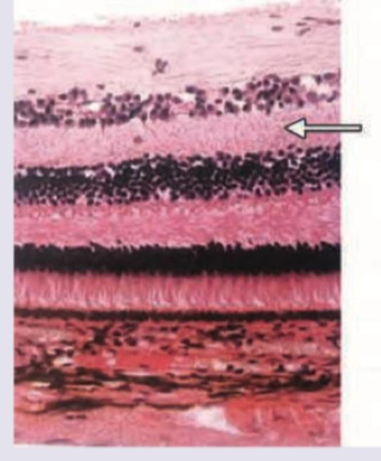
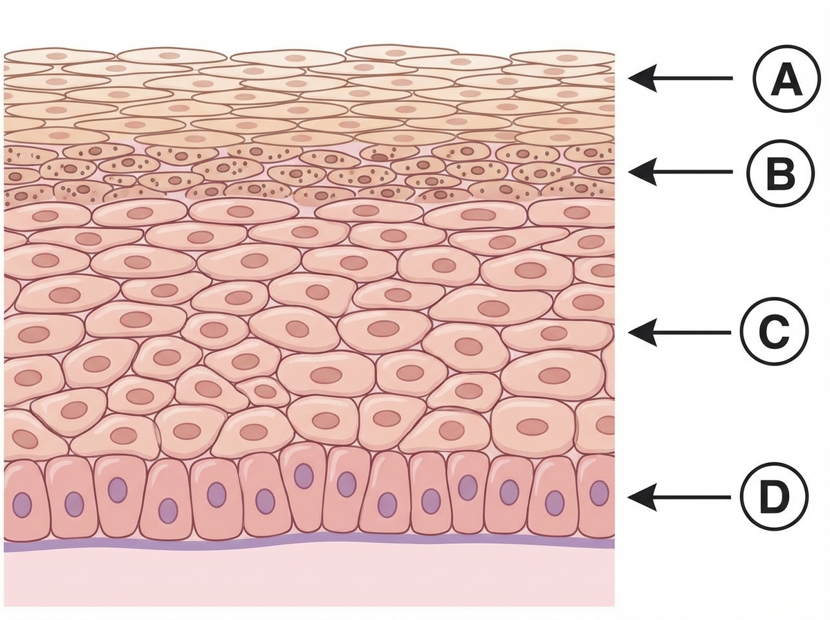
Identify the marked layer in the given histological section.
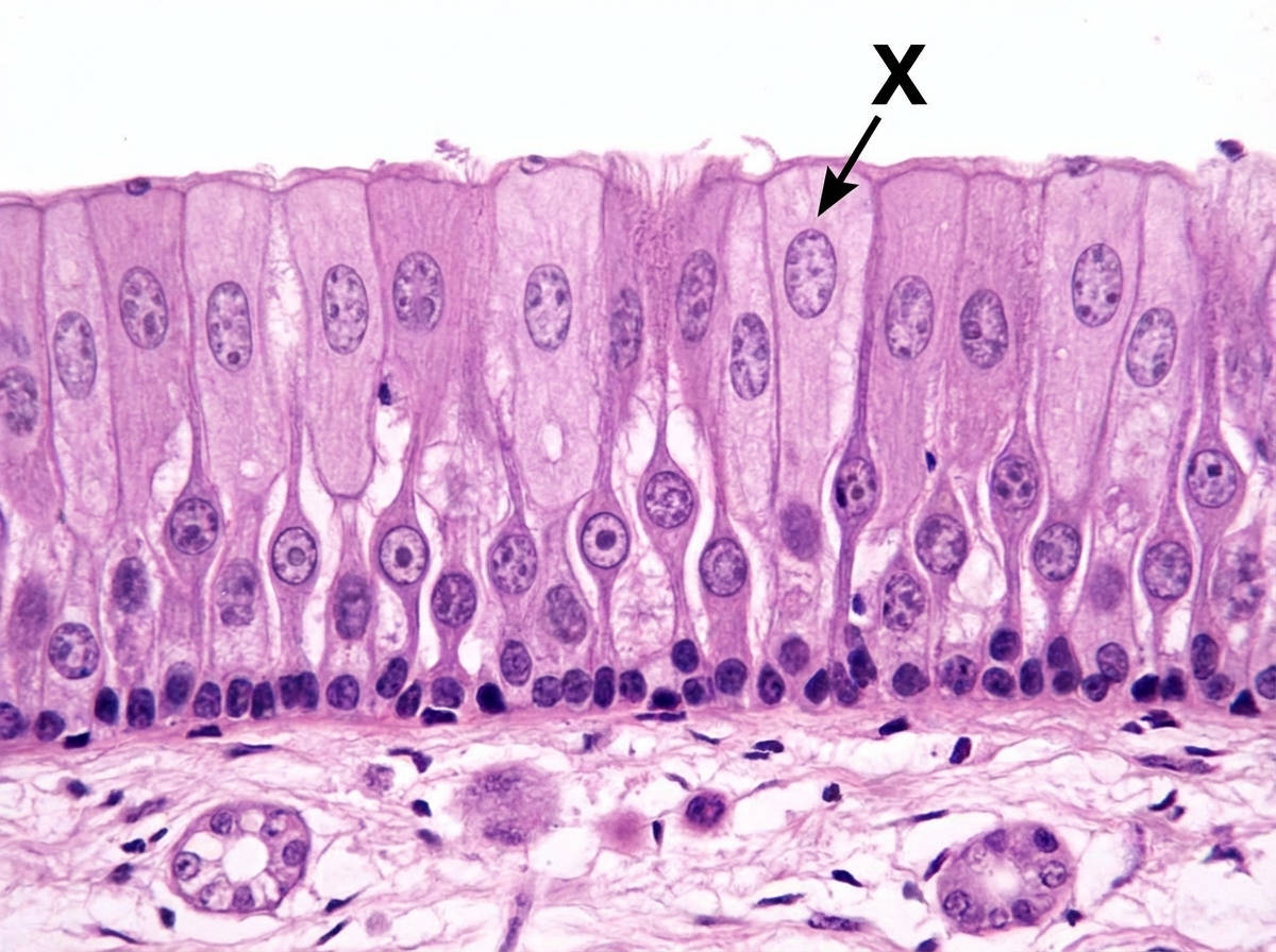
The cells marked as $X$ are called:
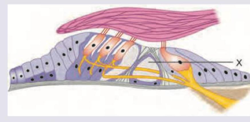
The area marked as $X$ is called:
Which of the following layers has abundance of desmosomes? (AIIMS Nov 2017)
Which of the following layers contains Odland bodies?
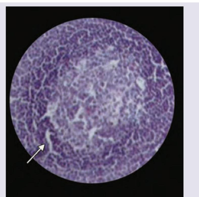
Which of the following area is marked in the histology of lymph node? (AIIMS May 2017)
Which one of the following is the myelinating cell of Central Nervous System?
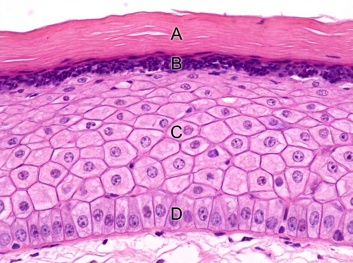
Identify the structures labeled A, B, C, and D in the given histological section of skin
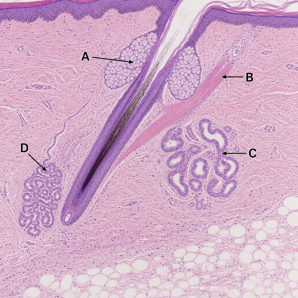
Identify which of the following structure is a sebaceous gland:
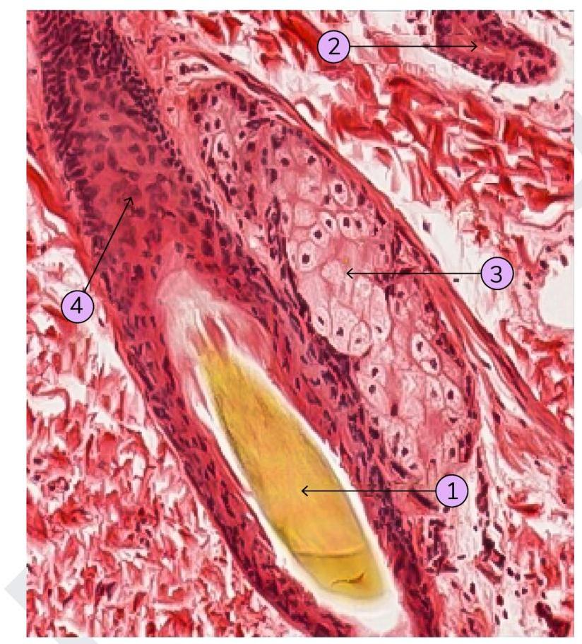
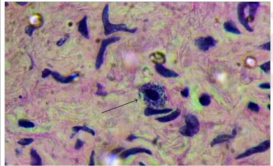
Identify the cell marked in the image below
Practice by Chapter
Basic Tissue Types
Practice Questions
Cell Biology and Organelles
Practice Questions
Epithelial Tissue
Practice Questions
Connective Tissue
Practice Questions
Muscular Tissue
Practice Questions
Nervous Tissue
Practice Questions
Cardiovascular System Histology
Practice Questions
Lymphoid Organs and Immune System
Practice Questions
Endocrine System Histology
Practice Questions
Respiratory System Histology
Practice Questions
Digestive System Histology
Practice Questions
Urinary and Reproductive System Histology
Practice Questions
Want unlimited practice?
Get full access to all questions, explanations, and performance tracking.
Scan to download app