
Histology — MCQs

On this page
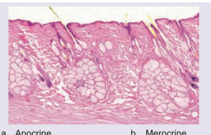
Which type of gland is depicted here?
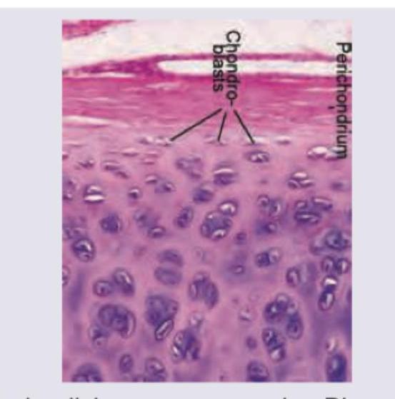
The lower two thirds of the following hematoxylin and eosin stained specimen is similar in appearance to which of the following structures?
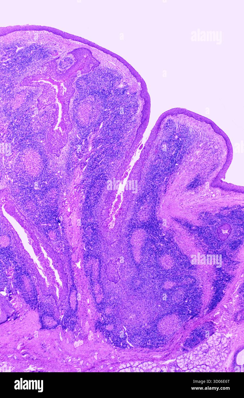
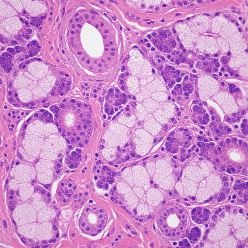
The hematoxylin and eosin stained biopsy shown below is taken from which tissue?
Which of the cells labelled below secrete hydrochloric acid?
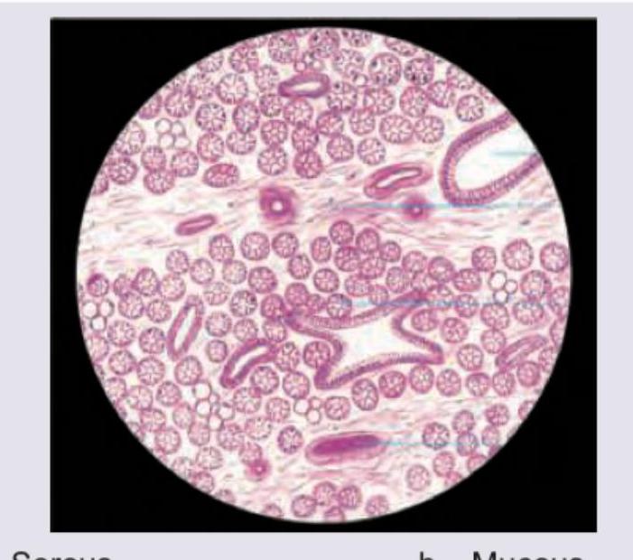
Which type of salivary glands is shown in the image?
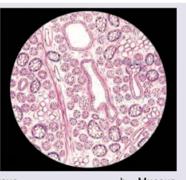
Which type of salivary gland is shown in the image?
Which type of salivary glands is shown in the image?
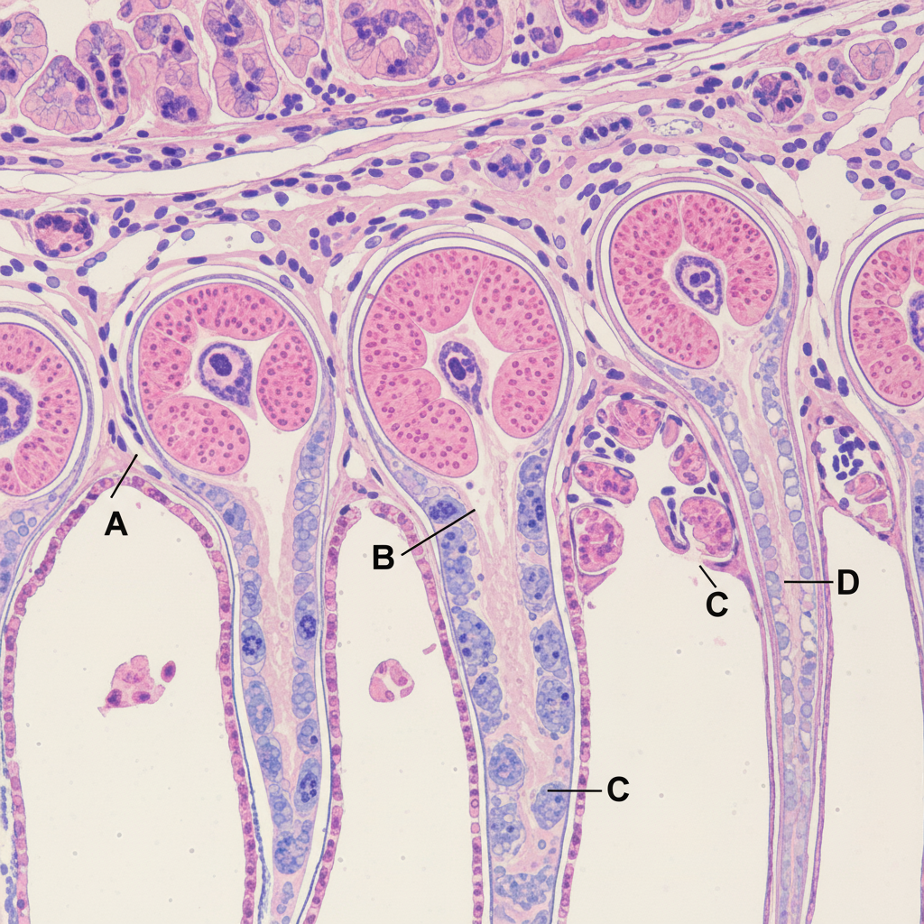
Identify the structure:
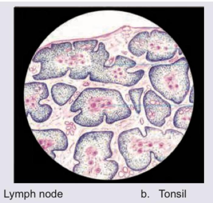
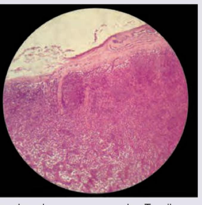
Identify the tissue shown in the histopathological image:
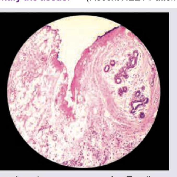
Identify the tissue
Practice by Chapter
Basic Tissue Types
Practice Questions
Cell Biology and Organelles
Practice Questions
Epithelial Tissue
Practice Questions
Connective Tissue
Practice Questions
Muscular Tissue
Practice Questions
Nervous Tissue
Practice Questions
Cardiovascular System Histology
Practice Questions
Lymphoid Organs and Immune System
Practice Questions
Endocrine System Histology
Practice Questions
Respiratory System Histology
Practice Questions
Digestive System Histology
Practice Questions
Urinary and Reproductive System Histology
Practice Questions
Want unlimited practice?
Get full access to all questions, explanations, and performance tracking.
Scan to download app