
Histology — MCQs

On this page
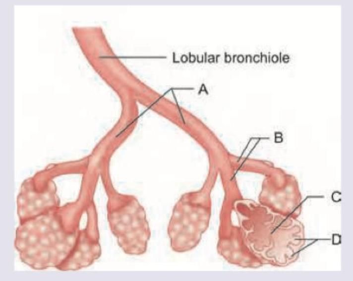
Which of the following has maximum smooth muscle as compared to wall thickness?
Which is correct about structures marked as "X" found in smooth muscle?

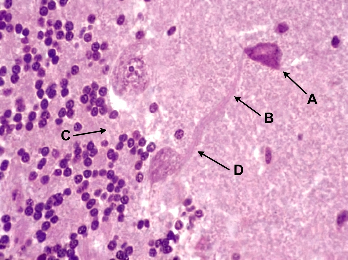
The image shows histological section of cerebellum. Identify the structures labeled in the image:
Identify the cell in the given histological image which does not migrate from the base of the crypt to ends of villi: (Recent NEET Pattern 2016-17)
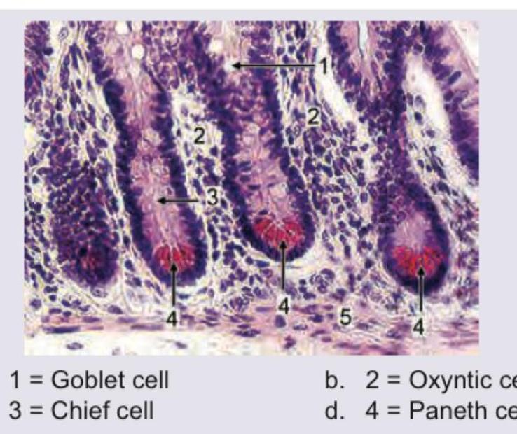
Which of the following is Paneth cell?
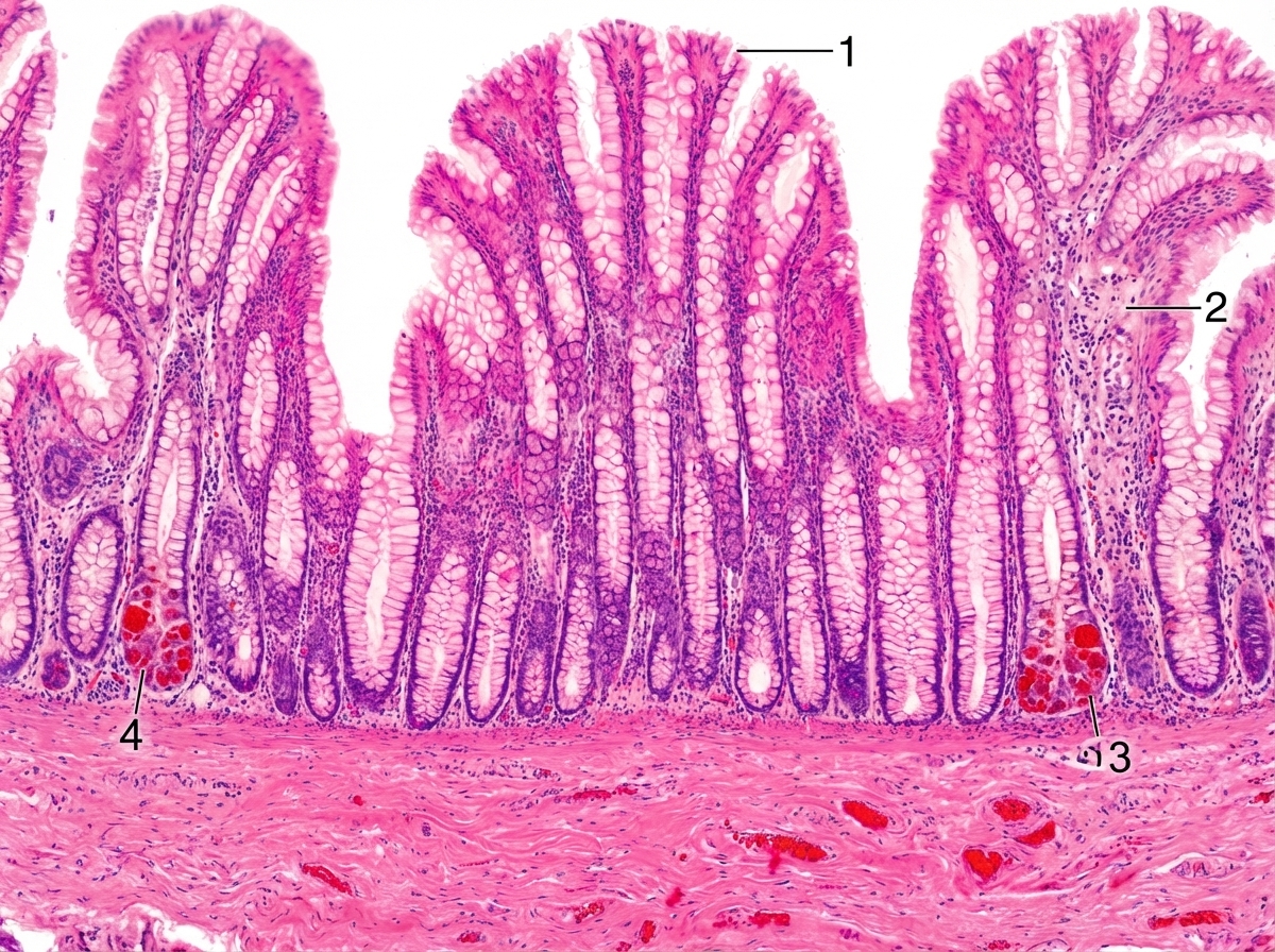
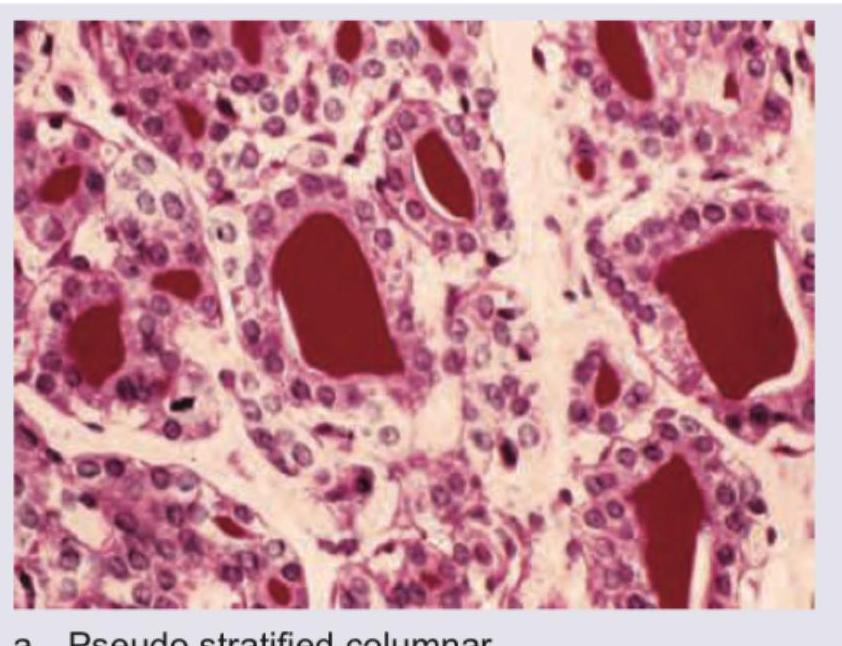
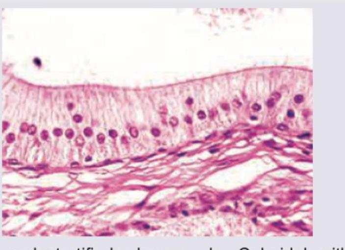
Name the epithelium shown in the picture:
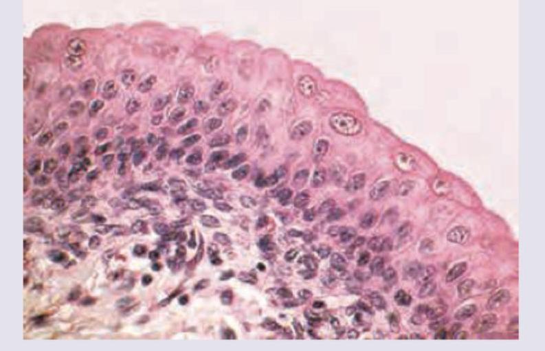
Identify the epithelium shown in the picture.
Name the epithelium, and the area where it is found:
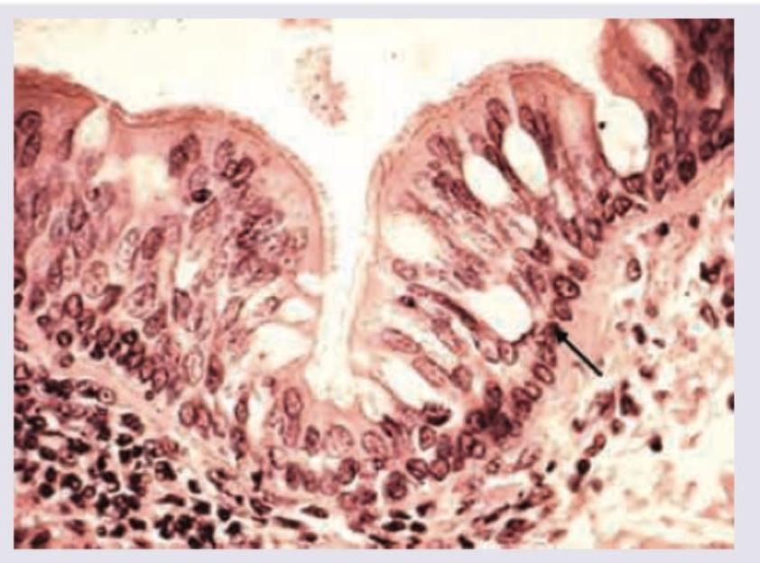
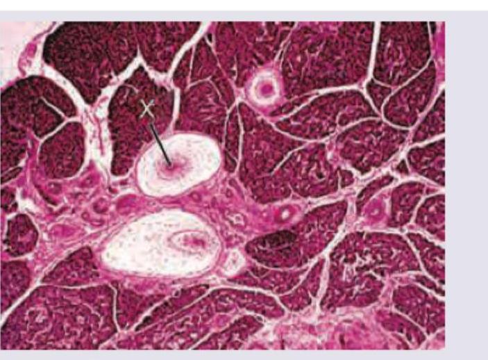
Name the structure marked as $X$ :
Identify the type of epithelium shown in the image:
Practice by Chapter
Basic Tissue Types
Practice Questions
Cell Biology and Organelles
Practice Questions
Epithelial Tissue
Practice Questions
Connective Tissue
Practice Questions
Muscular Tissue
Practice Questions
Nervous Tissue
Practice Questions
Cardiovascular System Histology
Practice Questions
Lymphoid Organs and Immune System
Practice Questions
Endocrine System Histology
Practice Questions
Respiratory System Histology
Practice Questions
Digestive System Histology
Practice Questions
Urinary and Reproductive System Histology
Practice Questions
Want unlimited practice?
Get full access to all questions, explanations, and performance tracking.
Scan to download app