
Histology — MCQs

On this page
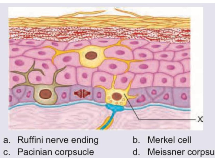
Identify the somato-receptor marked as $X$.
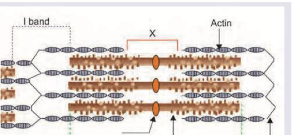
In the sarcomere diagram shown below, what do the marked areas X and Y represent?
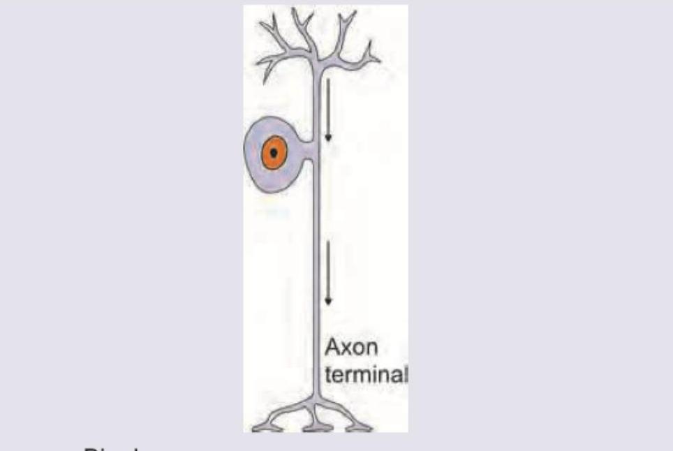
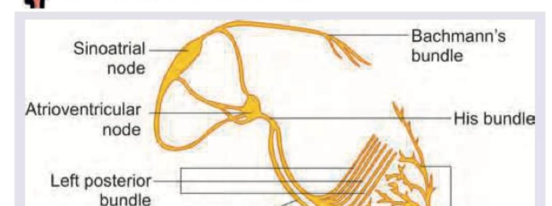
Which of the following is correct about the type of neuron shown below?
Which of the following is correct about the image shown below?
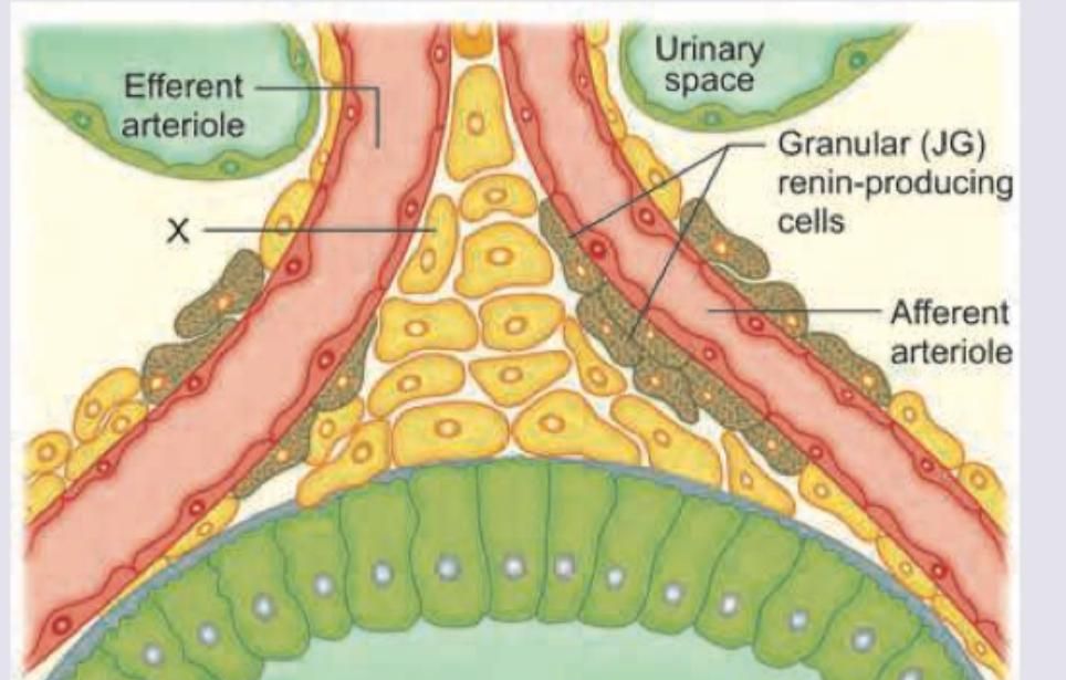
Identify the cells marked X in the image shown below.
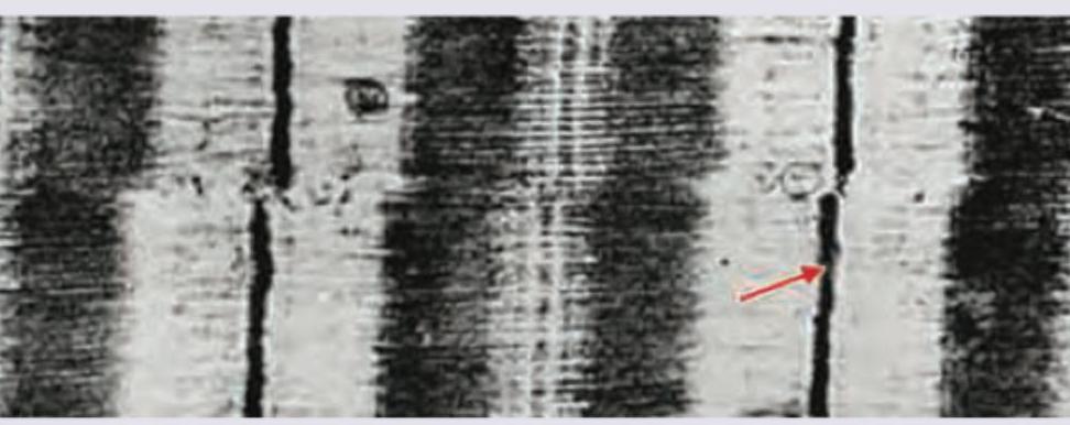
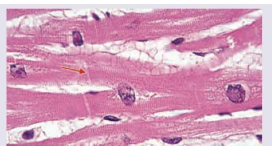
The fiber marked as X is:
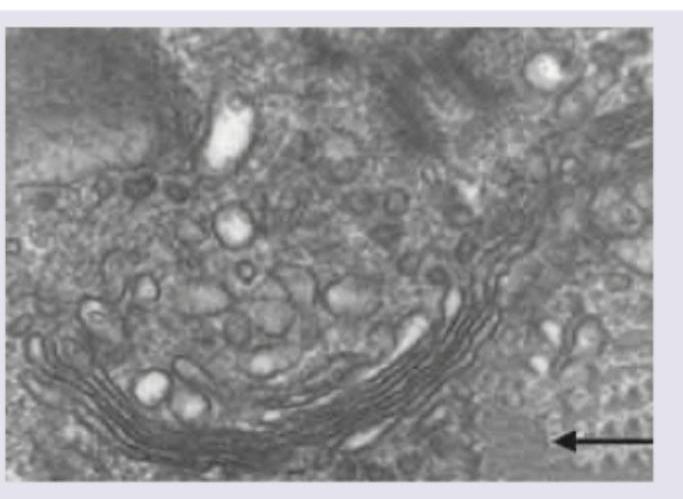
In this electron micrograph, identify the structure marked with arrow.
Which of the following is the constituent of the marked area in the given electron microscope picture of the muscle?
What is incorrect about the shown basement membrane?
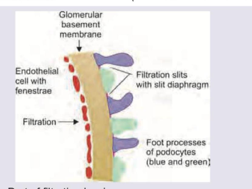
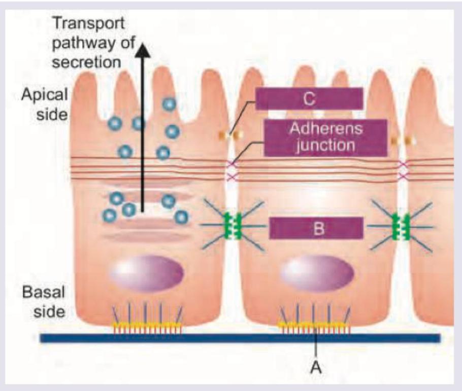
Which of the following proteins are not seen in the region marked in the image?
Practice by Chapter
Basic Tissue Types
Practice Questions
Cell Biology and Organelles
Practice Questions
Epithelial Tissue
Practice Questions
Connective Tissue
Practice Questions
Muscular Tissue
Practice Questions
Nervous Tissue
Practice Questions
Cardiovascular System Histology
Practice Questions
Lymphoid Organs and Immune System
Practice Questions
Endocrine System Histology
Practice Questions
Respiratory System Histology
Practice Questions
Digestive System Histology
Practice Questions
Urinary and Reproductive System Histology
Practice Questions
Want unlimited practice?
Get full access to all questions, explanations, and performance tracking.
Scan to download app