
Histology — MCQs

On this page
What is the type of cartilage present in the histology section given below?
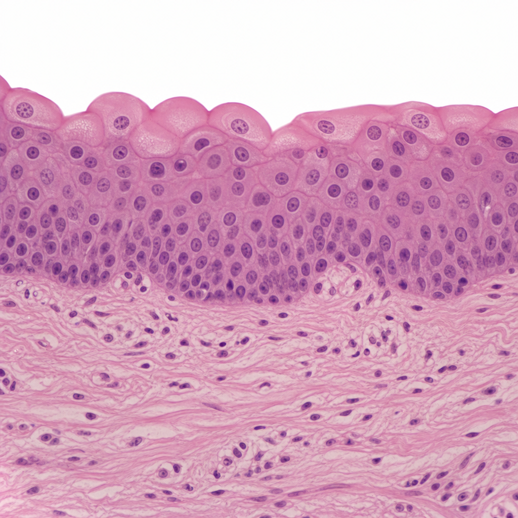
Identify the given histology slide:
Identify the histological image below.
Which of the following skin layers contains melanocytes responsible for skin pigmentation?
Tight junctions are primarily located at which part of the cell?
Identify the structure labeled in the histology image shown.
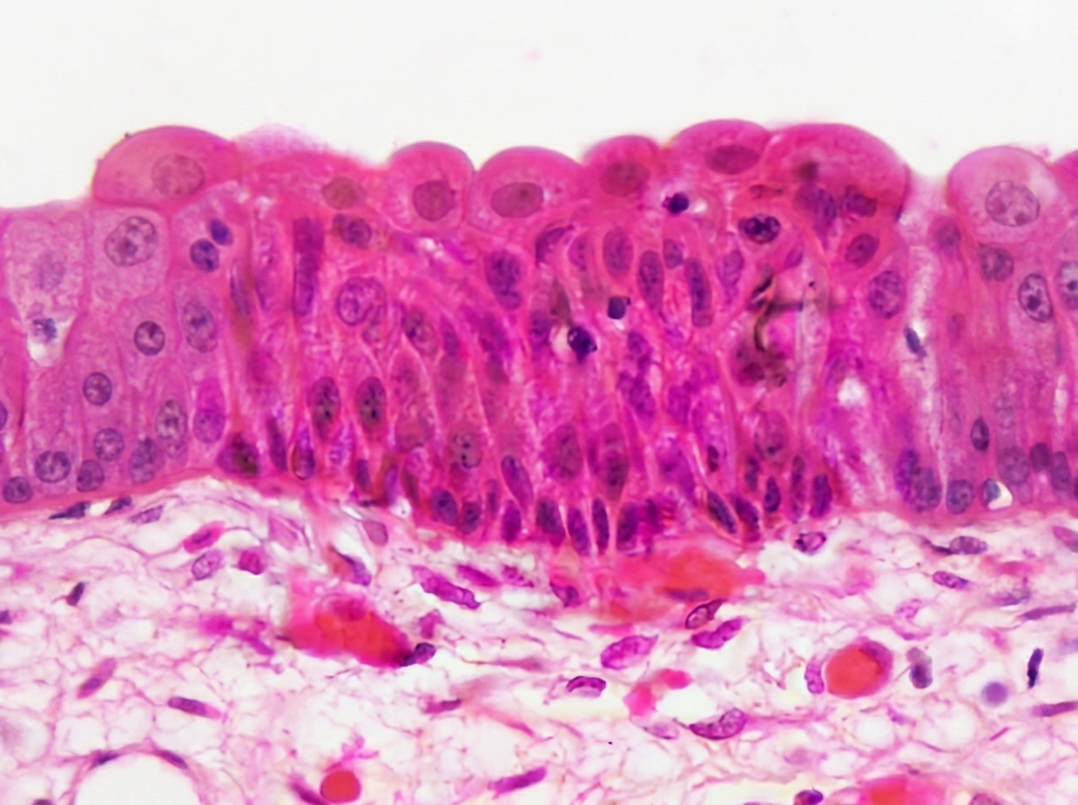
This type of epithelium is most likely found lining which of the following structures?
Which of the following organs does this epithelium most likely belong to?
Out of the following, which statements are correct? 1. Epidermis can regenerate from hair bulbs and sebaceous glands 2. Stratum corneum is the outermost cellular layer 3. Epidermis originates from the ectoderm 4. Dermis and hypodermis originate from the endoderm
Tight junctions are primarily located at which part of the cell?
Practice by Chapter
Basic Tissue Types
Practice Questions
Cell Biology and Organelles
Practice Questions
Epithelial Tissue
Practice Questions
Connective Tissue
Practice Questions
Muscular Tissue
Practice Questions
Nervous Tissue
Practice Questions
Cardiovascular System Histology
Practice Questions
Lymphoid Organs and Immune System
Practice Questions
Endocrine System Histology
Practice Questions
Respiratory System Histology
Practice Questions
Digestive System Histology
Practice Questions
Urinary and Reproductive System Histology
Practice Questions
Want unlimited practice?
Get full access to all questions, explanations, and performance tracking.
Scan to download app