
Histology — MCQs

On this page
A 35-year-old male presents for an infertility evaluation. A biopsy of his testis is performed to assess sperm production and maturation. A microscopic section reveals only a few germ cells near the basal lamina in the seminiferous tubule. Which of the following cells is the germ cell closest to the basal lamina in the seminiferous tubule?
Which cells are located outside the blood-testis barrier?
In a sperm cell, where are the mitochondria located?
What is the germinal cell layer surrounding the oocyte before ovulation called?
A 70-year-old man presents with urinary retention and back pain. Which investigation should be performed?
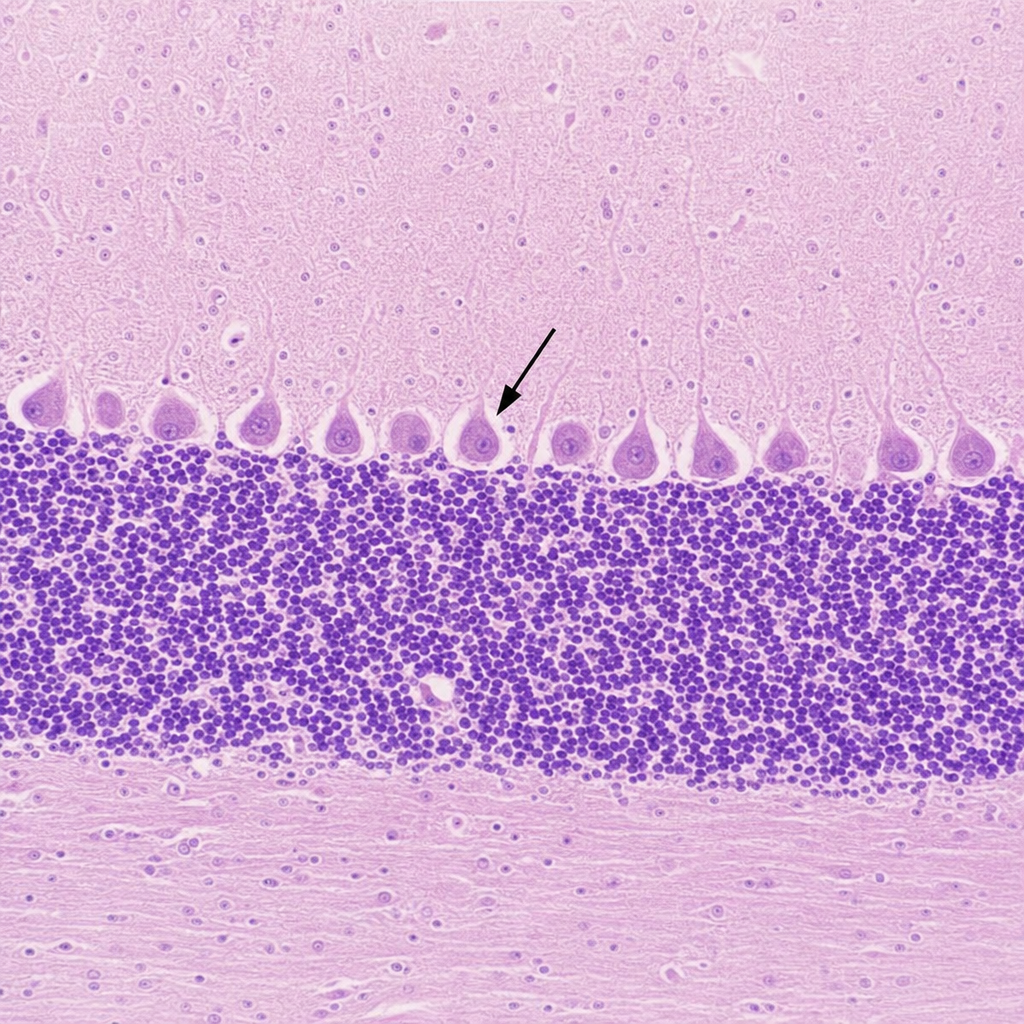
Histological section is given below. Identify the marked cell.
Histological section is given below. Identify the marked cell. 
Which of the following images displays the characteristic histology of the Thymus?
Identify the lymphatic structure from the histology section given below?
A renal biopsy image is shown. Identify the cells marked in the histological section.
Practice by Chapter
Basic Tissue Types
Practice Questions
Cell Biology and Organelles
Practice Questions
Epithelial Tissue
Practice Questions
Connective Tissue
Practice Questions
Muscular Tissue
Practice Questions
Nervous Tissue
Practice Questions
Cardiovascular System Histology
Practice Questions
Lymphoid Organs and Immune System
Practice Questions
Endocrine System Histology
Practice Questions
Respiratory System Histology
Practice Questions
Digestive System Histology
Practice Questions
Urinary and Reproductive System Histology
Practice Questions
Want unlimited practice?
Get full access to all questions, explanations, and performance tracking.
Scan to download app