A midline protrusion in the maxillary region is mostly due to which of the following?
The term Stratum Malpighi refers to which of the following layers of the epidermis?
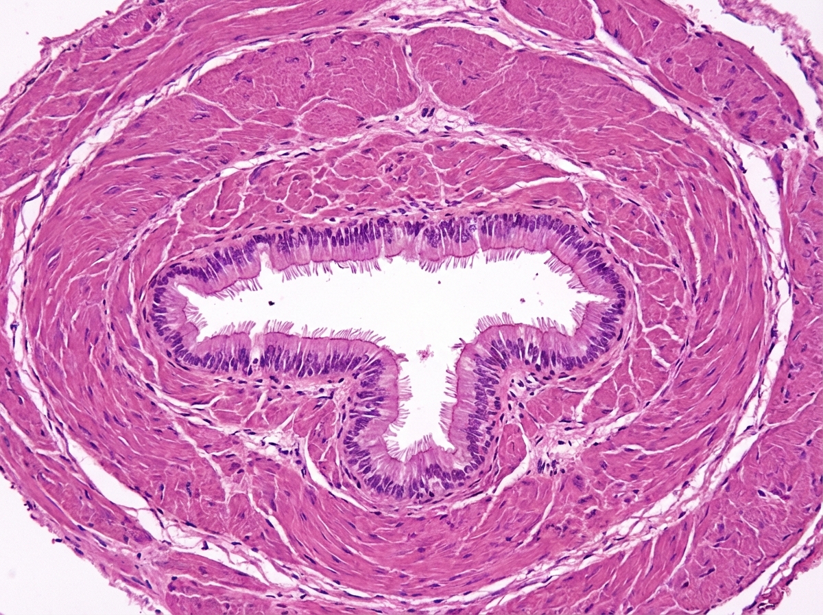
The following histological sample belongs to which of the following tissues?

Mean corpuscular hemoglobin concentration (MCHC) is increased in which of the following conditions?
Which type of collagen is present in the basement membrane?
Which stain is used for fat cells?
Small intestine and colon are lined by which type of epithelium?
What type of collagen is predominantly found in the ovary?
Circumcision is included in the management of Ca penis at which site?
Paneth cells are known to contain which of the following trace elements?
Explanation: **Explanation:** The correct answer is **Torus palatinus**. A torus is a benign, non-neoplastic bony exostosis (outgrowth) that occurs in specific locations of the facial skeleton. **1. Why Torus palatinus is correct:** Torus palatinus is a slow-growing, sessile bony mass that occurs specifically at the **midline of the hard palate**. It is located along the median palatine suture where the horizontal plates of the palatine bones and the palatine processes of the maxilla meet. It is the most common bony outgrowth of the oral cavity, more prevalent in females, and is typically asymptomatic unless it interferes with the fitting of a maxillary denture. **2. Why the other options are incorrect:** * **Torus maxillaris:** This is not a standard anatomical term. While exostoses can occur on the buccal or facial aspects of the maxilla, they are simply referred to as "maxillary exostoses" and are rarely strictly midline. * **Torus mandibularis:** This refers to a bony outgrowth on the **lingual surface** of the mandible, typically in the premolar region. It is usually bilateral rather than midline. * **Torus tubarius:** This is a mucosal elevation in the **nasopharynx** caused by the underlying medial end of the cartilaginous portion of the Eustachian tube. It is not a bony protrusion of the maxillary region. **Clinical Pearls for NEET-PG:** * **Prevalence:** Torus palatinus is found in approximately 20-30% of the population. * **Radiology:** On X-rays, they appear as well-circumscribed radiopaque (white) masses. * **Surgical Significance:** They are usually left alone unless they cause surface ulceration, speech impediments, or require removal for **prosthodontic reasons** (denture stability). * **Histology:** They consist of dense, cortical (lamellar) bone with a small amount of fatty marrow.
Explanation: The **Stratum Malpighi** (also known as the Malpighian layer or *stratum germinativum* in broader contexts) refers to the combination of the **Stratum basale** and the **Stratum spinosum**. [1] **Why the correct answer is right:** The epidermis is composed of five layers (from deep to superficial: Basale, Spinosum, Granulosum, Lucidum, and Corneum). The Stratum Malpighi represents the **mitotically active** part of the skin. [1] The Stratum basale contains the stem cells that undergo division, while the Stratum spinosum contains cells that are still metabolically active and capable of some mitosis. Together, these two layers are responsible for the constant renewal of keratinocytes. **Analysis of Incorrect Options:** * **Options A & B:** These include the **Stratum corneum**, which consists of dead, flattened keratinocytes (corneocytes) lacking nuclei. [1] This layer is involved in barrier function, not cellular proliferation. * **Option C:** While it includes the Stratum basale, the **Stratum granulosum** is a layer where cells begin to undergo apoptosis and accumulate keratohyalin granules. It is not considered part of the Malpighian proliferative unit. **NEET-PG High-Yield Pearls:** * **Desmosomes:** The Stratum spinosum is characterized by prominent desmosomes, which give the cells a "prickly" appearance (Prickle cell layer). * **Melanocytes:** These are primarily located in the **Stratum basale**. [1] * **Clinical Correlation:** Psoriasis involves an accelerated cell cycle in the Stratum Malpighi, leading to epidermal hyperplasia (acanthosis) and a thickened stratum corneum (parakeratosis). * **Bullous Diseases:** Pemphigus vulgaris involves the destruction of desmosomes specifically within the Stratum spinosum.
Explanation: ***Vas deferens*** - Characterized by a **thick 3-layered smooth muscle wall** (inner longitudinal, middle circular, outer longitudinal) surrounding a narrow lumen. - Lined with **pseudostratified columnar epithelium with stereocilia** for sperm maturation and transport. *Urethra* - Lined with **transitional epithelium** (urothelium) that can stretch, unlike the pseudostratified epithelium seen here. - Has a **much thinner muscular wall** and larger lumen compared to vas deferens. *Vermiform appendix* - Contains abundant **lymphoid tissue** and **Peyer's patches** in the submucosa, which are absent in this sample. - Lined with **simple columnar epithelium with goblet cells** for mucus production, not pseudostratified with stereocilia. *Stenson's duct* - This **parotid gland duct** is lined with **stratified columnar epithelium**, not pseudostratified. - Has a **thinner muscular wall** and is associated with **serous acinar tissue**, unlike the thick smooth muscle seen here.
Explanation: **Explanation:** **Mean Corpuscular Hemoglobin Concentration (MCHC)** represents the average concentration of hemoglobin in a given volume of packed red blood cells (RBCs). It is calculated as: $MCHC = (Hemoglobin / Hematocrit) \times 100$. **Why Spherocytosis is correct:** In **Hereditary Spherocytosis**, there is a defect in RBC membrane proteins (like spectrin or ankyrin), leading to the loss of membrane fragments. This results in a decreased surface-area-to-volume ratio, forcing the cell into a spherical shape. Because the cell shrinks (decreased volume) while the hemoglobin content remains relatively constant, the hemoglobin becomes more "concentrated," leading to an **increased MCHC** (typically >36 g/dL). This is one of the few clinical conditions where MCHC is elevated. **Why other options are incorrect:** * **Iron Deficiency Anemia (IDA):** This is a microcytic hypochromic anemia. Since hemoglobin synthesis is impaired more than the cell size reduction, the MCHC is **decreased**. * **Thalassemia:** Similar to IDA, Thalassemia involves defective globin chain synthesis, leading to microcytic hypochromic cells with a **decreased** MCHC. **High-Yield Clinical Pearls for NEET-PG:** * **Normal MCHC Range:** 32–36 g/dL. * **Hyperchromia:** True hyperchromia is rare; Spherocytosis is the classic example. * **Other causes of high MCHC:** Cold agglutinin disease (due to RBC clumping) and severe dehydration (spurious). * **Diagnostic Triad for Spherocytosis:** Splenomegaly, jaundice, and spherocytes on peripheral smear with a positive Osmotic Fragility Test.
Explanation: **Explanation:** The correct answer is **Type IV Collagen**. **Why Type IV is correct:** Type IV collagen is a specialized **non-fibrillar** collagen that forms a multi-dimensional meshwork or "chicken-wire" framework. Unlike fibrillar collagens, it retains its pro-peptide ends, allowing it to associate laterally into sheets. This structure provides the essential scaffolding for the **basal lamina** (a layer of the basement membrane), supporting epithelial and endothelial cells and acting as a selective filtration barrier [1]. **Analysis of Incorrect Options:** * **Type I:** The most abundant collagen in the body. It forms thick, tough fibers found in **B**one, **B**ow-string (tendons), and skin. (Mnemonic: Type **One** is for **Bone**). * **Type II:** Found primarily in **C**artilage (hyaline and elastic) and the vitreous humor. (Mnemonic: Type **Two** is for **Car-two-lage**). * **Type III:** Also known as **reticular fibers**. It forms a delicate supporting meshwork in highly cellular organs like the liver, spleen, and lymph nodes. It is also the first collagen deposited during wound healing before being replaced by Type I. **High-Yield Clinical Pearls for NEET-PG:** * **Alport Syndrome:** A genetic defect in **Type IV collagen** chains (α3, α4, α5) leading to hereditary nephritis, sensorineural deafness, and ocular defects ("Can't see, can't pee, can't hear high-C"). * **Goodpasture Syndrome:** Characterized by autoantibodies against the non-collagenous (NC1) domain of **Type IV collagen**, affecting the basement membranes of the glomerulus (kidney) and alveoli (lungs). * **Mnemonic for Collagen Types:** * Type I: Bone, Skin, Tendon * Type II: Cartilage * Type III: Reticulin (Blood vessels, fetal skin) * Type IV: Basement Membrane (Floor)
Explanation: **Explanation:** **Sudan IV** is a lipid-soluble dye used specifically for staining neutral triglycerides and lipids. In histology, routine processing (using alcohol and xylene) dissolves fat, leaving "empty" spaces in adipocytes. To visualize fat, tissue must be prepared using **frozen sections** and stained with lysochrome dyes like Sudan IV, Sudan Black B, or Oil Red O. These dyes work by being more soluble in the lipid droplets than in the solvent, thereby coloring the fat cells (Sudan IV specifically stains them **red-orange**). **Analysis of Incorrect Options:** * **PAS (Periodic Acid-Schiff):** Used to demonstrate **glycogen** and carbohydrate-rich structures like the basement membrane and mucin. It stains them magenta. * **Alcian Blue:** Used to identify **acidic mucopolysaccharides** and glycosaminoglycans. It is commonly used to highlight cartilage or goblet cells. * **Masson’s Trichrome:** A three-color protocol used to differentiate **collagen fibers** (blue/green) from muscle fibers and cytoplasm (red/pink). It is the gold standard for assessing liver fibrosis or cirrhosis. **High-Yield Clinical Pearls for NEET-PG:** * **Oil Red O** is the most common stain used to diagnose **Fat Embolism Syndrome** in lung or kidney tissue samples. * **Osmium Tetroxide** is another specialized stain that colors fat **black** and is unique because it also fixes the lipid, preventing it from being dissolved during routine processing. * For identifying **Glycogen**, the **Best’s Carmine** stain is a specific alternative to PAS.
Explanation: **Explanation:** The primary function of the small intestine and colon is the **absorption** of nutrients and water, as well as the **secretion** of enzymes and mucus [1]. To facilitate these processes, the gastrointestinal tract (from the stomach to the rectum) is lined by **Simple Columnar Epithelium**. These tall, narrow cells provide a large surface area for transport mechanisms [1]. In the small intestine, these cells are specifically called enterocytes and feature a "brush border" (microvilli) to further increase absorptive capacity [2], [4]. **Analysis of Options:** * **Simple Squamous (A):** These are thin, flat cells designed for rapid diffusion or filtration. They are found in the alveoli of lungs, endothelium of blood vessels, and the mesothelium [3] of serous membranes. * **Simple Cuboidal (B):** These box-like cells are typically involved in secretion and absorption in smaller ducts, such as the thyroid follicles, surface of the ovary, and renal tubules. * **Stratified Squamous (D):** This multi-layered epithelium is designed for protection against mechanical stress/abrasion. It lines the esophagus and the anal canal (below the pectinate line), but not the absorptive segments of the gut [1]. **High-Yield Clinical Pearls for NEET-PG:** * **Transition Zone:** The most high-yield site is the **Anorectal Junction**, where the epithelium abruptly changes from simple columnar (rectum) to stratified squamous (anal canal) [1]. * **Goblet Cells:** These mucus-secreting cells are interspersed within the columnar epithelium. Their density **increases** distally, being most numerous in the colon [2]. * **Celiac Disease:** Characterized by "villous atrophy" and "crypt hyperplasia" of the simple columnar lining in the small intestine [2].
Explanation: The correct answer is **Type XXVI (D)**. This is a relatively recent discovery in histology and a high-yield fact for competitive exams like NEET-PG. **1. Why Type XXVI is correct:** Type XXVI collagen is a non-fibrillar collagen belonging to the subfamily of **FACITs** (Fibril-Associated Collagens with Interrupted Triple helices). It is uniquely and predominantly expressed in the **reproductive tissues**, specifically the **ovary** and the testis. In the ovary, it is localized within the extracellular matrix of the developing follicles and plays a crucial role in follicular maturation and structural integrity during the ovulatory cycle. **2. Why other options are incorrect:** * **Type I:** This is the most abundant collagen in the body, found in "hard" structures like bone, tendons [1, 3], and dermis. While present in the ovarian stroma/tunica albuginea, it is not the *predominant* or specific collagen type that defines ovarian follicular histology in recent literature. * **Type IV:** This is the primary collagen of the **basal lamina** (basement membrane). In the ovary, it is found in the follicular basement membrane (separating the granulosa cells from the theca cells), but it is not the predominant type overall. * **Type XXIV:** This is a fibrillar collagen mainly expressed in developing **bone and cartilage**. It is not a major constituent of ovarian tissue. **Clinical Pearls & High-Yield Facts for NEET-PG:** * **Type I:** Bone, Tendon, Skin (90% of body collagen) [1, 3]. * **Type II:** Cartilage (Hyaline and Elastic), Vitreous body. * **Type III:** Reticular fibers, Blood vessels, Granulation tissue. * **Type IV:** Basement membrane (Alport syndrome involves a mutation here). * **Type XXVI:** Specifically associated with **Ovarian Folliculogenesis**. Memory aid: "Type 26 is for the Sex (Reproductive) organs."
Explanation: **Explanation:** **Why Prepuce is Correct:** Carcinoma of the penis most commonly affects the glans (approx. 48%) and the prepuce (approx. 21%). When the malignancy is strictly confined to the **prepuce (foreskin)**, a wide local excision in the form of **radical circumcision** is the definitive surgical management. This procedure removes the primary tumor while preserving the functional integrity of the glans and the shaft. It is only indicated when the lesion is small, localized, and does not involve the coronal sulcus or the glans. **Analysis of Incorrect Options:** * **Glans (A):** Tumors of the glans usually require more extensive surgery, such as a glansectomy or partial penectomy, to ensure adequate oncological margins (usually 1-2 cm). Circumcision alone would leave residual malignant tissue. * **Glandulo-prepucial (C):** If the tumor involves the junction (coronal sulcus), it has likely invaded deeper tissues or the glans itself. This necessitates a partial penectomy rather than a simple circumcision. * **Shaft of penis (D):** Lesions on the shaft require wide local excision with skin grafting or, more commonly, partial or total penectomy depending on the depth of invasion into the corpora cavernosa. **High-Yield Clinical Pearls for NEET-PG:** * **Risk Factor:** The single most significant risk factor for Ca Penis is **phimosis** (present in 25-75% of cases), which leads to the accumulation of smegma and chronic inflammation. * **Protective Factor:** Neonatal circumcision is highly protective against the later development of squamous cell carcinoma of the penis. * **Histology:** Over 95% of penile cancers are **Squamous Cell Carcinomas**. * **Lymphatic Spread:** The primary nodal drainage is to the **Inguinal lymph nodes** (Vertical group of superficial inguinal nodes). The "Sentinel node" of Cabanas is located near the epigastric-saphenous junction.
Explanation: **Explanation:** **Paneth cells** are specialized secretory cells located at the bases of the **Crypts of Lieberkühn** in the small intestine [1]. They play a critical role in innate mucosal immunity. 1. **Why Zinc is Correct:** Paneth cells contain prominent eosinophilic apical granules. These granules are rich in antimicrobial substances like **lysozyme, alpha-defensins (cryptidins), and phospholipase A2**. Crucially, these cells contain a high concentration of **Zinc**, which acts as a cofactor for several enzymes and stabilizes the storage of these antimicrobial peptides within the secretory granules. The presence of zinc can be histologically demonstrated using specific stains like dithizone. 2. **Why Other Options are Incorrect:** * **Copper:** Primarily associated with Menkes disease (deficiency) or Wilson’s disease (overload in liver/basal ganglia), but not a characteristic feature of Paneth cell granules. * **Molybdenum:** A cofactor for enzymes like xanthing oxidase and sulfite oxidase; it does not have a specific localization in the intestinal crypts. * **Selenium:** An essential component of glutathione peroxidase (antioxidant system), but not the trace element defining Paneth cell histology. **High-Yield Clinical Pearls for NEET-PG:** * **Location:** Paneth cells are most numerous in the **ileum** and are absent in the large intestine (except in pathological states like "Paneth cell metaplasia" in IBD). * **Function:** They regulate the gut microbiome by secreting antimicrobial peptides [1]. * **Stem Cell Niche:** They provide essential growth factors (like Wnt) to the neighboring intestinal stem cells [1]. * **Zinc Deficiency:** Can lead to **Acrodermatitis Enteropathica**, which may impair Paneth cell function and mucosal immunity.
Basic Tissue Types
Practice Questions
Cell Biology and Organelles
Practice Questions
Epithelial Tissue
Practice Questions
Connective Tissue
Practice Questions
Muscular Tissue
Practice Questions
Nervous Tissue
Practice Questions
Cardiovascular System Histology
Practice Questions
Lymphoid Organs and Immune System
Practice Questions
Endocrine System Histology
Practice Questions
Respiratory System Histology
Practice Questions
Digestive System Histology
Practice Questions
Urinary and Reproductive System Histology
Practice Questions
Get full access to all questions, explanations, and performance tracking.
Start For Free