Oncocytes are found in all of the following organs, except:
What is the cell lining of the common bile duct?
Aggrecan is a component of which of the following?
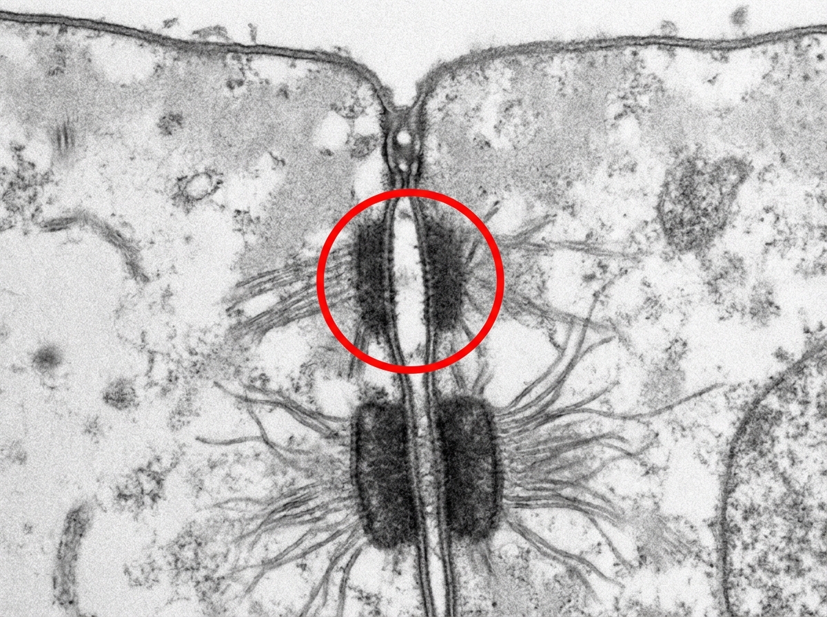
What type of collagen is predominantly found in hyaline cartilage?
All of the following statements about Paneth cells are true, EXCEPT?
Which of the following is the most important prognostic factor in ALL?
Nissl substance is found in:
During laparoscopic hernia surgery, the triangle of doom is visualized. Which of the following is NOT a boundary of this anatomical space?
Ectocervix is predominantly lined by which of the following type of epithelium?
Iron overload occurs in all of the following conditions except:
Explanation: **Explanation:** **Oncocytes** (also known as oxyphil cells or Askanazy cells) are large, polygonal epithelial cells characterized by an abundant, granular, eosinophilic cytoplasm. This distinct appearance is due to the presence of a massive number of **mitochondria**. 1. **Why Pineal Body is the correct answer:** The pineal body consists primarily of pinealocytes and glial cells. It does not contain oncocytes. While it may contain "brain sand" (corpora arenacea) with age, oncocytic transformation is not a feature of this neuroendocrine gland. 2. **Analysis of Incorrect Options:** * **Thyroid (Option A):** Oncocytes in the thyroid are known as **Hürthle cells**. They are commonly seen in Hashimoto’s thyroiditis and Hürthle cell tumors. * **Kidneys (Option B):** Oncocytes are found in the renal tubules. A benign tumor arising from these cells is called a **Renal Oncocytoma**, which typically shows a characteristic "central stellate scar" on imaging. * **Pituitary (Option C):** Oncocytic changes can occur in the anterior pituitary gland, particularly in older individuals or within specific adenomas (Oncocytomas). **High-Yield Clinical Pearls for NEET-PG:** * **Salivary Glands:** The **Warthin’s tumor** (Adenolymphoma) of the parotid gland is classically composed of a double layer of oncocytic epithelium. * **Parathyroid:** Oxyphil cells appear after puberty and increase with age; they are larger and darker than chief cells but their exact function remains unknown [1]. They are characterized by being tightly packed with mitochondria [1]. * **Staining:** Due to high mitochondrial content, oncocytes stain strongly with **phosphotungstic acid-hematoxylin (PTAH)**. * **Mnemonic:** Oncocytes are common in **"T-P-K-S"** (Thyroid, Parathyroid/Pituitary, Kidney, Salivary glands).
Explanation: ### Explanation The correct answer is **D. Simple columnar**. **1. Why Simple Columnar is Correct:** The entire extrahepatic biliary tree, including the hepatic ducts, cystic duct, and the **common bile duct (CBD)**, is lined by a **simple columnar epithelium**. These cells, often called cholangiocytes, are specialized for the transport of water and electrolytes, helping to modify the bile as it flows toward the duodenum. The tall, columnar shape provides a protective barrier against the detergent properties of bile while allowing for active secretion and absorption. **2. Analysis of Incorrect Options:** * **A. Stratified columnar:** This is a rare epithelium found only in specific transition zones (e.g., parts of the male urethra or conjunctiva). It is not found in the biliary system. * **B. Stratified squamous:** This epithelium is designed for protection against mechanical friction (e.g., esophagus, skin). If found in the CBD, it would represent **squamous metaplasia**, usually due to chronic irritation from gallstones. * **C. Simple cuboidal:** While the smaller, intrahepatic bile ductules (Canals of Hering) are lined by simple cuboidal epithelium, the epithelium becomes taller (columnar) as the ducts increase in diameter toward the CBD. **3. Clinical Pearls for NEET-PG:** * **The Mucosa:** Unlike the intestine, the CBD mucosa lacks a muscularis mucosae and a distinct submucosa. * **Rokistansky-Aschoff Sinuses:** These are mucosal herniations into the muscular wall, specifically characteristic of the gallbladder (often seen in chronic cholecystitis), not the CBD. * **Ampulla of Vater:** At its distal end, the CBD joins the pancreatic duct; the epithelium remains simple columnar but transitions to the intestinal type at the Major Duodenal Papilla. * **High-Yield Fact:** The gallbladder also shares this **simple columnar** lining, but it is characterized by prominent microvilli for concentrating bile.
Explanation: **Explanation:** **Aggrecan** is the most abundant **proteoglycan** found in the extracellular matrix (ECM) of **cartilage** (specifically hyaline and fibrocartilage) [1]. It consists of a core protein to which numerous glycosaminoglycan (GAG) chains—primarily **chondroitin sulfate** and **keratan sulfate**—are covalently attached. These aggrecan molecules bind to a long strand of hyaluronic acid to form massive proteoglycan aggregates [1]. Due to its high negative charge, aggrecan attracts water, providing cartilage with its essential osmotic properties and the ability to resist compressive forces (load-bearing capacity) [1]. **Analysis of Incorrect Options:** * **Option A:** Receptors on platelets are typically glycoproteins (e.g., GpIIb/IIIa or GpIb-IX-V), not large proteoglycans like aggrecan. * **Option B:** While osteoid (unmineralized bone matrix) contains proteoglycans, its primary components are Type I collagen and non-collagenous proteins like Osteocalcin and Osteopontin [2]. Aggrecan is specific to the cartilaginous matrix. * **Option C:** Leukocyte granules contain enzymes (like myeloperoxidase), histamine, or heparin (in basophils), but not aggrecan. **High-Yield Facts for NEET-PG:** * **Cartilage Composition:** Type II collagen provides tensile strength, while Aggrecan provides compressive strength [1]. * **Link Protein:** This protein stabilizes the interaction between the aggrecan core protein and hyaluronan. * **Clinical Correlation:** Degradation of aggrecan by enzymes known as **ADAMTS** (aggrecanases) is a hallmark of early **Osteoarthritis**. * **Marker:** Aggrecan is often used as a marker for chondrocyte differentiation in histology.
Explanation: **Explanation:** The correct answer is **Type II Collagen**. Hyaline cartilage, the most common type of cartilage in the body (found in articular surfaces, the respiratory tract, and the fetal skeleton), consists of a dense extracellular matrix [1]. The primary structural framework of this matrix is composed of **Type II collagen fibers**, which provide tensile strength and help anchor the proteoglycan aggregates (like aggrecan) that give the cartilage its weight-bearing properties [1]. **Analysis of Options:** * **Type I Collagen (Option A):** This is the strongest collagen, found in tissues requiring high tensile strength such as **bone, tendons, ligaments, and skin**. It is also the predominant collagen in **fibrocartilage** (e.g., intervertebral discs). * **Type III Collagen (Option B):** Also known as **reticular fibers**, these form a supportive meshwork in highly cellular organs like the liver, spleen, and lymph nodes. It is also involved in early wound healing (granulation tissue). * **Type IV Collagen (Option D):** This type does not form fibrils; instead, it forms a two-dimensional meshwork that is a key structural component of the **basal lamina** (basement membrane). **High-Yield NEET-PG Pearls:** * **Mnemonic for Collagen Types:** "Be (I) So (II) Totally (III) Cool (IV)" → **B**one (I), **S**oft tissue/Cartilage (II), **T**hree/Reticular (III), **C**olumn/Basal Lamina (IV). * **Clinical Correlation:** Mutations in Type II collagen lead to **Skeletal Dysplasias** (e.g., Stickler Syndrome). * **Articular Cartilage:** It is a specific type of hyaline cartilage that lacks a perichondrium, making its regeneration difficult after injury [1].
Explanation: Paneth cells are specialized secretory cells located at the **base of the Crypts of Lieberkühn** in the small intestine [1]. They play a crucial role in innate immunity and maintaining the gut microbiome. **Why "Foamy appearance" is the correct (False) statement:** Paneth cells do **not** have a foamy appearance. Instead, they are characterized by prominent, **large eosinophilic (acidophilic) apical granules**. A "foamy" or "vacuolated" appearance is characteristic of cells containing lipid droplets or mucus, such as **Goblet cells** [1] or certain macrophages (foam cells). Paneth cells are structurally dense due to their high protein-secretory activity. **Analysis of other options:** * **Rich in rough endoplasmic reticulum (RER):** True. As protein-synthesizing cells, they possess an extensive network of basal RER and a prominent Golgi apparatus to produce antimicrobial peptides. * **Rich in zinc:** True. Paneth cell granules contain a high concentration of zinc, which acts as a cofactor for various enzymes and stabilizes the stored secretory proteins. * **Contain lysozyme:** True. Their primary function is the secretion of antimicrobial substances, including **lysozyme**, **alpha-defensins** (cryptdins), and phospholipase A2, which digest bacterial cell walls. **High-Yield Clinical Pearls for NEET-PG:** * **Location:** Found only in the small intestine (duodenum, jejunum, ileum); their presence in the colon is pathological (Paneth cell metaplasia) [1]. * **Staining:** Their granules stain bright red with H&E and are **PAS-positive**. * **Function:** They regulate the intestinal flora and protect intestinal stem cells located nearby in the crypts [1]. * **Zinc deficiency:** Can lead to impaired Paneth cell function, contributing to the skin lesions and diarrhea seen in **Acrodermatitis enteropathica**.
Explanation: **Explanation:** In Acute Lymphoblastic Leukemia (ALL), the **response to initial therapy (specifically steroids)** is considered the single most important independent prognostic factor. This is because the "in-vivo" sensitivity of leukemic blasts to corticosteroids (prednisolone) within the first 7–14 days of treatment reflects the underlying biological aggressiveness and chemo-sensitivity of the disease. A poor response (high blast count after a steroid prophase) indicates a significantly higher risk of relapse and treatment failure. **Analysis of Options:** * **Response to Steroids (Correct):** Early clearance of peripheral blasts (prednisolone response) and achieving Minimal Residual Disease (MRD) negativity are the strongest predictors of long-term survival. * **Age (Incorrect):** While age is a major prognostic factor (favorable between 1–9 years; unfavorable <1 or >10 years), it is secondary to the dynamic response to treatment. * **Total Leukocyte Count (TLC) (Incorrect):** A TLC >50,000/µl is a poor prognostic marker (High-Risk category), but many patients with high TLC still achieve remission if they respond well to induction therapy. * **Hyperploidy (Incorrect):** Cytogenetics are vital for risk stratification (e.g., Hyperdiploidy >50 is favorable; Philadelphia chromosome t(9;22) is unfavorable), but clinical response remains the ultimate guide for prognosis. **High-Yield Clinical Pearls for NEET-PG:** * **Best Prognosis:** Age 1–9 years, TLC <50,000, Hyperdiploidy (>50 chromosomes), and t(12;21) (TEL-AML1). * **Worst Prognosis:** Age <1 year or >10 years, TLC >50,000, Hypodiploidy, and t(9;22) (BCR-ABL). * **L3 Subtype (Burkitt’s):** Associated with t(8;14) and has the worst prognosis among FAB classifications.
Explanation: **Explanation:** **1. Why the Correct Answer is Right:** Nissl substance (or Nissl bodies) refers to large, granular structures found in the cytoplasm of **neurons**. Ultrastructurally, they are composed of **Rough Endoplasmic Reticulum (RER)** and free ribosomes [1]. Their primary function is high-rate protein synthesis [4], necessary for maintaining the neuron's structure and producing neurotransmitters. They are located in the **cell body (soma)** and **dendrites**, but are characteristically **absent in the axon and the axon hillock** [2]. **2. Why the Other Options are Incorrect:** * **Hepatocytes (B):** While liver cells are rich in RER for albumin synthesis, these organelles are not organized into the specific "Nissl" configuration unique to nervous tissue. * **Cardiocytes (C):** Cardiac muscle cells contain abundant mitochondria and sarcoplasmic reticulum (smooth ER) for energy and calcium handling, but do not contain Nissl bodies. * **Enterocytes (D):** These intestinal epithelial cells are specialized for absorption and secretion; they possess standard organelles but lack the specialized Nissl substance. **3. Clinical Pearls & High-Yield Facts for NEET-PG:** * **Chromatolysis:** This is a high-yield clinical phenomenon where Nissl bodies disperse and disappear from the soma following axonal injury (retrograde degeneration) [3]. It indicates an active attempt at protein synthesis for repair. * **Staining:** Nissl bodies are highly "basophilic" and are best visualized using basic dyes like **Cresyl Violet** or **Methylene Blue**. * **Distribution:** Remember the "Axon Hillock Rule"—the absence of Nissl substance in the axon hillock helps histologists distinguish the axon from dendrites under a microscope [2].
Explanation: The **Triangle of Doom** is a critical anatomical landmark visualized during laparoscopic inguinal hernia repair (TEP/TAPP). It is an inverted V-shaped area located between the pelvic floor and the anterior abdominal wall. [1] ### Why "Inguinal Ligament" is the Correct Answer The **Inguinal ligament** does not form a boundary of the Triangle of Doom. Instead, it serves as the **superior boundary** of the **Triangle of Pain**. In laparoscopic surgery, the Triangle of Doom is defined by structures located *inferior* to the internal inguinal ring. [1] ### Analysis of Boundaries (Incorrect Options) The Triangle of Doom is bounded by: * **Medially:** The **Vas deferens** (Option A) in males (or the round ligament in females). [1] * **Laterally:** The **Gonadal vessels** (Option B) (testicular artery and vein). [1] * **Superiorly/Apex:** The internal inguinal ring. [1] * **Base:** The **Reflected peritoneum** (Option C) or the iliac vessels. [1] ### Clinical Pearls for NEET-PG * **Contents:** The most critical structure within this triangle is the **External Iliac Artery and Vein**. [1] * **Surgical Significance:** Surgeons must avoid placing tacks, staples, or sutures in this zone to prevent life-threatening hemorrhage. [1] * **Triangle of Pain:** Located lateral to the Triangle of Doom (lateral to the gonadal vessels). Its boundaries are the gonadal vessels (medially), the iliopubic tract/inguinal ligament (superiorly), and the reflected peritoneum (inferiorly). It contains the **Femoral nerve**, **Genitofemoral nerve (genital branch)**, and **Lateral femoral cutaneous nerve**. [1] * **Trapezoid of Disaster:** The combined area of the Triangle of Doom and the Triangle of Pain.
Explanation: **Explanation:** The cervix is divided into two distinct anatomical and histological regions: the **endocervix** (inner canal) and the **ectocervix** (the portion projecting into the vagina). The **ectocervix** is exposed to the acidic environment and mechanical friction of the vaginal vault. To withstand this "wear and tear," it is lined by **Nonkeratinized Stratified Squamous Epithelium** [2]. This multi-layered epithelium provides a protective barrier, similar to the lining of the vagina and esophagus. **Analysis of Incorrect Options:** * **Option A (Cuboidal):** This is typically found in kidney tubules or glandular ducts, not the cervix. * **Option B (Mucin-secreting columnar):** This is the characteristic lining of the **Endocervix**. These cells secrete mucus to form a cervical plug. * **Option C (Pseudostratified columnar):** This "respiratory-type" epithelium is found in the trachea and bronchi, not the female reproductive tract. **Clinical Pearls for NEET-PG:** 1. **Squamocolumnar Junction (SCJ):** The point where the endocervical columnar epithelium meets the ectocervical squamous epithelium. 2. **Transformation Zone:** The area where columnar cells undergo **metaplasia** into squamous cells [1]. This is the most common site for **Cervical Cancer (Squamous Cell Carcinoma)** and is the area sampled during a **Pap smear** [2]. 3. **Nabothian Cysts:** Formed when squamous epithelium overgrows and blocks the orifices of endocervical columnar glands.
Explanation: **Explanation:** Iron overload (hemosiderosis) typically occurs due to two main mechanisms: repeated blood transfusions or ineffective erythropoiesis leading to increased intestinal iron absorption. **Why Polycythemia Vera is the correct answer:** Polycythemia Vera (PV) is a myeloproliferative neoplasm characterized by the autonomous overproduction of red blood cells. Because the body is rapidly utilizing iron to synthesize hemoglobin for the massive excess of erythrocytes, patients with PV are actually prone to **iron deficiency**, not overload. Therapeutic phlebotomy (the standard treatment for PV) further depletes iron stores. **Analysis of Incorrect Options:** * **Thalassemia:** Patients develop iron overload due to both chronic blood transfusions and increased dietary iron absorption triggered by "ineffective erythropoiesis" (erythroid hyperplasia). * **Myelodysplastic Syndrome (MDS):** These patients often suffer from refractory anemia requiring lifelong blood transfusions. Each unit of packed RBCs contains ~200–250 mg of iron, which the body cannot actively excrete. * **Sideroblastic Anemia:** This condition involves a defect in porphyrin synthesis, preventing iron from being incorporated into hemoglobin. Iron accumulates in the mitochondria of erythroblasts (forming ringed sideroblasts), leading to systemic overload. **High-Yield Clinical Pearls for NEET-PG:** * **Prussian Blue Stain:** The gold standard histological stain to visualize iron (hemosiderin) in tissues or bone marrow. * **Hereditary Hemochromatosis:** An autosomal recessive disorder (HFE gene mutation) causing primary iron overload. * **Classic Triad:** Bronze skin, Cirrhosis, and Diabetes mellitus ("Bronze Diabetes"). * **Hepcidin:** The key regulatory hormone produced by the liver that inhibits iron absorption; it is suppressed in states of ineffective erythropoiesis.
Explanation: The **Prostate** is a unique accessory male reproductive organ characterized by its **fibromuscular stroma** [1]. Histologically, the prostate is composed of approximately 30–50 tubuloalveolar glands embedded in a dense stroma. This stroma consists of a mixture of **collagenous connective tissue** and a significant amount of **smooth muscle fibers**. During ejaculation, the contraction of this smooth muscle helps squeeze the prostatic secretions into the prostatic urethra. **Analysis of Options:** * **Testis (A):** The stroma of the testis is primarily fibrous (tunica albuginea) and contains interstitial cells (Leydig cells), but it lacks a significant smooth muscle component within the parenchyma. * **Liver (B):** The liver has a connective tissue framework (Glisson’s capsule and septa), but the stroma is purely fibroelastic, not muscular. * **Urinary Bladder (D):** While the bladder contains a massive amount of smooth muscle (detrusor muscle), this muscle forms the **wall (muscularis propria)** of the organ rather than a "stroma" that supports glandular elements. **NEET-PG High-Yield Pearls:** 1. **Prostatic Concretions (Corpora Amylacea):** These are calcified proteinaceous bodies found in the lumina of prostatic glands, increasing with age. 2. **Zonal Anatomy:** Benign Prostatic Hyperplasia (BPH) typically occurs in the **Transitional Zone**, whereas Prostatic Carcinoma usually arises in the **Peripheral Zone**. 3. **Epithelium:** The glands are lined by a pseudostratified columnar epithelium. 4. **PSA:** Prostate-Specific Antigen is a serine protease produced by these glands to liquefy the semen.
Explanation: **Explanation:** Testicular tumors are broadly classified into **Germ Cell Tumors (GCTs)**, which account for approximately 95% of cases, and Sex Cord-Stromal Tumors. **Why Seminoma is correct:** Seminoma is the **most common single histological type** of testicular germ cell tumor, accounting for about 50% of all cases. It typically occurs in the 4th decade of life (ages 30–40). Histologically, it is characterized by large, uniform cells with clear cytoplasm (“fried-egg appearance”) and distinct cell borders, separated by fibrous septa containing lymphocytic infiltrates [1]. It is highly radiosensitive and has an excellent prognosis. **Why the other options are incorrect:** * **Teratoma:** While common in children (pre-pubertal), in adults, it usually occurs as a component of a "Mixed Germ Cell Tumor" rather than as a pure, isolated entity [2]. * **Sertoli Cell Tumor:** These fall under Sex Cord-Stromal Tumors. They are rare (approx. 1%) and are generally benign. * **Choriocarcinoma:** This is the most aggressive but **least common** pure germ cell tumor [2]. It is characterized by high levels of hCG and early hematogenous spread. **High-Yield Clinical Pearls for NEET-PG:** * **Most common testicular tumor in infants/children:** Yolk Sac Tumor (Endodermal Sinus Tumor); look for **Schiller-Duval bodies**. * **Most common testicular tumor in elderly (>60 years):** Spermatocytic Tumor (formerly Spermatocytic Seminoma) or Lymphoma (secondary). * **Tumor Marker:** Seminomas may show elevated **hCG** (in 10-15% of cases) but **never** elevated AFP. If AFP is raised, it indicates a non-seminomatous component. * **Risk Factor:** Cryptorchidism (undescended testis) is the most significant risk factor for developing Seminoma.
Explanation: **Explanation:** **Aggrecan** is a high-molecular-weight proteoglycan that serves as a critical structural component of the extracellular matrix (ECM) in **cartilage** [1]. 1. **Why Option D is Correct:** Aggrecan consists of a core protein with numerous attached glycosaminoglycan (GAG) chains, primarily **chondroitin sulfate** and **keratan sulfate**. In cartilage, multiple aggrecan molecules bind to a single long strand of **hyaluronic acid** to form massive proteoglycan aggregates [1]. These aggregates are highly hydrophilic; they trap water molecules, providing cartilage with its essential **osmotic swelling pressure** and the ability to resist compressive loads (load-bearing capacity) [1]. 2. **Why Other Options are Incorrect:** * **Option A:** Receptors on platelets are typically glycoproteins (e.g., GpIb, GpIIb/IIIa), not large proteoglycans like aggrecan. * **Option B:** While osteoid contains proteoglycans (like decorin and biglycan), aggrecan is specifically characteristic of the cartilaginous matrix, not the organic bone matrix (osteoid) [2]. * **Option C:** Granules in leukocytes contain enzymes (like myeloperoxidase) or specific proteoglycans like **serglycin**, but not aggrecan. **High-Yield Clinical Pearls for NEET-PG:** * **Cartilage Composition:** Type II Collagen provides tensile strength, while Aggrecan provides compressive strength [1]. * **Link Protein:** This protein stabilizes the non-covalent bond between aggrecan and hyaluronic acid. * **Clinical Correlation:** Degradation of aggrecan by enzymes known as **ADAMTS** (aggrecanases) is a hallmark of early **Osteoarthritis**. * **Marker:** Aggrecan is often used as a marker for chondrocyte differentiation in histological studies.
Explanation: **Explanation:** The **Juxtaglomerular (JG) cells** are specialized cells located primarily in the wall of the **afferent arteriole** (and occasionally the efferent arteriole) [1]. They are derived from **modified smooth muscle cells** of the tunica media [1]. These cells act as baroreceptors (pressure sensors) and are responsible for the synthesis, storage, and release of the enzyme **renin** in response to low blood pressure [1]. **Analysis of Options:** * **Option B (Correct):** JG cells are modified smooth muscle cells specifically located in the afferent arteriole [2]. They contain prorenin granules and form a crucial part of the Juxtaglomerular Apparatus (JGA) [1]. * **Option A:** The **Macula densa** consists of specialized columnar epithelial cells in the **Distal Convoluted Tubule (DCT)**. They act as chemoreceptors sensing sodium chloride (NaCl) concentrations, not as the JG cells themselves [3]. * **Option C:** While a few JG cells may be found in the efferent arteriole, they are predominantly and characteristically located in the **afferent arteriole** [1]. * **Option D:** This is a distractor. The JGA does include "Polkissen cells" (Lacis cells/Extraglomerular mesangial cells), but these are not described as islets of epithelial cells. **High-Yield Clinical Pearls for NEET-PG:** 1. **Components of JGA:** 1. Macula densa (DCT), 2. JG cells (Afferent arteriole), 3. Lacis cells (Extraglomerular mesangial cells). 2. **Renin Release:** Stimulated by decreased renal perfusion pressure (detected by JG cells), decreased NaCl (detected by Macula densa), and Sympathetic stimulation ($\beta_1$ receptors) [3]. 3. **Histology Tip:** JG cells contain membrane-bound granules that stain with **Bowie’s stain** or periodic acid-Schiff (PAS) [1].
Explanation: ### Explanation The small intestinal mucosa contains a specialized component of the mucosal immune system known as **Gut-Associated Lymphoid Tissue (GALT)**. Within the epithelial layer itself, there are specialized cells called **Intraepithelial Lymphocytes (IELs)** [1]. **1. Why T lymphocytes are correct:** The vast majority (over 90%) of intraepithelial lymphocytes are **T lymphocytes** [1]. Specifically, these are predominantly **CD8+ cytotoxic T cells**. They serve as the first line of immunological defense, patrolling the epithelial barrier to eliminate infected or transformed (cancerous) cells. Unlike the lamina propria, which contains a mix of immune cells, the intra-epithelial space is uniquely dominated by these T cells [1]. **2. Why the other options are incorrect:** * **Basophils:** These are granulocytes primarily found in the peripheral blood. They are involved in systemic allergic reactions and are not a resident feature of the intestinal epithelium. * **Plasma cells:** While plasma cells are abundant in the **lamina propria** (the connective tissue layer beneath the epithelium) where they secrete IgA, they are not typically found within the intra-epithelial region [1]. * **B lymphocytes:** These are primarily located in organized lymphoid follicles like **Peyer’s patches** or within the lamina propria. They do not migrate into the epithelial layer in significant numbers [1]. **High-Yield Clinical Pearls for NEET-PG:** * **IEL Count & Celiac Disease:** An increase in the number of intraepithelial lymphocytes (specifically >25 IELs per 100 enterocytes) is a hallmark histological finding in **Celiac Disease** (Marsh Grade 1). * **M Cells:** These are specialized epithelial cells located over Peyer's patches that sample antigens from the lumen and deliver them to underlying lymphoid tissue. * **Paneth Cells:** Found at the base of the Crypts of Lieberkühn; they secrete antimicrobial peptides like **defensins** and **lysozyme** [2].
Explanation: **Explanation:** The **Tympanic Membrane** is the correct answer because it possesses a unique trilaminar structure rather than being solely lined by non-keratinized stratified squamous epithelium. Its three layers are: 1. **Outer layer:** Cuticular layer (Keratinized stratified squamous epithelium, continuous with the external auditory canal). 2. **Middle layer:** Fibrous layer (Lamina propria containing collagen and elastic fibers). 3. **Inner layer:** Mucous layer (Simple cuboidal/low columnar epithelium, continuous with the middle ear mucosa). **Analysis of Incorrect Options:** * **Hypopharynx and Laryngopharynx:** These areas are part of the upper digestive tract and are subject to mechanical friction from food boluses. Therefore, they are lined by **non-keratinized stratified squamous epithelium** for protection. * **Oesophagus:** This is a classic high-yield example of **non-keratinized stratified squamous epithelium**. It protects the tube from abrasion during swallowing (Note: The lining changes to simple columnar at the gastroesophageal junction—the "Z-line"). * **Cornea:** The anterior surface of the cornea is lined by a specialized, highly regular **non-keratinized stratified squamous epithelium** that must remain moist and transparent for optical clarity. **High-Yield Clinical Pearls for NEET-PG:** * **Barrett’s Oesophagus:** Metaplasia where the normal squamous epithelium of the esophagus changes to simple columnar (intestinal metaplasia) due to chronic acid reflux. * **Vitamin A Deficiency:** Can lead to squamous metaplasia and keratinization of the corneal epithelium (Xerophthalmia). * **Rule of Thumb:** Non-keratinized stratified squamous epithelium is found on "moist" surfaces subject to wear and tear (Oral cavity, Vagina, Oesophagus, Anal canal below the pectinate line).
Explanation: The correct answer is **Phosphate (Option A)**, specifically in the form of **Struvite stones** (Magnesium Ammonium Phosphate). These are classically known as "infection stones." **Why Phosphate is correct:** Urinary tract infections (UTIs) caused by **urease-producing bacteria** (most commonly *Proteus mirabilis*, but also *Klebsiella* and *Staphylococcus saprophyticus*) lead to the hydrolysis of urea into ammonia. This process increases the urinary pH (alkalinization). In an alkaline environment, the solubility of phosphate decreases, leading to the precipitation of magnesium ammonium phosphate and carbonate apatite crystals. These stones can grow rapidly and often form large **Staghorn calculi** that fill the renal pelvis and calyces. **Why the other options are incorrect:** * **B. Urate (Uric Acid):** These stones form in **acidic urine** (low pH). They are associated with gout, high purine intake, or rapid cell turnover (e.g., leukemia). They are typically radiolucent. * **C. Cysteine:** These are rare genetic stones caused by a defect in the transport of dibasic amino acids (COAL: Cysteine, Ornithine, Arginine, Lysine). They form in acidic urine and have a characteristic "hexagonal" shape. * **D. Calcium Oxalate:** This is the **most common type of kidney stone overall**, but it is primarily associated with metabolic factors (hypercalciuria, hyperoxaluria) and dehydration, rather than infection. **High-Yield Clinical Pearls for NEET-PG:** * **Morphology:** Struvite stones show a characteristic **"Coffin-lid"** appearance under microscopy. * **Radiology:** Struvite and Calcium stones are **Radio-opaque**, whereas Uric acid stones are **Radiolucent**. * **Staghorn Calculus:** While most staghorn stones are Struvite, they can occasionally be composed of Cystine or Calcium oxalate. * **Management:** Treatment requires complete surgical removal of the stone and eradication of the underlying infection to prevent recurrence [1].
Explanation: The growth plate, or **epiphyseal plate**, is a classic example of a **Primary Cartilaginous Joint (Synchondrosis)**. **1. Why it is correct:** Primary cartilaginous joints are characterized by bones united by a plate of **hyaline cartilage**. These joints are typically temporary; the cartilage eventually ossifies (synostosis) once skeletal maturity is reached [1]. The growth plate connects the epiphysis and diaphysis of a long bone, allowing for longitudinal bone growth through endochondral ossification [2]. **2. Analysis of Incorrect Options:** * **Fibrous Joint (A):** Here, bones are joined by dense connective tissue (e.g., sutures of the skull, gomphosis, or syndesmosis). There is no cartilage involved, and they allow negligible movement. * **Secondary Cartilaginous Joint (C):** Also known as **Symphysis**, these occur in the midline of the body (e.g., Pubic symphysis, Intervertebral discs). They consist of a coating of hyaline cartilage on the bone ends with a thick pad of **fibrocartilage** in between. Unlike primary joints, these are permanent and do not ossify with age. * **Plane Joint (D):** This is a type of **Synovial joint** (e.g., intercarpal joints) characterized by flat articular surfaces that allow gliding movements. **High-Yield Clinical Pearls for NEET-PG:** * **Mnemonic for Primary Cartilaginous:** "1st Rib and Growth Plate." (The joint between the 1st rib and the manubrium is also primary cartilaginous). * **Salter-Harris Classification:** Used to describe fractures involving the growth plate in children. * **Key Difference:** Primary = Hyaline cartilage only (temporary); Secondary = Fibrocartilage + Hyaline (permanent/midline).
Explanation: **Explanation:** The thyroid gland is unique because its histology reflects its functional state. The thyroid follicles are the structural units, and their lining epithelium changes based on the activity of the gland. **1. Why Simple Columnar is correct:** Under normal physiological conditions, the thyroid follicles are lined by **simple cuboidal epithelium** [2]. However, in states of **hyperactivity** (hyperthyroidism), such as Graves' disease, the follicular cells become highly active under the influence of TSH or TSH-receptor antibodies [1]. To accommodate increased protein synthesis and secretion of thyroid hormones (T3 and T4), the cells increase in height, transforming into **simple columnar epithelium** [1]. **2. Analysis of Incorrect Options:** * **Simple Squamous:** This is seen in **hypoactive** or resting follicles. When the gland is inactive, the colloid distends the follicle, flattening the epithelial cells. * **Simple Cuboidal:** This is the **normal/inactive** state of the thyroid gland [2]. While it is the "standard" answer for thyroid histology, the question specifically mentions a hyperthyroid disorder. * **Ciliated Columnar:** This epithelium is typically found in the respiratory tract or fallopian tubes to move particles or ova; it is never found in the thyroid gland. **NEET-PG High-Yield Pearls:** * **Active Thyroid:** Tall columnar cells, scanty colloid, and presence of **scalloped edges** (resorption lacunae) at the periphery of the colloid [1]. * **Inactive Thyroid:** Flattened/Squamous cells with abundant, dense colloid. * **Parafollicular (C-cells):** Derived from the **ultimobranchial body** (neural crest cells), they secrete Calcitonin and are located between follicles. * **Embryology:** The thyroid gland is the first endocrine gland to develop (around day 24) from the endoderm of the floor of the primitive pharynx [3].
Explanation: **Explanation:** The correct answer is **Stomach (Option B)**. Extranodal lymphomas are those that arise from tissues other than the lymph nodes, spleen, or thymus. The **Gastrointestinal (GI) tract** is the most frequent site for extranodal lymphomas, accounting for approximately 30–40% of all cases. Within the GI tract, the **stomach** is the most common site (50–60%), followed by the small intestine and the ileocecal region. The majority of gastric lymphomas are Non-Hodgkin Lymphomas (NHL), specifically **MALToma** (Mucosa-Associated Lymphoid Tissue lymphoma) and **Diffuse Large B-Cell Lymphoma (DLBCL)** [1]. The strong association between *Helicobacter pylori* infection and the development of gastric MALToma is a key pathophysiological driver. **Analysis of Incorrect Options:** * **Option A (Esophagus):** Primary esophageal lymphoma is extremely rare, representing less than 1% of all GI lymphomas. * **Option C (Intestine):** While the small intestine is the second most common site in the GI tract, it lags significantly behind the stomach in overall frequency. * **Option D (Skin):** Cutaneous lymphomas (like Mycosis Fungoides) are the second most common group of extranodal lymphomas globally, but they occur less frequently than GI/gastric lymphomas. **High-Yield Clinical Pearls for NEET-PG:** * **Most common site for Extranodal Lymphoma:** Stomach. * **Most common histological type in the stomach:** DLBCL (most common overall) and MALToma [1]. * **Key Association:** *H. pylori* is linked to >90% of gastric MALTomas; eradication of the bacteria can lead to tumor regression in early stages. * **IPSID (Immunoproliferative Small Intestinal Disease):** A specific type of lymphoma involving the small intestine, often associated with *Campylobacter jejuni*.
Explanation: **Explanation:** **Kupffer cells** are specialized, stellate-shaped cells located within the **sinusoids of the liver**. They are the resident **macrophages** of the liver [2] and form part of the Mononuclear Phagocyte System (MPS). 1. **Why Macrophages is correct:** Kupffer cells are derived from monocyte precursors [2]. Their primary function is to filter the portal blood by phagocytosing aged red blood cells (recycling iron), bacteria, and cellular debris. They are strategically positioned attached to the endothelial lining of the sinusoids to monitor blood flowing from the gastrointestinal tract [2]. 2. **Why the other options are incorrect:** * **Lymphocytes:** While the liver contains "Pit cells" (Natural Killer cells), Kupffer cells are functionally and morphologically distinct from lymphocytes. * **Complement & Coagulation proteins:** These are plasma proteins synthesized by **Hepatocytes** (the parenchymal cells of the liver), not by the resident immune cells. **High-Yield Clinical Pearls for NEET-PG:** * **Location:** They are found in the **Space of Moll** (sinusoidal lumen), unlike Ito cells which are in the Space of Disse [1]. * **Staining:** They can be demonstrated using vital stains like **India ink** or Trypan blue, as they readily engulf the dye particles. * **Ito Cells (Stellate Cells):** Often confused with Kupffer cells; Ito cells are located in the Space of Disse [1] and are responsible for **Vitamin A storage** and collagen production (leading to liver cirrhosis). * **Function:** Kupffer cells play a critical role in the breakdown of hemoglobin into heme and globin.
Explanation: The correct answer is **Iron dextran**. [1] The risk of anaphylaxis with parenteral iron preparations is primarily linked to the **molecular weight and the nature of the carbohydrate shell** surrounding the iron core. Iron dextran contains a high-molecular-weight dextran polymer. Dextran is highly immunogenic and can trigger the formation of anti-dextran antibodies, leading to classic Type I IgE-mediated hypersensitivity or non-immunologic anaphylactoid reactions (complement activation). Due to this high risk, a **test dose** is mandatory before administering a full therapeutic dose of iron dextran. **Analysis of Incorrect Options:** * **Iron sucrose (B) and Iron gluconate (D):** These are "non-dextran" compounds. They have a much lower risk of serious allergic reactions because they lack the immunogenic dextran component. [1] They are generally considered safer and do not require a mandatory test dose. * **Ferumoxytol (C):** This is a semi-synthetic carbohydrate-coated iron oxide. While it carries a "Black Box Warning" for hypersensitivity, the statistical incidence of true anaphylaxis is lower than that of traditional high-molecular-weight iron dextran. **NEET-PG High-Yield Pearls:** * **Test Dose:** Iron dextran is the only parenteral iron preparation that strictly requires a 25 mg test dose. * **Mechanism:** Anaphylaxis with iron dextran is often due to pre-existing anti-dextran antibodies (often from cross-reactivity with bacterial polysaccharides). * **Modern Preparations:** Newer low-molecular-weight iron dextrans have a better safety profile than older high-molecular-weight versions, but "Iron Dextran" as a class remains the highest-risk option in exams. * **Management:** If anaphylaxis occurs, the first-line treatment is **Intramuscular Epinephrine (1:1000).**
Explanation: Legend: **Explanation:** **1. Why Weibel-Palade bodies is correct:** Endothelial cells are specialized epithelial cells lining the blood vessels. Ultra-structurally, their hallmark feature is the presence of **Weibel-Palade bodies**. These are rod-shaped, membrane-bound storage organelles. They primarily contain **von Willebrand factor (vWF)**—essential for platelet adhesion during clot formation—and **P-selectin**, which mediates leukocyte rolling during inflammation. They may also exhibit fenestrations, which are gaps 20–100 nm in diameter that permit the passage of larger molecules, particularly in specialized locations like the liver where the endothelium is discontinuous [1]. **2. Why the other options are incorrect:** * **Langerhans granules (Birbeck granules):** These are characteristic "tennis-racket" shaped granules found in **Langerhans cells** (dendritic cells of the skin), not endothelial cells. * **Abundant glycogen:** While many cells contain glycogen, it is the defining feature of cells with high metabolic storage needs, such as **hepatocytes** or **skeletal muscle fibers**. Endothelial cells do not store significant glycogen. * **Kallikrein:** This is an enzyme involved in the kinin system (producing bradykinin). While it circulates in the plasma and interacts with the endothelium, it is not an ultra-structural component or organelle of the endothelial cell itself. **3. NEET-PG High-Yield Clinical Pearls:** * **vWF Source:** Remember that vWF is synthesized by endothelial cells (stored in Weibel-Palade bodies) and **megakaryocytes** (stored in α-granules). * **Factor VIII:** vWF acts as a carrier protein for Factor VIII, protecting it from degradation. * **Tumor Marker:** In pathology, **CD31** and **Factor VIII-related antigen** are used as immunohistochemical markers to identify tumors of endothelial origin (e.g., Angiosarcoma). * **Location:** Weibel-Palade bodies are most abundant in arterial endothelium compared to veins.
Explanation: **Explanation:** The correct answer is **Type IV Collagen**. **Why Type IV is Correct:** Basement membranes (basal laminae) throughout the body, including the glomerular basement membrane (GBM) of the kidney, are primarily composed of **Type IV collagen** [1]. Unlike fibrillar collagens, Type IV collagen forms a multi-dimensional **meshwork or "chicken-wire" network**. This structure provides the structural scaffolding necessary for the filtration barrier, allowing the kidney to filter plasma while retaining large proteins. **Why Other Options are Incorrect:** * **Type I:** This is the most abundant collagen in the body. it forms thick, strong fibers found in **"BONE"** (Bone, bOne) [2], tendons, ligaments, and dermis. It provides tensile strength rather than filtration. * **Type II:** Found primarily in **"CAR-TWO-LAGE"** (Cartilage) [2]. It is the main component of hyaline and elastic cartilage and the vitreous humor. * **Type III:** Also known as **Reticular fibers**. It forms a delicate branching network in highly cellular organs like the liver, spleen, and lymph nodes. It is also prominent in blood vessels and early wound healing (granulation tissue). **Clinical Pearls for NEET-PG:** * **Alport Syndrome:** A genetic defect in the synthesis of **Type IV collagen** (specifically the α3, α4, or α5 chains) leading to a "split" basement membrane, resulting in hereditary nephritis, deafness, and ocular defects. * **Goodpasture Syndrome:** Characterized by antibodies against the non-collagenous (NC1) domain of **Type IV collagen**, affecting both the GBM (hematuria) and alveolar basement membrane (hemoptysis). * **Mnemonic for Collagen Types:** * Type **I**: **B**one * Type **II**: **C**artilage * Type **III**: **R**eticular fibers * Type **IV**: **F**loor (Basement membrane)
Explanation: **Explanation:** **Burr cells**, also known as **Echinocytes**, are characterized by small, uniform, evenly spaced thorny projections (spicules) across the surface of the red blood cell membrane. **Why Uremia is the Correct Answer:** The primary cause of Burr cell formation is an alteration in the extracellular environment, specifically changes in plasma pH or electrolyte balance. In **Uremia** (chronic kidney disease), the accumulation of nitrogenous waste products and metabolic toxins alters the osmotic pressure and damages the erythrocyte membrane. This leads to the reversible contraction of the cell membrane, resulting in the characteristic serrated appearance. **Analysis of Incorrect Options:** * **Hepatocellular Carcinoma:** While liver disease can cause RBC morphology changes, it typically presents with **Acanthocytes** (irregularly spaced, varying length projections) or **Target cells** (Codocytes) due to cholesterol loading. * **Gastric Carcinoma:** This is more commonly associated with **Microangiopathic Hemolytic Anemia (MAHA)**, where one might see **Schistocytes** (fragmented cells) rather than Burr cells. * **Ovarian Cancer:** Similar to other solid tumors, this does not typically present with Burr cells unless there is secondary renal failure or associated MAHA. **High-Yield Clinical Pearls for NEET-PG:** * **Burr Cells (Echinocytes):** Think **Uremia**, Hypophosphatemia, or Pyruvate Kinase deficiency. They are often reversible. * **Acanthocytes (Spur Cells):** Think **Abetalipoproteinemia** or severe **Liver disease**. These are irregular and non-reversible. * **Artifact Alert:** Burr cells are a common "glass effect" artifact in poorly prepared or old blood smears; always correlate with clinical history of renal dysfunction.
Explanation: **Explanation:** The correct answer is **Hemidesmosomes**. **1. Why Hemidesmosomes are correct:** Hemidesmosomes are specialized junctional complexes that anchor the basal domain of an epithelial cell to the underlying **basal lamina** [1]. Unlike desmosomes, which link cell to cell, hemidesmosomes use **integrins** as transmembrane receptors to bind to laminin and collagen type IV in the basement membrane. Internally, they link to the keratin intermediate filaments of the cytoskeleton. **2. Why the other options are incorrect:** * **Macula adherens (Desmosomes):** These provide strong **cell-to-cell** adhesion [1]. They link the intermediate filaments of adjacent cells using cadherin family proteins (desmogleins and desmocollins). * **Zonula adherens (Adherens junction):** These are **cell-to-cell** junctions located just below the tight junctions [1]. They connect the **actin microfilaments** of adjacent cells via cadherins. * **Zonula occludens (Tight junctions):** These are the most apical junctions [1]. Their primary function is to seal the intercellular space to prevent the paracellular passage of molecules, forming a selective barrier. **3. Clinical Pearls for NEET-PG:** * **Bullous Pemphigoid:** An autoimmune blistering disease where antibodies target **hemidesmosomes** (BP180/BP230), leading to subepidermal blisters (tense blisters) [2]. * **Pemphigus Vulgaris:** An autoimmune disease where antibodies target **desmosomes** (Desmoglein 3/1), leading to intraepidermal blisters (flaccid blisters and positive Nikolsky sign) [2]. * **Key Protein Summary:** * Hemidesmosomes = Integrins * Desmosomes/Adherens = Cadherins * Tight Junctions = Occludins/Claudins
Explanation: The question tests the knowledge of the Mononuclear Phagocyte System (MPS), a functional network of cells derived from monocytes in the bone marrow that migrate into various tissues to act as specialized phagocytes [1]. **Why Lymphocytes are the correct answer:** Lymphocytes (T-cells, B-cells, and NK cells) are **not** macrophages [1]. They are a type of leukocyte involved in adaptive and innate immunity, but they do not belong to the myeloid lineage. Unlike macrophages, which function primarily through phagocytosis and antigen presentation, lymphocytes function through antibody production or direct cell-mediated cytotoxicity. **Analysis of other options (The Macrophage System):** * **Histiocytes:** These are resident macrophages found in **connective tissue**. They are the classic example of tissue-fixed macrophages. * **Kupffer cells:** These are specialized macrophages located in the **sinusoids of the liver**. They play a critical role in clearing pathogens and aged red blood cells from the portal circulation [1]. * **Osteoclasts:** These are large, multinucleated cells found in **bone**. They are derived from the fusion of monocyte-macrophage precursors and are responsible for bone resorption. **High-Yield NEET-PG Clinical Pearls:** * **Microglia:** The resident macrophages of the **Central Nervous System (CNS)** [1]. * **Dust Cells:** Also known as Alveolar Macrophages, found in the **Lungs** [1]. * **Langerhans Cells:** Specialized dendritic cells (antigen-presenting cells) in the **Skin**. * **Mesangial Cells:** Macrophages located in the **Kidney (Glomerulus)**. * **Hofbauer Cells:** Macrophages found in the **Placenta**. * **Littoral Cells:** Macrophages found in the **Spleen**.
Explanation: The correct answer is **Type II Collagen**. In histology, collagen types are categorized based on their structural properties and tissue distribution. **Why Type II is correct:** Type II collagen consists of thin fibrils that provide structural support and pressure resistance. It is the hallmark of **cartilage** (hyaline, elastic, and fibrocartilage) [1]. In hyaline cartilage, these fibrils form a dense network that traps proteoglycans and water, allowing the tissue to withstand compressive forces. A helpful mnemonic is: *"Type **Two** is for Car-**two**-lage."* **Analysis of Incorrect Options:** * **Type I:** This is the most abundant collagen in the body [2]. It forms thick, tough bundles designed to resist tension. It is found in **Bone**, skin, tendons, and ligaments. (Mnemonic: *"Type **One** is for B-**one**."*) * **Type III:** Also known as **Reticular fibers**, it forms a delicate supporting meshwork in highly cellular organs like the liver, spleen, and lymph nodes. It is also prominent in granulation tissue during early wound healing. * **Type IV:** This type does not form fibrils; instead, it forms a 2D meshwork that constitutes the **Basal lamina** (part of the basement membrane). (Mnemonic: *"Type **Four** is under the **Floor**."*) **High-Yield Clinical Pearls for NEET-PG:** * **Osteogenesis Imperfecta:** Caused by a defect in Type I collagen. * **Alport Syndrome:** Caused by a defect in Type IV collagen (affects kidneys and ears). * **Ehlers-Danlos Syndrome (Vascular type):** Associated with Type III collagen deficiency. * **Stickler Syndrome:** A genetic disorder affecting Type II collagen, leading to joint and vision problems.
Explanation: ### Explanation **Correct Answer: D. Anemia in chronic renal failure** **1. Why Anemia in Chronic Renal Failure is the Correct Answer:** Reticulocytes are immature red blood cells (RBCs) that indicate the bone marrow's regenerative response to anemia. In **Chronic Renal Failure (CRF)**, the primary cause of anemia is the **deficiency of Erythropoietin (EPO)**, a hormone produced by the peritubular interstitial cells of the kidney. Without sufficient EPO, the bone marrow is not stimulated to produce new RBCs. Therefore, the reticulocyte count remains low (hypoproliferative anemia), making it a non-regenerative anemia. **2. Analysis of Incorrect Options:** * **A. Paroxysmal Nocturnal Hemoglobinuria (PNH):** This is an acquired hemolytic anemia. In response to the destruction of RBCs, a healthy bone marrow compensates by increasing production, leading to **reticulocytosis**. * **B. Following Acute Bleeding:** After sudden blood loss, the body senses hypoxia, triggering a surge in EPO. This stimulates the marrow to release immature RBCs into the circulation, causing a peak in reticulocytes within 5–7 days. * **C. Hereditary Spherocytosis:** This is a congenital hemolytic anemia caused by membrane defects. The constant destruction of spherocytes in the spleen triggers a compensatory **erythroid hyperplasia** in the bone marrow, resulting in high reticulocyte counts. **3. NEET-PG High-Yield Pearls:** * **Reticulocyte Count:** The best indicator of **bone marrow activity/responsiveness**. * **Normal Range:** 0.5% to 2.5% in adults. [1] * **Staining:** Reticulocytes are visualized using **Supravital stains** (e.g., New Methylene Blue or Brilliant Cresyl Blue), which highlight the ribosomal RNA (reticulum). * **Corrected Reticulocyte Count (CRC):** In anemic patients, always use the CRC formula: *Observed Reticulocyte % × (Patient's Hct / Normal Hct)*. A CRC < 2% suggests inadequate marrow response.
Explanation: **Explanation:** The clinical presentation of bilateral renal masses (8 cm on the right and 3 cm on the left) in a young male is highly suggestive of **Bilateral Renal Cell Carcinoma (RCC)**. The primary goal of surgical management in bilateral disease is to achieve oncological clearance while preserving as much renal function as possible to avoid long-term dialysis. 1. **Why Option C is correct:** The standard of care for bilateral synchronous renal tumors is **Nephron-Sparing Surgery (NSS)** whenever feasible. The 8 cm mass on the right is large and likely requires a **Radical Nephrectomy** (removal of the entire kidney). However, the 3 cm mass on the left is small and localized, making it an ideal candidate for **Partial Nephrectomy** (NSS) [1]. This approach ensures the removal of all malignant tissue while preserving the left kidney's functional parenchyma. 2. **Why other options are incorrect:** * **Option A:** Bilateral radical nephrectomy would render the patient anephric, necessitating lifelong dialysis or a transplant, which is avoided if the tumor is resectable via NSS. * **Option B:** Biopsy is generally not indicated if imaging is classic for RCC and surgery is already planned; it does not treat the contralateral mass. * **Option D:** Leaving the 3 cm mass untreated would allow for disease progression and metastasis. **Clinical Pearls for NEET-PG:** * **T1a tumors (<4 cm):** Partial nephrectomy is the gold standard [1]. * **Hereditary Syndromes:** Bilateral or multifocal RCC in a young patient should raise suspicion for **Von Hippel-Ludlau (VHL) syndrome** (associated with *VHL* gene on Chromosome 3p). * **Triad of RCC:** Flank pain, hematuria, and palpable mass (seen in only 10% of cases). * **Investigation of Choice:** Contrast-Enhanced CT (CECT) abdomen.
Explanation: **Explanation:** **Hassall’s corpuscles** (also known as thymic corpuscles) are the pathognomonic histological feature of the **Thymus**. They are located specifically in the **thymic medulla**. Structurally, they consist of concentric layers of flattened epithelial reticular cells (Type VI) that have undergone keratinization and sometimes calcification. Their primary function is the production of cytokines (like TSLP) that aid in the development of regulatory T-cells. **Analysis of Options:** * **Spleen (B):** Characterized by Red Pulp (sinusoids) and White Pulp (Periarteriolar Lymphoid Sheaths - PALS). A key histological marker here is the **Central Arteriole**. [3] * **Lymph Node (C):** Distinguished by an outer cortex containing lymphoid follicles (with germinal centers), a paracortex (T-cell zone with High Endothelial Venules), and a medulla with medullary cords and sinuses. [2] * **Appendix (D):** Identified by its colonic-like mucosa with crypts of Lieberkühn and a heavy infiltration of **lymphoid follicles** in the submucosa that often form a continuous ring. **High-Yield Clinical Pearls for NEET-PG:** * **Thymic Involution:** With age, the thymus undergoes "fatty involution," where lymphoid tissue is replaced by adipose tissue, but Hassall’s corpuscles often persist. [1] * **DiGeorge Syndrome:** Results from the failure of the 3rd and 4th pharyngeal pouches to develop, leading to thymic aplasia and a lack of Hassall’s corpuscles. * **Blood-Thymus Barrier:** Found only in the **cortex**, not the medulla. This prevents premature exposure of developing T-cells to blood-borne antigens.
Explanation: The thyroid gland is a unique endocrine organ composed of follicles, where the height of the lining follicular epithelium serves as a direct indicator of the gland's functional activity [1]. ### **1. Why Columnar is Correct** In an **active (secreting) state**, the thyroid gland is under the influence of Thyroid Stimulating Hormone (TSH). To meet the demand for hormone synthesis, the follicular cells undergo hypertrophy, increasing in height to become **Simple Columnar** [2]. This increased cytoplasmic volume allows for enhanced synthesis of thyroglobulin and efficient endocytosis of colloid for conversion into T3 and T4 [1]. ### **2. Why Other Options are Incorrect** * **Cuboidal:** This is the "resting" or **normal** state of the thyroid follicle [1]. While the gland is functional, it is not in its peak secretory phase. * **Squamous:** Simple squamous epithelium lines **inactive** or "hypofunctioning" follicles. In conditions like colloid goitre, the follicles are distended with stored colloid, which flattens the lining cells. * **Pseudostratified squamous:** This is not a standard histological classification. Pseudostratified epithelium is typically columnar (e.g., respiratory tract), and squamous epithelium is either simple or stratified. ### **3. NEET-PG High-Yield Pearls** * **The Rule of Height:** Epithelial height is proportional to activity: Squamous (Inactive) → Cuboidal (Normal) → Columnar (Active). * **Scalloping Sign:** In active states (like Graves' disease), "scalloping" or "moth-eaten" appearance of the colloid edges is seen due to rapid reabsorption by the tall columnar cells [2]. * **Parafollicular Cells (C-cells):** These are derived from the **ultimobranchial body** (Neural crest cells) and secrete Calcitonin; they are located between the follicles and do not reach the lumen [3].
Explanation: **Explanation:** The investigation of choice for ureteric stones is a **Non-Contrast Computed Tomography (NCCT) of the Kidney, Ureter, and Bladder (KUB)**. **Why CT is the Correct Answer:** NCCT KUB is considered the gold standard because it has the highest sensitivity (95-97%) and specificity (96-98%) for detecting urolithiasis. It can detect almost all types of stones, including radiolucent stones (like uric acid stones) which are invisible on plain X-rays. It also provides crucial information regarding stone size, precise location, and secondary signs of obstruction like hydroureteronephrosis or "rim signs." **Analysis of Incorrect Options:** * **CECT (Contrast-Enhanced CT):** Contrast is generally avoided in the initial diagnosis of stones because the hyperdense (white) contrast material in the collecting system can mask the stone, making it difficult to visualize. * **USG (Ultrasonography):** While safe and radiation-free, USG is operator-dependent and often misses small ureteric stones, especially in the mid-ureter, due to overlying bowel gas. It is, however, the investigation of choice in **pregnant women** and children. * **IVP (Intravenous Pyelogram):** Formerly the gold standard, it has been replaced by CT. It is time-consuming, requires contrast injection (risk of anaphylaxis), and provides less anatomical detail than CT. **Clinical Pearls for NEET-PG:** * **Gold Standard/IOC:** NCCT KUB. * **IOC in Pregnancy:** Ultrasonography (MRI is the second line). * **Most Radio-opaque stone:** Calcium Oxalate (most common overall). * **Purely Radiolucent stones:** Uric acid, Xanthine, and 2,8-dihydroxyadenine stones (though they appear "opaque" on CT). * **Hounsfield Units (HU):** CT can predict stone composition; for example, Uric acid stones typically have low HU (<500), while Calcium stones have high HU (>1000).
Explanation: **Explanation:** **Marginal Zone Lymphoma (MZL)** is a group of indolent (slow-growing) Non-Hodgkin Lymphomas (NHL) that originate from **B-lymphocytes** located in the marginal zone of secondary lymphoid follicles. 1. **Why Option A is Correct:** The marginal zone is a distinct anatomical region of the spleen, lymph nodes, and mucosa-associated lymphoid tissue (MALT). It is populated by **post-germinal center B cells**. These cells undergo malignant transformation, leading to the three subtypes of MZL: Extranodal (MALToma), Nodal, and Splenic marginal zone lymphoma. All these subtypes express B-cell markers such as **CD19, CD20, and CD22**. 2. **Why Other Options are Incorrect:** * **Option B & C:** T-cell and NK-cell lymphomas arise from different lineages. Common T-cell lymphomas include Mycosis Fungoides or Peripheral T-cell Lymphoma, which have distinct clinical presentations and immunophenotypes (e.g., CD3+). * **Option D:** Hodgkin Lymphoma is characterized by the presence of Reed-Sternberg cells in a rich inflammatory background, whereas MZL is a subtype of Non-Hodgkin Lymphoma. **High-Yield Clinical Pearls for NEET-PG:** * **MALToma Association:** Extranodal MZL (MALToma) is strongly associated with chronic inflammation or infection, most notably ***Helicobacter pylori*** in the stomach. Treatment of the infection can often lead to regression of the lymphoma. * **Other Associations:** Splenic MZL is frequently associated with **Hepatitis C virus (HCV)**. * **Immunophenotype:** MZL cells are typically **CD5 negative** and **CD10 negative**, which helps differentiate them from Mantle Cell Lymphoma (CD5+) and Follicular Lymphoma (CD10+). * **Translocation:** The most common translocation in MALToma is **t(11;18)**.
Explanation: The cellular composition of adult bone marrow is a dynamic balance between **hematopoietic (red) marrow** and **yellow (fatty) marrow**. In a healthy adult, the standard ratio of fat cells to hematopoietic cells in the active marrow is approximately **1:1 (50% cellularity)**. **Why 1:1 is correct:** Bone marrow cellularity is age-dependent. A common clinical rule of thumb is: **Cellularity % = 100 – Age**. For an average adult (middle age), the marrow is roughly 50% hematopoietic cells and 50% adipocytes. Therefore, the ratio of fat to blood-forming cells is 1:1. **Analysis of Incorrect Options:** * **A (1:4) and B (1:2):** These ratios represent **hypercellular marrow** (80% and 66% cellularity, respectively). This is typically seen in children, where almost all marrow is red marrow, or in adults with compensatory hyperplasia (e.g., chronic anemia or leukemia). * **D (2:1):** This represents **hypocellular marrow** (approx. 33% cellularity). This is more characteristic of the elderly or pathological states like aplastic anemia, where fat cells replace hematopoietic tissue. **High-Yield Clinical Pearls for NEET-PG:** * **Marrow Conversion:** At birth, all bone marrow is hematopoietic (red). With age, it is progressively replaced by yellow marrow (centripetal replacement—starting from distal limbs toward the axial skeleton). * **Biopsy Site:** In adults, the **posterior superior iliac spine** is the preferred site for marrow aspiration/biopsy because it retains a 1:1 ratio (active marrow) longer than long bones. * **Aplastic Anemia:** Characterized by a "dry tap" or biopsy showing >90% fat cells (severely hypocellular).
Explanation: Explanation: The skin (epidermis) is a dynamic tissue characterized by the process of **keratinization** (cornification). This process involves the transformation of living granular cells into dead, flattened squames of the stratum corneum [1]. **Why Option D is the Correct Answer (The False Statement):** Keratinization is a process of **dehydration**, not hydration. As cells move from the stratum granulosum to the stratum corneum, they undergo programmed cell death (apoptosis). During this transition, the nucleus and organelles are lost, and the cytoplasm is replaced by keratin filaments and filaggrin. This results in the loss of intracellular water, making the cells dry and tough to provide a protective barrier. **Analysis of Other Options:** * **Option A:** The epidermis is histologically classified as **keratinized stratified squamous epithelium**, designed to withstand mechanical friction [1]. * **Option B:** Melanocytes (from neural crest), Merkel cells (from neural crest/epidermal lineage), and Langerhans cells (from bone marrow) are termed **"immigrant cells"** because they migrate into the epithelium during development, unlike keratinocytes which are native [1]. * **Option C:** **Keratin (intermediate filaments)** is the structural hallmark of all epithelial cells. In the skin, they provide structural integrity via desmosomes. **High-Yield NEET-PG Pearls:** * **Keratohyalin granules:** Found in the stratum granulosum; contain profilaggrin. * **Birbeck Granules:** "Tennis-racket" shaped granules found in Langerhans cells (CD1a positive) [1]. * **Melanosomes:** The site of melanin synthesis; transferred to keratinocytes via "cytocrine secretion." * **Layers of Epidermis (Deep to Superficial):** Stratum Basale $\rightarrow$ Spinosum $\rightarrow$ Granulosum $\rightarrow$ Lucidum (only in thick skin) $\rightarrow$ Corneum [1].
Explanation: **Explanation:** **Chronic Lymphocytic Leukemia (CLL)** is the correct answer. Smudge cells (also known as **Basket cells**) are remnants of fragile, mature-appearing lymphocytes that rupture during the preparation of a peripheral blood smear. In CLL, the neoplastic B-cells have an altered cytoskeleton, making them abnormally delicate. When the blood is spread on a glass slide, the mechanical pressure causes these cells to burst, leaving behind a smudged nucleus without a defined cytoplasm or cell membrane. **Analysis of Incorrect Options:** * **Chronic Myelogenous Leukemia (CML):** Characterized by a "left shift" in the myeloid series (neutrophils, myelocytes, metamyelocytes) and prominent basophilia. Smudge cells are not a feature. * **Acute Myelogenous Leukemia (AML):** Defined by the presence of myeloblasts. The hallmark finding is **Auer rods** (pink, needle-like inclusions in the cytoplasm). * **Acute Lymphoblastic Leukemia (ALL):** Characterized by lymphoblasts with scant cytoplasm and condensed chromatin. While cells are immature, they do not typically exhibit the specific fragility seen in CLL. **High-Yield Clinical Pearls for NEET-PG:** * **CLL Immunophenotype:** CD5+, CD19+, CD20+, and CD23+. Note that CD5 is usually a T-cell marker, but its expression on B-cells is characteristic of CLL. * **Prognostic Tip:** To prevent smudge cell formation in the lab, an **albuminized smear** (adding a drop of bovine albumin) can be used to stabilize the cells. * **Richter Transformation:** The sudden transformation of CLL into a high-grade Large B-cell Lymphoma. * **Most Common Leukemia:** CLL is the most common leukemia in the elderly in Western countries.
Explanation: **Explanation:** The management of suspected urethral injury is a high-yield topic in NEET-PG. The correct answer is **A** because **immediate catheterization is strictly contraindicated** in patients with suspected urethral trauma [1]. Attempting to pass a catheter can convert a partial urethral tear into a complete transection and introduce infection into a perivesical hematoma. **Analysis of Options:** * **Option A (Correct):** In the presence of signs like blood at the meatus or a high-riding prostate, the gold standard initial investigation is a **Retrograde Urethrogram (RUG)** [1]. Catheterization should only be attempted after urethral integrity is confirmed. * **Option B:** Posterior urethral injuries (specifically the membranous urethra) are highly associated with **pelvic fractures** (up to 10% of cases) due to the shearing forces acting on the puboprostatic ligaments [1]. * **Option C:** Severe blunt trauma causing pelvic fractures often results in concomitant injuries to the **bladder** (extraperitoneal or intraperitoneal rupture) along with the posterior urethra [2]. * **Option D:** **Blood at the external urethral meatus** is the most reliable clinical sign of urethral injury and necessitates an immediate RUG. **Clinical Pearls for NEET-PG:** * **Triad of Urethral Injury:** Blood at the meatus, inability to void, and a palpable distended bladder. * **Posterior Urethra:** Most commonly injured in pelvic fractures (Membranous part) [1]. Look for a "high-riding prostate" (also known as pie-in-the-sky bladder on imaging) on DRE [3]. * **Anterior Urethra:** Most commonly injured in "straddle injuries" (Bulbar part) [1]. Look for a "butterfly-shaped" perineal hematoma if Buck’s fascia is ruptured. * **Initial Management:** If the patient cannot void, a **Suprapubic Cystostomy (SPC)** is the preferred method to divert urine before definitive repair.
Explanation: In the histology of the gastrointestinal tract (GIT), the submucosa typically consists of loose connective tissue containing blood vessels and nerves (Meissner’s plexus) [1]. However, there are only two locations in the entire alimentary canal where glands are found in the submucosal layer: the **Esophagus** (Esophageal glands proper) and the **Duodenum** (**Brunner’s glands**). **1. Why Duodenum is Correct:** The duodenum contains **Brunner’s glands** in its submucosa. These are branched tubuloalveolar glands that secrete an alkaline fluid (rich in bicarbonate and mucus). This secretion serves two vital functions: it neutralizes the acidic chyme entering from the stomach and provides an optimal pH for the activation of pancreatic enzymes. **2. Analysis of Incorrect Options:** * **Stomach:** Glands (Gastric glands) are located strictly in the **mucosa** (lamina propria), not the submucosa [2]. * **Colon:** Contains deep intestinal crypts (Crypts of Lieberkühn) in the **mucosa** characterized by numerous goblet cells, but the submucosa is devoid of glands. * **Anal Canal:** Contains mucosal glands and circumanal glands in the skin, but lacks the characteristic submucosal glandular architecture found in the duodenum. **High-Yield Clinical Pearls for NEET-PG:** * **Brunner’s Glands:** They are most numerous in the proximal (first) part of the duodenum and gradually decrease toward the jejunum. * **Hyperplasia of Brunner’s Glands:** Can occur in response to chronic gastric hyperacidity (e.g., Peptic Ulcer Disease). * **Differentiation Tip:** If a histology slide shows glands "breaching" the muscularis mucosae into the submucosa, it is a hallmark of the duodenum. * **Mnemonic:** "Submucosal glands are **B.E.**st" (**B**runner’s in Duodenum, **E**sophageal in Esophagus). [1]
Explanation: **Explanation:** The primary goal of an irrigant during **Transurethral Resection of the Prostate (TURP)** is to provide a clear surgical field while remaining **non-conductive**. **Why Normal Saline is NOT used (Correct Answer):** Normal Saline is an isotonic, electrolyte-containing solution. Because it contains ions ($Na^+$ and $Cl^-$), it is **highly conductive**. During TURP, monopolar cautery is traditionally used to resect tissue. If a conductive fluid like Normal Saline is used, the electrical current would dissipate into the fluid rather than focusing on the tissue, leading to ineffective cutting and potential thermal injury to surrounding structures. (Note: Saline *can* be used in newer Bipolar TURP systems, but for standard NEET-PG questions, it remains the contraindicated irrigant). **Analysis of Other Options:** * **1.5% Glycine:** The most commonly used irrigant. It is non-conductive and transparent. However, its absorption can lead to "TURP Syndrome" (hyponatremia and ammonia toxicity). * **5% Dextrose:** Non-conductive and isotonic; however, it is rarely used because it can cause hyperglycemia and makes the surgical field "sticky." * **Distilled Water:** Non-conductive and provides excellent visibility. It is rarely used now because it is hypotonic, leading to hemolysis if absorbed into the circulation. **High-Yield Clinical Pearls for NEET-PG:** * **TURP Syndrome:** Occurs due to systemic absorption of glycine. Key features include **dilutional hyponatremia**, confusion, visual disturbances (due to glycine acting as an inhibitory neurotransmitter in the retina), and fluid overload. * **Ideal Irrigant Properties:** Isotonic, non-conductive, transparent, non-toxic, and inexpensive. * **Bipolar TURP:** This newer technology allows the use of **Normal Saline**, significantly reducing the risk of TURP syndrome.
Explanation: **Explanation:** The classification of exocrine glands based on their **mode of secretion** is a high-yield topic in histology. **1. Why Sebaceous Gland is Correct:** **Holocrine secretion** occurs when the entire secretory cell matures, dies, and ruptures to release its contents (the sebum). In this process, the cell itself becomes the secretory product. Sebaceous glands, located in the skin, are the classic example of this mechanism [1]. **2. Analysis of Incorrect Options:** * **Salivary Glands (Option A):** These are primarily **Merocrine (Eccrine)** glands. Secretion occurs via exocytosis from membrane-bound vesicles, leaving the cell membrane and cytoplasm completely intact. * **Mammary Glands (Option B):** These exhibit **Apocrine** secretion for the lipid component of milk. In apocrine secretion, the apical portion of the cell cytoplasm is pinched off along with the secretory product. (Note: The protein component of milk is secreted via the merocrine method). * **Gastric Glands (Option D):** These glands (including Chief cells and Parietal cells) utilize **Merocrine** secretion to release digestive enzymes and HCl into the stomach lumen. **3. NEET-PG Clinical Pearls & High-Yield Facts:** * **Mnemonic for Secretion Types:** * **M**erocrine = **M**erely exocytosis (Cell stays intact). * **A**pocrine = **A**pical part lost. * **H**olocrine = **H**ole (Whole) cell lost. * **Sebaceous Glands:** They are usually associated with hair follicles (pilosebaceous unit), except in specific locations like Meibomian glands (eyelids), Fordyce spots (lips/buccal mucosa), and Tyson’s glands (prepuce) [1]. * **Clinical Correlation:** Acne vulgaris is a chronic inflammation of the pilosebaceous unit, often triggered by increased sebum production during puberty.
Explanation: **Explanation:** **1. Why Duodenum is Correct:** Brunner’s glands (also known as duodenal glands) are the histological hallmark of the **duodenum**. Their defining characteristic is that they are located in the **submucosa**. These are branched tubuloalveolar glands that secrete an alkaline fluid (rich in bicarbonate and mucus). This secretion serves two vital functions: neutralizing the highly acidic chyme entering from the stomach and providing an optimal pH for the activation of pancreatic enzymes [1]. **2. Why Other Options are Incorrect:** * **Lungs:** The respiratory tract contains seromucous glands in the submucosa of the trachea and bronchi, but these are not Brunner’s glands. * **Stomach:** The stomach contains gastric pits and glands (cardiac, fundic, and pyloric) located primarily in the **mucosa**, not the submucosa [3]. * **Ileum:** The distinguishing histological feature of the ileum is **Peyer’s patches** (lymphoid aggregates) located in the lamina propria and submucosa. The ileum lacks Brunner’s glands [2]. **3. High-Yield Clinical Pearls for NEET-PG:** * **Location:** They are most numerous in the proximal duodenum (first part) and gradually decrease toward the duodenojejunal junction. * **Histological Identification:** In a slide of the GI tract, if you see glands in the **submucosa**, the answer is either Duodenum (Brunner’s) or Esophagus (Esophageal glands proper) [2]. * **Urogastrone:** Brunner’s glands also secrete human epidermal growth factor (urogastrone), which inhibits gastric acid secretion and promotes epithelial cell proliferation. * **Hyperplasia:** Brunner’s gland adenoma (Hamartoma) is a rare benign lesion usually found in the second part of the duodenum.
Explanation: ### Explanation The correct answer is **CD117 (c-kit)**. **Why CD117 is the Correct Answer:** CD117, also known as c-kit, is a receptor tyrosine kinase. In the context of hematopoiesis, it is highly expressed on **hematopoietic stem cells** and is considered the **most specific marker for the myeloid series** (specifically early myeloid precursors). It is a crucial diagnostic marker in pathology to identify **Acute Myeloid Leukemia (AML)** and is also the hallmark marker for **Gastrointestinal Stromal Tumors (GIST)**. **Analysis of Incorrect Options:** * **CD34:** This is a marker for **multipotent hematopoietic stem cells** and vascular endothelium. While it is found on early myeloid cells, it is not specific to the myeloid series as it is also expressed on lymphoid progenitors and various non-hematopoietic stem cells. * **CD45:** Known as the **Leukocyte Common Antigen (LCA)**. It is a pan-leukocyte marker expressed on almost all white blood cells (lymphocytes, monocytes, granulocytes). It is used to differentiate hematologic malignancies from carcinomas but lacks lineage specificity. * **CD99 (MIC2):** This is a cell surface glycoprotein. While it can be seen in some lymphoblastic leukemias, its high-yield association for NEET-PG is as a diagnostic marker for **Ewing’s Sarcoma** and PNET. **High-Yield Clinical Pearls for NEET-PG:** * **Myeloperoxidase (MPO):** The most specific *enzyme* marker for the myeloid lineage (used in IHC and flow cytometry). * **CD13 & CD33:** Other common markers used to identify the myeloid series. * **CD3:** Specific for T-cells. * **CD19, CD20, CD22:** Specific for B-cells. * **CD15 & CD30:** Characteristic markers for Reed-Sternberg cells in Hodgkin Lymphoma.
Explanation: **Explanation:** The question asks for the "except" statement regarding Sickle Cell Anemia (SCA). While SCA is indeed caused by a mutation in the beta chain [1], the phrasing of the question suggests a technicality often tested in NEET-PG: **Option A is the "correct" answer because it is an incomplete or technically flawed statement in the context of a "true/false" selection, or more likely, it is the intended answer if the question implies the mutation occurs in the alpha chain (which is false).** In SCA, there is a **point mutation** in the **6th codon of the β-globin gene** on chromosome 11, where Glutamic acid (hydrophilic) is replaced by Valine (hydrophobic) [1]. This leads to the formation of HbS. * **Option B (Symptoms are ameliorated by HbF):** This is **true**. Fetal hemoglobin (HbF) inhibits the polymerization of HbS. This is the pharmacological basis for using **Hydroxyurea**, which increases HbF levels to reduce the frequency of crises. * **Option C (Veno-occlusive crises):** This is **true**. Sickled RBCs are rigid and obstruct microvasculature, leading to tissue ischemia, infarction, and significant morbidity (e.g., Acute Chest Syndrome, Stroke) [2]. * **Option D (Bone pain):** This is **true**. Bone pain (Vaso-occlusive crisis) is the most common presenting feature, often manifesting as **Dactylitis** (Hand-foot syndrome) in infants. **High-Yield Clinical Pearls for NEET-PG:** * **Inheritance:** Autosomal Recessive. * **Blood Film:** Sickle cells and **Howell-Jolly bodies** (due to autosplenectomy). * **Diagnosis:** Hb Electrophoresis (HbS moves slowest towards the anode; "Fat Santa" mnemonic: HbA > HbF > HbS) [2]. * **Protective Effect:** Heterozygotes (Sickle cell trait) are protected against *Plasmodium falciparum* malaria.
Explanation: **Explanation:** The correct answer is **Enamel**. Ameloblasts are specialized epithelial cells derived from the **inner enamel epithelium** of the enamel organ. During the process of amelogenesis, these cells secrete enamel matrix proteins (like amelogenin), which later mineralize to form tooth enamel—the hardest substance in the human body. **Analysis of Options:** * **Option A (Cementum):** This is produced by **cementoblasts**, which are derived from the dental follicle. Cementum covers the root of the tooth. * **Option B (Dentin):** This is produced by **odontoblasts**, which are derived from the dental papilla (neural crest origin). Dentin forms the bulk of the tooth structure and lies beneath the enamel. * **Option D:** Incorrect, as enamel is the specific product of ameloblasts. **High-Yield Clinical Pearls for NEET-PG:** * **Embryological Origin:** Ameloblasts are **ectodermal** in origin, whereas odontoblasts (dentin) and cementoblasts (cementum) are **mesenchymal/neural crest** in origin. * **Tomes' Process:** The apical projection of an ameloblast that secretes the enamel matrix is called Tomes' process. * **Life Cycle:** Unlike dentin, which can be produced throughout life (secondary dentin), ameloblasts are lost after the tooth erupts. Therefore, **enamel cannot regenerate** or repair itself biologically. * **Clinical Correlation:** **Amelogenesis Imperfecta** is a genetic disorder leading to abnormal enamel formation, making teeth small, discolored, and prone to rapid wear.
Explanation: **Explanation:** **Correct Option: A. Hodgkin’s Disease** Eosinophilia is a classic paraneoplastic manifestation of **Hodgkin’s Lymphoma (HL)**. The underlying mechanism involves the secretion of **Interleukin-5 (IL-5)** by Reed-Sternberg cells and surrounding T-cells. IL-5 is the primary cytokine responsible for the recruitment, activation, and proliferation of eosinophils. Approximately 15% of HL patients exhibit peripheral eosinophilia. **Analysis of Incorrect Options:** * **B. Filariasis:** While parasitic infections (like Filariasis) are a major cause of eosinophilia, this question specifically tests the association with hematological malignancies. In many standardized exams, if both a malignancy and a parasite are listed, the specific cytokine-mediated link in Hodgkin's is often the intended high-yield focus. However, note that Tropical Pulmonary Eosinophilia is a specific manifestation of Filariasis. * **C. Myocardial Infarction:** MI typically presents with **Neutrophilia** (due to acute inflammation and tissue necrosis), not eosinophilia. * **D. HIV Infection:** Advanced HIV/AIDS is generally associated with **Lymphopenia** and sometimes pancytopenia. While certain opportunistic infections in HIV can cause eosinophilia, the virus itself does not. **NEET-PG High-Yield Pearls:** * **Causes of Eosinophilia (Mnemonic: NAACP):** **N**eoplasm (Hodgkin’s), **A**llergy (Asthma/Hay fever), **A**ddison’s disease, **C**onnective tissue disorders (Churg-Strauss), **P**arasites (Strongyloides, Ascaris). * **IL-5** is the "Eosinophil growth factor." * In Hodgkin’s Lymphoma, the **Mixed Cellularity** subtype is most frequently associated with a high number of eosinophils in the lymph node biopsy.
Explanation: **Explanation:** **Carcinoma of the bladder** is a high-yield topic in NEET-PG, primarily focusing on **Urothelial (Transitional Cell) Carcinoma**, which accounts for over 90% of cases. 1. **Why Option A is correct:** Smoking is the most significant risk factor for bladder cancer, increasing the risk by 3–4 times. Carcinogens in cigarette smoke (like alpha and beta-naphthylamine) are absorbed into the blood, filtered by the kidneys, and stored in the bladder, where they cause chronic irritation and DNA damage to the urothelium. 2. **Why Option B is incorrect:** The most common histological type is **Transitional Cell Carcinoma (TCC)**. Adenocarcinoma is rare (<2%) and usually associated with urachal remnants or cystitis glandularis. Squamous cell carcinoma is more common in regions with endemic Schistosomiasis. 3. **Why Option C is incorrect:** Bladder cancer shows a strong male predilection, being **3 to 4 times more common in males** than in females. 4. **Why Option D is incorrect:** The classic "first" and most common symptom is **painless, intermittent gross hematuria**. Pain (suprapubic or dysuria) usually signifies advanced disease or secondary infection. **Clinical Pearls for NEET-PG:** * **Occupational Risks:** Exposure to **Azo dyes**, rubber, and leather industries (Benzidine and 2-Naphthylamine). * **Drug-induced:** Long-term use of **Cyclophosphamide** (causes hemorrhagic cystitis and TCC). * **Schistosoma haematobium:** Specifically associated with **Squamous Cell Carcinoma**, not TCC. * **Field Cancerization:** The entire urothelium (from renal pelvis to urethra) is at risk due to exposure to the same urinary carcinogens.
Explanation: ### Explanation The correct answer is **Fallopian tubes** (also known as uterine tubes or oviducts). **1. Why Fallopian tubes is correct:** The mucosal lining of the Fallopian tube consists of a **simple columnar epithelium** composed of two distinct types of cells: * **Ciliated cells:** These possess kinocilia that beat toward the uterus, assisting in the transport of the ovum or zygote. * **Peg cells (Non-ciliated secretory cells):** These are narrow, "peg-like" cells interspersed between ciliated cells. They secrete a nutrient-rich fluid that provides nourishment to the spermatozoa and the pre-implantation zygote. Their height varies with the menstrual cycle, becoming more prominent during the luteal phase under the influence of progesterone. **2. Why the other options are incorrect:** * **Ovary:** The surface is covered by a "germinal epithelium," which is typically simple cuboidal or squamous. It does not contain peg cells. * **Vulva:** This is lined by stratified squamous epithelium (keratinized on the labia majora and non-keratinized in the vestibule). * **Vagina:** The vaginal mucosa is lined by **non-keratinized stratified squamous epithelium**, which is rich in glycogen but lacks glands and peg cells. **3. High-Yield Clinical Pearls for NEET-PG:** * **Epithelium Transition:** The transition from the simple columnar epithelium of the Fallopian tube/Uterus to the stratified squamous epithelium of the Vagina occurs at the **Squamocolumnar Junction** (Transformation Zone) of the cervix—a high-yield site for cervical cancer. * **Kartagener Syndrome:** In this condition, ciliary dyskinesia affects the ciliated cells of the Fallopian tube, leading to an increased risk of **ectopic pregnancy** and infertility. * **Histology Tip:** If a question mentions "ciliated columnar epithelium" in the female reproductive tract, the Fallopian tube is the most likely answer.
Explanation: **Explanation:** **Oxyntic cells** (also known as **Parietal cells**) are responsible for secreting Hydrochloric Acid (HCl) and Intrinsic Factor [1, 2]. Their unique ultrastructure is characterized by two key features: **Intracellular Canaliculi** and the **Tubulovesicular System** [2]. * **Mechanism:** In the resting state, the cytoplasm contains numerous membrane-bound tubulovesicles [2]. Upon stimulation (by histamine, gastrin, or acetylcholine), these vesicles fuse with the apical plasma membrane to form extensive microvilli within the canaliculi [2, 3]. This process significantly increases the surface area for the H+/K+ ATPase pump to actively secrete acid. **Analysis of Incorrect Options:** * **Zymogen cells (Chief cells):** These cells are characterized by abundant **Rough Endoplasmic Reticulum (RER)** and apical **zymogen granules** containing pepsinogen. They lack a tubulovesicular system. * **Goblet cells:** These are unicellular glands found in the intestines (not the stomach proper) characterized by large, apical **mucinogen droplets** that displace the nucleus to the base. * **Enteroendocrine cells (G cells, ECL cells):** These belong to the APUD system and contain small, membrane-bound **secretory granules** located basally, as they release hormones into the bloodstream rather than the lumen [5]. **High-Yield Clinical Pearls for NEET-PG:** * **Location:** Parietal cells are primarily located in the **body and fundus** of the stomach (absent in the antrum) [4]. * **Staining:** They are highly **acidophilic** (eosinophilic) due to an abundance of mitochondria required for active transport. * **Clinical Correlation:** In **Pernicious Anemia**, autoimmune destruction of parietal cells leads to achlorhydria and Vitamin B12 deficiency.
Explanation: The bone matrix consists of an organic component (Type I collagen and ground substance) and an inorganic mineral component [1]. The **chief mineral of bone is Hydroxyapatite**, a crystalline form of calcium phosphate with the chemical formula **$Ca_{10}(PO_4)_6(OH)_2$**. These crystals are deposited within the organic framework, providing bone with its characteristic hardness, compressive strength, and acting as a reservoir for calcium and phosphorus. **Analysis of Options:** * **Calcium oxalate (A):** This is the primary constituent of most **kidney stones** (nephrolithiasis), not bone. * **Calcium carbonate (C):** While found in trace amounts in bone and as a major component in the shells of marine organisms, it is not the primary mineral phase in human bone. * **Calcite (D):** This is a stable polymorph of calcium carbonate found in limestone and the otoliths of the inner ear, but it is not the structural mineral of the skeletal system. **High-Yield Clinical Pearls for NEET-PG:** * **Composition:** Bone is approximately 65% inorganic (mineral) and 35% organic (osteoid) by weight [1]. * **Osteoid:** The organic part is 90% **Type I Collagen** [1]. * **Mineralization:** This process is initiated by **Osteoblasts** through the release of matrix vesicles containing alkaline phosphatase [1]. * **Osteomalacia/Rickets:** These conditions occur due to deficient mineralization of the osteoid, often resulting from Vitamin D deficiency, leading to "soft" bones. * **Fluoride:** In cases of skeletal fluorosis, fluoride ions can replace the hydroxyl ions in hydroxyapatite to form **fluoroapatite**, which is more resistant to acid but can make bones more brittle.
Explanation: **Explanation:** The correct answer is **C (No secretory function)** because it is a false statement. Type II pneumocytes are highly active metabolic cells whose primary function is the **secretion of pulmonary surfactant** [1]. **1. Why Option C is the correct answer (False statement):** Type II pneumocytes contain characteristic membrane-bound organelles called **lamellar bodies** [1]. These bodies store and secrete surfactant (a mixture of phospholipids like DPPC and proteins), which reduces surface tension in the alveoli, preventing lung collapse during expiration [2]. **2. Analysis of Incorrect Options (True statements):** * **Option A (Progenitor cells):** Type II pneumocytes act as the "stem cells" of the alveolar epithelium [1]. When Type I cells are damaged (e.g., by toxic gases or inflammation), Type II cells undergo mitosis to replace themselves and differentiate into Type I cells. * **Option B (Septal cells):** They are also known as septal cells or Great Alveolar cells. They are typically found at the alveolar angles (junctions) and occupy about 5% of the alveolar surface area, despite being more numerous than Type I cells. * **Option D (Lamellar bodies):** These are the histological hallmark of Type II pneumocytes [1]. On electron microscopy, they appear as concentric layers of lipid membranes. **High-Yield Clinical Pearls for NEET-PG:** * **Development:** Surfactant production begins around **24–26 weeks** of gestation, but reaches adequate levels only after **35 weeks**. * **Clinical Correlation:** Deficiency of surfactant in premature neonates leads to **Infant Respiratory Distress Syndrome (IRDS)** or Hyaline Membrane Disease [2]. * **Morphology:** Type I cells are squamous (flat) for gas exchange, while Type II cells are **cuboidal** with apical microvilli.
Explanation: The correct answer is **D. Membranous urethra**. **Underlying Medical Concept:** The urinary tract is lined by different types of epithelium depending on the location and functional requirements. **Urothelium (Transitional Epithelium)** is a specialized stratified epithelium unique to the urinary system [1]. It is designed to be impermeable to urine and capable of significant distension. It lines the urinary tract from the **renal pelvis down to the proximal part of the urethra.** **Why Membranous Urethra is the correct answer:** The male urethra is divided into four parts, and the lining changes progressively: 1. **Pre-prostatic & Prostatic:** Lined by urothelium. 2. **Membranous & Bulbar:** Lined by **stratified or pseudostratified columnar epithelium.** 3. **Distal (Navicular fossa):** Lined by non-keratinized stratified squamous epithelium. Therefore, the membranous urethra does not possess urothelium. **Analysis of Incorrect Options:** * **A. Ureters:** Entirely lined by urothelium to accommodate the bolus of urine via peristalsis. * **B. Minor Calyx:** Along with the major calyces and renal pelvis, these represent the beginning of the "conducting" portion of the urinary system and are lined by urothelium. * **C. Urinary Bladder:** Lined by a thick layer of urothelium (containing "umbrella cells") [2] that flattens as the bladder fills. **High-Yield Clinical Pearls for NEET-PG:** * **Umbrella Cells:** The most superficial layer of urothelium contains large, dome-shaped cells with "plagues" (uroplakins) that provide the osmotic barrier. * **Schistosomiasis:** Chronic infection can cause squamous metaplasia of the bladder urothelium, leading to **Squamous Cell Carcinoma** (rather than the more common Transitional Cell Carcinoma). * **Female Urethra:** Primarily lined by stratified squamous epithelium, with some areas of pseudostratified columnar epithelium.
Explanation: The skin is histologically divided into three primary layers: the epidermis, the dermis, and the hypodermis (subcutaneous tissue) [1]. **1. Why Sub-cutaneous tissue is correct:** The **subcutaneous tissue (hypodermis)** is the primary site for fat storage in the integumentary system. It consists of loose connective tissue and **adipose tissue** (organized into lobules by fibrous septa) [2]. Its physiological roles include thermal insulation, mechanical cushioning, and acting as an energy reservoir [2]. **2. Why the other options are incorrect:** * **Epidermis:** This is the outermost, avascular layer composed of stratified squamous keratinized epithelium [1]. It contains keratinocytes, melanocytes, Langerhans cells, and Merkel cells [1], but **no fat**. * **Dermis:** This layer consists of dense irregular connective tissue containing collagen, elastic fibers, blood vessels, nerves, and hair follicles [1]. While it supports the skin, it does not contain adipose tissue layers. * **Nail bed:** This is the specialized epithelial surface (sterile matrix) upon which the nail plate rests. It is highly vascular but lacks a subcutaneous fat layer to ensure the nail plate remains firmly attached to the underlying distal phalanx. **Clinical Pearls & High-Yield Facts:** * **Panniculus Adiposus:** The technical term for the fatty layer of the subcutaneous tissue. * **Injection Site:** The subcutaneous layer is the target for insulin and heparin injections due to its vascularity and predictable absorption rates. * **Skin Ligaments (Retinacula cutis):** These fibrous bands extend from the dermis through the subcutaneous fat to the deep fascia; their length and density determine skin mobility. * **Brown Fat:** In neonates, specialized brown adipose tissue is found in the subcutaneous layer (interscapular region) for non-shivering thermogenesis [2].
Explanation: The fundamental concept in cartilage histology is the composition of the extracellular matrix. **Elastic cartilage** is characterized by a dense network of branching elastic fibers in addition to Type II collagen. This provides the tissue with significant flexibility and the ability to recoil after deformation. The classic locations for elastic cartilage follow the **"3 E’s" rule**: **E**piglottis, **E**xternal ear (pinna/auditory canal), and **E**ustachian tube (also the corniculate and cuneiform cartilages of the larynx). **Analysis of Incorrect Options:** * **A. Articular cartilage:** This is **Hyaline cartilage** [1]. It lacks elastic fibers and is designed to provide a smooth, low-friction surface for joints. It is the most common type of cartilage in the body. * **B. Costal cartilage:** This is also **Hyaline cartilage**. It connects the ribs to the sternum, providing structural support with limited flexibility. * **D. Intervertebral disc:** This is **Fibrocartilage**. It contains thick bundles of **Type I collagen**, making it the strongest type of cartilage, designed to withstand heavy pressure and shearing forces. It is also found in the pubic symphysis and knee menisci. **High-Yield NEET-PG Pearls:** * **Staining:** Elastic cartilage is best visualized using special stains like **Orcein** or **Verhoeff-Van Gieson (VVG)**, which turn the elastic fibers black/purple. * **Perichondrium:** Both Hyaline (except articular) and Elastic cartilage possess a perichondrium. **Fibrocartilage lacks a perichondrium.** * **Calcification:** Unlike hyaline cartilage, elastic cartilage **does not calcify** with age.
Explanation: **Explanation:** Extragonadal Germ Cell Tumors (EGGCTs) are rare neoplasms that arise from primordial germ cells that failed to migrate to the gonadal ridges during embryogenesis. They are typically found in midline structures such as the mediastinum, retroperitoneum, and pineal gland. **Why Chemotherapy is Correct:** The primary treatment modality for EGGCTs is **platinum-based chemotherapy** (typically the BEP regimen: Bleomycin, Etoposide, and Cisplatin) [1]. Germ cell tumors are highly chemosensitive. Because EGGCTs are often bulky, located in surgically challenging midline areas, and have a high propensity for systemic micrometastasis, systemic chemotherapy is required as the first-line treatment to downstage the tumor and address potential spread [1]. **Why Other Options are Incorrect:** * **Radiotherapy:** While some dysgerminomas/seminomas are radiosensitive, radiation is generally reserved for specific sites (like intracranial germinomas) or palliative care [1]. It is not the primary modality for systemic or bulky EGGCTs. * **Surgery:** In EGGCTs, surgery is rarely the primary treatment. It is usually performed **post-chemotherapy** to resect residual masses (especially in non-seminomatous types) to check for viable tumor or teratoma [1]. * **Immunotherapy:** This is currently not a standard first-line treatment for germ cell tumors, though it is being investigated for refractory cases. **High-Yield Clinical Pearls for NEET-PG:** * **Most common site:** The mediastinum is the most common extragonadal site in adults [1]. * **Tumor Markers:** Always check **AFP** (elevated in Yolk Sac Tumors) and **β-hCG** (elevated in Choriocarcinomas). * **Association:** Mediastinal non-seminomatous GCTs are associated with hematologic malignancies (e.g., Acute Myeloid Leukemia). * **Klinefelter Syndrome (47, XXY):** There is a strong clinical association between Klinefelter syndrome and the development of mediastinal EGGCTs.
Explanation: Cartilage is classified into three types based on the composition of its matrix: Hyaline, Elastic, and Fibrocartilage. **Hyaline cartilage** is the most abundant type, characterized by a glassy, homogeneous matrix containing Type II collagen fibers [1]. **Why Thyroid Cartilage is Correct:** The laryngeal skeleton is composed of both hyaline and elastic cartilages. The **Thyroid, Cricoid, and the base of the Arytenoid cartilages** are composed of hyaline cartilage. A key clinical characteristic of hyaline cartilage in the larynx is its tendency to undergo **calcification and ossification** with advancing age (usually starting after 25 years), which is visible on radiological imaging. **Analysis of Incorrect Options:** * **A. Epiglottis:** This is composed of **Elastic cartilage**. Elastic cartilage contains a dense network of elastic fibers, allowing for flexibility and recoil, which is essential for the epiglottis to seal the laryngeal inlet during swallowing. * **C. Apex of Arytenoid:** While the base of the arytenoid is hyaline, the **Apex and Vocal process** are made of **Elastic cartilage**. Other small laryngeal cartilages, the **Cuneiform and Corniculate**, are also elastic. * **D. Pinna (Auricle):** The external ear is a classic example of **Elastic cartilage**, providing the structural integrity and flexibility required for the ear flap. **High-Yield NEET-PG Pearls:** * **Mnemonic for Elastic Cartilage:** The "7 E’s" — **E**piglottis, **E**xternal Ear (Pinna), **E**xternal Auditory Meatus, **E**ustachian Tube, and the small laryngeal cartilages (**E**pices of arytenoids, Corniculat**e**, Cun**e**iform). * **Articular Cartilage:** This is a specialized form of hyaline cartilage that **lacks a perichondrium**. * **Fibrocartilage:** Found in the intervertebral discs, pubic symphysis, and TMJ; it contains Type I collagen and is the strongest type.
Explanation: **Explanation:** **1. Why Bielschowsky stain is correct:** The **Bielschowsky stain** is a silver impregnation technique used primarily to visualize nerve fibers, neurofibrils, and **myelin** in the central nervous system [3]. It utilizes silver nitrate to highlight axons and is particularly useful in identifying pathological changes like senile plaques and neurofibrillary tangles in Alzheimer’s disease. While Luxol Fast Blue is the most specific stain for myelin [2], silver stains like Bielschowsky and Bodian are high-yield alternatives frequently tested in histology. **2. Analysis of Incorrect Options:** * **A. Warthin-Starry stain:** This is a silver nitrate-based stain used specifically for detecting **Spirochetes** (e.g., *Treponema pallidum*), *H. pylori*, and *Bartonella henselae*. * **B. Von Kossa stain:** This is the gold standard for demonstrating **Calcium** deposits (mineralization) in tissue sections. It works by transforming calcium salts into silver salts. * **C. Romanowsky stain:** This is a group of stains (including Leishman, Giemsa, and Wright stains) used primarily for **Peripheral Blood Smears** and bone marrow aspirates to differentiate blood cells. **3. Clinical Pearls & High-Yield Facts for NEET-PG:** * **Specific Myelin Stain:** **Luxol Fast Blue (LFB)** is the most specific stain for myelin [2]. * **Peripheral Nerve Myelin:** Formed by Schwann cells; **CNS Myelin:** Formed by Oligodendrocytes [1]. * **Demyelinating Disease:** Multiple Sclerosis (CNS) and Guillain-Barré Syndrome (PNS) [4]. * **Other Silver Stains:** * *Gomori’s Methenamine Silver (GMS):* Fungi and *Pneumocystis jirovecii*. * *Masson’s Fontana:* Melanin and Argentaffin cells.
Explanation: The correct answer is **Langerhans cells** because they are specialized dendritic (antigen-presenting) cells primarily found in the **stratum spinosum of the epidermis** [2]. They should not be confused with **Langhans giant cells** (found in granulomas like Tuberculosis) or **Dust cells** (alveolar macrophages found in the lungs) [3]. ### Explanation of Options: * **Kulchitsky cells (Enterochromaffin cells):** These are neuroendocrine cells located in the bronchial epithelium. They belong to the APUD system and are the precursors to **Small Cell Carcinoma** of the lung and bronchial carcinoid tumors [4]. * **Clara cells (Club cells):** These are non-ciliated, dome-shaped cells found in the **terminal and respiratory bronchioles** [1]. They secrete surfactant-like lipoproteins, detoxify inhaled toxins via Cytochrome P450 enzymes, and act as stem cells to regenerate ciliated epithelium. * **Brush cells:** These are columnar cells with microvilli found throughout the tracheobronchial tree. They function as chemoreceptors (sensory receptors) associated with afferent nerve endings. ### High-Yield Clinical Pearls for NEET-PG: * **Langerhans Cell Histiocytosis (LCH):** Characterized by the presence of **Birbeck granules** (tennis-racket shaped) on electron microscopy and positivity for **S100 and CD1a** markers. * **Type II Pneumocytes:** These are the "caretakers" of the alveoli; they produce surfactant (DPPC) and act as stem cells for Type I pneumocytes [3]. * **Blood-Air Barrier:** Composed of Type I pneumocytes, fused basal laminae, and capillary endothelial cells [3].
Explanation: The correct answer is **Stomach (Option D)**. The stomach is lined by **simple columnar epithelium** composed of **surface mucous cells**. Unlike goblet cells, which are unicellular glands scattered among other cell types, every cell on the surface of the stomach is a mucus-secreting cell. These cells form a continuous protective layer against gastric acid. The presence of true goblet cells in the stomach is considered pathological and is a hallmark of **intestinal metaplasia**, often associated with chronic gastritis or a precursor to gastric adenocarcinoma. **Analysis of other options:** * **Small Intestine (Option A):** Goblet cells are abundant here, interspersed among enterocytes to lubricate the passage of chyme and protect the mucosa from enzymatic digestion [1]. * **Large Intestine (Option B):** The density of goblet cells increases distally from the duodenum to the rectum. The large intestine has the highest concentration of goblet cells to facilitate the movement of increasingly solid fecal matter. * **Esophagus (Option C):** While the normal esophagus is lined by non-keratinized stratified squamous epithelium (lacking goblet cells), the question asks for locations where they are *normally* absent. However, in the context of standard histology questions, the stomach is the classic "exception" because it has its own unique secretory surface. (Note: The presence of goblet cells in the esophagus is diagnostic of **Barrett’s Esophagus**). **High-Yield NEET-PG Pearls:** 1. **Staining:** Goblet cells contain mucin, which stains positive with **PAS (Periodic Acid-Schiff)** and **Alcian Blue**. 2. **Respiratory System:** Goblet cells are present in the trachea and bronchi but disappear at the level of the **terminal bronchioles** (replaced by Clara/Club cells). 3. **Key Distinction:** The stomach has "Mucous Neck Cells" and "Surface Mucous Cells," but **never** histological goblet cells under physiological conditions.
Explanation: ### Explanation The peripheral smear description of **small mature lymphocytes without blast forms** is the classic hematological hallmark of **Chronic Lymphocytic Leukemia (CLL)**. **1. Why 65 years is correct:** CLL is primarily a disease of the elderly, with a median age at diagnosis of approximately 70 years. It is characterized by the clonal proliferation of morphologically mature but immunologically incompetent B-lymphocytes. On a peripheral smear, these cells appear as small, round lymphocytes with scant cytoplasm and "block-like" chromatin. A key diagnostic feature often seen alongside these cells is **Smudge cells** (Gumprecht shadows), which are fragile lymphocytes ruptured during slide preparation. **2. Why other options are incorrect:** * **1 year (Option A):** This age group is most commonly associated with **Acute Lymphoblastic Leukemia (ALL)**. ALL presents with "blasts" (large cells with high N:C ratio and nucleoli), not mature lymphocytes. * **20 years (Option B):** While lymphomas can occur in young adults, a leukemia of mature lymphocytes is extremely rare in this age group. * **45 years (Option C):** This age is more characteristic of **Chronic Myeloid Leukemia (CML)**, which would show a full spectrum of myeloid cells (myelocytes, metamyelocytes) rather than mature lymphocytes. **3. High-Yield Clinical Pearls for NEET-PG:** * **Most common leukemia:** CLL is the most common leukemia in adults in Western countries. * **Immunophenotype:** CLL cells characteristically express **CD5** (a T-cell marker) along with B-cell markers **CD19, CD20, and CD23**. * **Richter Transformation:** In about 5-10% of cases, CLL can transform into a high-grade Diffuse Large B-cell Lymphoma (DLBCL). * **Pathognomonic sign:** Look for the mention of "Smudge cells" or "Basket cells" in the question stem to confirm CLL.
Explanation: The **interdigitating dendritic cell (IDC)** is the characteristic dendritic cell of the **thymus**, primarily located in the **medulla** and at the corticomedullary junction. These cells are highly specialized Antigen Presenting Cells (APCs) that express high levels of MHC Class I and II molecules. Their primary role in the thymus is **Negative Selection**: they present self-antigens to developing T-cells; those T-cells that bind too strongly to self-antigens undergo apoptosis, ensuring central self-tolerance and preventing autoimmunity. **Analysis of Incorrect Options:** * **A. Interstitial dendritic cells:** These are found in the connective tissue of most organs (e.g., heart, lungs, liver) but are not the characteristic functional cells of the thymic parenchyma. * **C. Circulating dendritic cells:** These are immature dendritic cells (veiled cells) found in the blood or lymph, currently in transit to peripheral tissues or secondary lymphoid organs. * **D. Follicular dendritic cells (FDCs):** These are found in the **germinal centers of B-cell follicles** in the lymph nodes and spleen. They trap antigen-antibody complexes to present to B-cells. Since the thymus is a primary lymphoid organ for T-cell maturation and lacks B-cell follicles, FDCs are not typically found there. **High-Yield Facts for NEET-PG:** * **Hassall’s Corpuscles:** These are characteristic acidophilic structures in the thymic medulla formed by Type VI epithelioreticular cells. * **Blood-Thymus Barrier:** Found only in the **cortex**, preventing premature exposure of developing T-cells to blood-borne antigens. * **DiGeorge Syndrome:** Failure of the 3rd and 4th pharyngeal pouches to develop, leading to thymic aplasia and T-cell deficiency [1].
Explanation: Articular cartilage is a specialized form of **hyaline cartilage** that covers the weight-bearing surfaces of joints [1]. Its primary structural framework consists of **Type II collagen** fibers (comprising about 90-95% of the total collagen content) [1]. These fibers form a dense network that provides tensile strength and anchors the proteoglycan matrix, allowing the cartilage to withstand shear forces and pressure during joint movement [1]. **Analysis of Options:** * **Type I Collagen (Option A):** Found in "tough" tissues like **bone, skin, tendons, and fibrocartilage** (e.g., intervertebral discs, pubic symphysis). It is designed to resist tension rather than compression. * **Type III Collagen (Option B):** Also known as **reticular fibers**. It forms a supportive meshwork in soft organs like the liver, spleen, and lymph nodes, and is prominent during the early stages of wound healing (granulation tissue). * **Type IV Collagen (Option D):** This type does not form fibrils; instead, it forms a two-dimensional mesh that is a critical structural component of the **basal lamina** (basement membrane). **High-Yield Clinical Pearls for NEET-PG:** 1. **Mnemonic for Collagen:** * Type **I**: **B**one (and Skin/Tendon) * Type **II**: **C**artilage (Hyaline and Elastic) * Type **III**: **R**eticular fibers (Blood vessels/Spleen) * Type **IV**: Under the **F**loor (Basement membrane) 2. **Osteoarthritis:** Characterized by the progressive degradation of Type II collagen and proteoglycans in articular cartilage. 3. **Cartilage Types:** While hyaline and elastic cartilage contain Type II, **fibrocartilage** is unique because it contains a significant amount of **Type I collagen** to handle heavy tensile loads.
Explanation: **Explanation:** The correct answer is **Stellate cells of Ito** (also known as Hepatic Stellate Cells or Lipocytes) [1]. **1. Why Stellate cells of Ito is correct:** These cells are located in the **Space of Disse** (the perisinusoidal space between hepatocytes and sinusoidal endothelium) [1]. Their primary physiological function is the **storage of exogenous Vitamin A** (retinoids) and other fat-soluble vitamins like **Vitamin D** in the form of cytoplasmic lipid droplets. In a healthy liver, they store approximately 80% of the body's total Vitamin A. **2. Why other options are incorrect:** * **Kupffer cells:** These are specialized **macrophages** located within the liver sinusoids [1]. Their primary role is phagocytosis of pathogens, cell debris, and old red blood cells; they do not store vitamins. * **Stem cells:** In the liver, these are often referred to as "Oval cells" (located in the Canals of Hering). They function in regeneration and differentiation into hepatocytes or cholangiocytes during severe injury, not in vitamin metabolism. * **Sinusoids:** These are low-pressure vascular channels (capillaries) that allow for the exchange of substances between blood and hepatocytes [2]. They are a structural space, not a cell type responsible for vitamin storage. **3. Clinical Pearls for NEET-PG:** * **Fibrosis:** In chronic liver injury (e.g., cirrhosis), Ito cells lose their vitamin-storing capacity and transform into **myofibroblasts**, which secrete excess **Type I collagen**, leading to liver fibrosis [1]. * **Marker:** Desmin is a common histological marker for these cells. * **Space of Disse:** It is the site where nutrient exchange occurs and where lymph formation begins [2].
Explanation: **Explanation:** **1. Why Frozen Section is Correct:** Oil Red O is a lysochrome (fat-soluble) dye used to demonstrate **neutral lipids and triglycerides**. The fundamental principle of lipid staining is that the dye must be more soluble in the lipid than in its solvent. In routine histological processing (paraffin embedding), specimens undergo dehydration with **alcohol** and clearing with **xylene**. These organic solvents dissolve lipids, leaving behind empty vacuoles (as seen in adipocytes). To preserve lipids within the tissue, the specimen must bypass these solvents. **Frozen sections** (using a cryostat) are the gold standard because they maintain the chemical integrity of lipids, allowing the Oil Red O to physically dissolve into the fat droplets, staining them bright red. **2. Why Other Options are Incorrect:** * **Alcohol & Formalin fixed specimens (C & D):** While formalin itself does not dissolve lipids, the subsequent processing steps required for paraffin embedding (dehydration via graded alcohols and clearing agents) remove the lipids entirely. * **Glutaraldehyde fixed specimen (B):** This is primarily used for Electron Microscopy to provide superior protein cross-linking but does not prevent lipid loss during the routine dehydration required for light microscopy. **High-Yield Clinical Pearls for NEET-PG:** * **Sudan Black B:** Another common lipid stain; it is more sensitive for phospholipids and myelin. * **Clinical Application:** Oil Red O is used to diagnose **Fat Embolism Syndrome** (in lung/kidney biopsies) and to identify lipid-rich tumors like liposarcomas or xanthomas. * **Osmium Tetroxide:** The only fixative that also acts as a stain for lipids, turning them black (used in EM). * **Mounting:** Always use **aqueous mounting media** (e.g., glycerin jelly) for lipid stains, as organic mounting media will dissolve the stain.
Explanation: ### Explanation **Ito cells**, also known as **Hepatic Stellate Cells (HSCs)**, are specialized perisinusoidal cells located in the **Space of Disse** (the area between the hepatocytes and the sinusoidal endothelium) in the liver [1]. **Why Vitamin A is correct:** The primary physiological function of Ito cells in a healthy liver is the **storage of Vitamin A (Retinoids)**. They contain characteristic lipid droplets in their cytoplasm where approximately 80% of the body's total Vitamin A is stored in the form of retinyl esters. **Why other options are incorrect:** * **Vitamin C:** This is a water-soluble vitamin primarily absorbed in the distal ileum and utilized as a cofactor for collagen synthesis; it is not stored in specialized hepatic cells. * **Vitamin D:** While the liver performs the first hydroxylation of Vitamin D (converting it to 25-hydroxyvitamin D), the vitamin itself is primarily stored in adipose tissue and skeletal muscle, not specifically within Ito cells. **High-Yield Clinical Pearls for NEET-PG:** * **Fibrosis:** In response to chronic liver injury (e.g., alcohol, viral hepatitis), Ito cells lose their Vitamin A droplets and undergo "activation." They transform into **myofibroblasts**, which secrete excessive Type I and Type III collagen, leading to **liver cirrhosis**. * **Markers:** Activated Ito cells express **alpha-smooth muscle actin (α-SMA)** and Desmin. * **Location:** Always remember they are found in the **Space of Disse** [1]. * **Function:** They also play a role in regulating sinusoidal blood flow and secreting growth factors for hepatocyte regeneration.
Explanation: ### Explanation **Thrombospondin-1 (TSP-1)** is a multi-domain glycoprotein found in the extracellular matrix and stored within the alpha-granules of platelets. In the context of histology and tumor biology, it is primarily recognized as a potent **endogenous inhibitor of angiogenesis**. **1. Why Option D is Correct:** Thrombospondin-1 inhibits angiogenesis through a dual mechanism: * **Direct Effect:** It binds to the **CD36 receptor** on endothelial cells, triggering apoptosis (programmed cell death). * **Indirect Effect:** It binds to and sequesters Vascular Endothelial Growth Factor (**VEGF**), preventing it from interacting with its receptors, thereby halting the formation of new blood vessels. This makes it a key player in the "angiogenic switch" during tumor progression. **2. Why Other Options are Incorrect:** * **Option A & B:** While the name "thrombospondin" suggests a role in thrombosis, it is not a primary coagulation factor (like Fibrinogen or Prothrombin). While it is released during platelet activation and aids in platelet aggregation, its defining physiological role in competitive exams is its anti-angiogenic property. * **Option C:** Contractile proteins refer to Actin and Myosin found in muscle fibers. Thrombospondin is an adhesive glycoprotein of the extracellular matrix, not a contractile element. **3. High-Yield Clinical Pearls for NEET-PG:** * **Tumor Suppressor Link:** The expression of Thrombospondin-1 is positively regulated by the **p53 tumor suppressor gene**. Loss of p53 leads to decreased TSP-1 levels, favoring tumor angiogenesis. * **Alpha-Granules:** Remember that TSP-1 is stored in **alpha-granules** of platelets (along with PDGF, TGF-β, and PF4), not dense granules. * **Wound Healing:** Beyond angiogenesis, it plays a role in cell-to-cell and cell-to-matrix interactions during tissue repair. ### Available References (numbered 1 to 5)
Explanation: ### Explanation The skin is divided into the **epidermis** (outer epithelial layer) and the **dermis** (inner connective tissue layer). Most specialized sensory receptors are located within the dermis; however, the **Merkel disc** is a notable exception. **1. Why Merkel Disc is Correct:** Merkel discs (or Merkel cell-neurite complexes) are located in the **basal layer (stratum basale) of the epidermis** [1]. They consist of a specialized epithelial cell (Merkel cell) in close contact with an expanded nerve terminal. They are **slowly adapting Type I mechanoreceptors** responsible for sensing fine touch, pressure, and texture (e.g., reading Braille). **2. Analysis of Incorrect Options:** * **Meissner’s corpuscles:** These are rapidly adapting mechanoreceptors for fine touch and low-frequency vibration. They are located in the **dermal papillae** (just beneath the epidermis), not within the epidermis itself. * **Ruffini endings:** These are slowly adapting receptors that detect skin stretch and torque. They are located deep within the **dermis**. * **Pacinian corpuscles:** These are large, onion-like structures that detect deep pressure and high-frequency vibration. They are found in the **deep dermis and hypodermis** (subcutaneous tissue). **Clinical Pearls & High-Yield Facts:** * **Free Nerve Endings:** Along with Merkel discs, these are the only other receptors that penetrate the epidermis. * **Merkel Cell Carcinoma:** A rare but highly aggressive neuroendocrine skin cancer derived from these cells. * **Location Density:** Meissner’s and Merkel receptors are most numerous in hairless (glabrous) skin, such as fingertips and lips. * **Mnemonics:** Remember "**M**erkel and **M**eissner are for **M**apping touch," but only **M**erkel stays in the **M**alpighian (basal) layer of the epidermis.
Explanation: Explanation: **Cryptorchidism** (undescended testis) is the most significant risk factor for the development of testicular germ cell tumors [1]. Approximately **10%** of all cases of testicular cancer occur in men with a history of cryptorchidism. 1. **Why 10% is correct:** While the relative risk of developing cancer in an undescended testis is 3 to 4 times higher than in the general population, the absolute prevalence of the condition is low enough that it accounts for only about 1 in 10 (10%) of total testicular cancer cases. The risk is highest for intra-abdominal testes and is significantly reduced if orchiopexy is performed before puberty [1]. 2. **Why other options are incorrect:** * **30%, 50%, and 70%:** These values are overestimates. While cryptorchidism is a major risk factor, the vast majority (90%) of testicular cancers occur in men with normally descended testes. High percentages like 50-70% would imply that cryptorchidism is a prerequisite for the disease, which is clinically inaccurate. **High-Yield Clinical Pearls for NEET-PG:** * **Most Common Tumor:** The most common histological type associated with cryptorchidism is **Seminoma**. * **Contralateral Risk:** In patients with unilateral cryptorchidism, there is also an increased risk of cancer in the **contralateral, normally descended testis** (suggesting a possible underlying dysgenesis). * **Orchiopexy:** Surgical correction (orchiopexy) does not completely eliminate the cancer risk but makes the testis accessible for clinical examination and self-monitoring [1]. * **Location:** The higher the testis is located (e.g., abdominal vs. inguinal), the higher the risk of malignancy.
Explanation: The cervix is divided into two distinct anatomical and histological regions: the **endocervix** and the **ectocervix**. **1. Why Simple Columnar Epithelium is Correct:** The endocervical canal (the inner part of the cervix) is lined by a **simple columnar epithelium**. These cells are tall, mucus-secreting, and form deep invaginations known as cervical glands (crypts). This epithelium is responsible for producing cervical mucus, which changes in consistency during the menstrual cycle under hormonal influence. **2. Analysis of Incorrect Options:** * **Simple Squamous Epithelium (A):** This is found in areas requiring passive diffusion (e.g., alveoli, endothelium) and is not present in the female reproductive tract. * **Stratified Squamous Epithelium (C):** This lines the **ectocervix** (the portion projecting into the vagina) and the vagina itself [1]. It is non-keratinized and provides protection against mechanical stress. * **Ciliated Columnar Epithelium (D):** This is the characteristic lining of the **Fallopian tubes**, where cilia help transport the ovum. While some ciliated cells may be found in the endocervix, the predominant type is secretory simple columnar. **3. High-Yield Clinical Pearls for NEET-PG:** * **Squamocolumnar Junction (SCJ):** The point where the simple columnar epithelium (endocervix) meets the stratified squamous epithelium (ectocervix). * **Transformation Zone:** The area where columnar epithelium undergoes metaplasia into squamous epithelium [2]. This is the **most common site for Cervical Cancer (Squamous Cell Carcinoma)** and is the area sampled during a Pap smear [2]. * **Nabothian Cysts:** Formed when the squamous epithelium overgrows and blocks the orifices of the endocervical columnar glands.
Explanation: **Explanation:** The correct answer is **Vas deferens**. **1. Why Vas deferens is correct:** Pseudostratified columnar epithelium consists of a single layer of cells that appear stratified because their nuclei are located at different levels [1]. All cells touch the basement membrane, but not all reach the luminal surface. In the **Vas deferens** and the **Epididymis**, this epithelium is specifically characterized by the presence of **stereocilia** (long, non-motile microvilli) which aid in the absorption of fluid and the maturation of sperm. **2. Why the other options are incorrect:** * **Esophagus:** Lined by **Non-keratinized stratified squamous epithelium**, which is designed to withstand the mechanical stress and friction of swallowing food. * **Cornea:** The anterior surface of the cornea is lined by **Non-keratinized stratified squamous epithelium**, while the posterior surface (endothelium) is simple squamous. * **Thyroid:** The thyroid follicles are lined by **Simple cuboidal epithelium**. These cells can become columnar when highly active or squamous when inactive. **3. High-Yield Clinical Pearls for NEET-PG:** * **Respiratory Type:** Pseudostratified ciliated columnar epithelium with goblet cells is the hallmark of the **Trachea** and larger bronchi (Respiratory Epithelium) [2]. * **Non-ciliated Type:** Found in the **male urethra** and large ducts of parotid glands. * **Stereociliated Type:** Found exclusively in the **Epididymis** and **Vas deferens**. * **Exam Tip:** If a question mentions "Pseudostratified" and "Male Reproductive System," always look for the Epididymis or Vas deferens. If it mentions "Respiratory System," look for the Trachea.
Explanation: **Explanation:** The correct answer is **Type II Collagen**. Hyaline cartilage, the most common type of cartilage in the body (found in articular surfaces, costal cartilages, and the respiratory tract), consists of a matrix dominated by Type II collagen fibers [1]. These fibers are extremely fine and have the same refractive index as the ground substance, making the matrix appear clear or "glass-like" (Greek: *hyalos*) under a light microscope. **Analysis of Options:** * **Type I Collagen (Option A):** This is the strongest collagen type, found in tissues requiring high tensile strength like **bone, tendons, ligaments, and fibrocartilage**. * **Type III Collagen (Option B):** Also known as **reticular fibers**, these form a supportive meshwork in soft organs like the liver, spleen, and lymph nodes. * **Type IV Collagen (Option D):** This type does not form fibrils; instead, it forms a two-dimensional network that is a key structural component of the **basal lamina** (basement membrane). **High-Yield Clinical Pearls for NEET-PG:** * **Mnemonic for Collagen Types:** * **Type I:** **B**one (and Skin/Tendon) * **Type II:** **C**artilage (Hyaline and Elastic) * **Type III:** **R**eticular fibers (Blood vessels/Spleen) * **Type IV:** **B**asement membrane ("4 is on the floor") * **Cartilage Exceptions:** While Hyaline and Elastic cartilage contain Type II, **Fibrocartilage** (e.g., intervertebral discs, pubic symphysis) contains **Type I** collagen to withstand heavy pressure. * **Clinical Correlation:** Mutations in Type II collagen genes (COL2A1) lead to **Skeletal Dysplasias** and Achondrogenesis.
Explanation: Explanation: Nephroblastomatosis refers to the presence of multiple or diffuse nephrogenic rests (persistent foci of embryonal cells) in the kidney. These rests are considered the precursor lesions of Wilms tumor (nephroblastoma). 1. Why Option C is correct: Nephrogenic rests are found in approximately 35% of unilateral Wilms tumors but are present in nearly 100% of bilateral cases [1]. If a biopsy of a unilateral tumor (e.g., the left kidney) shows nephroblastomatosis, it indicates that the "field" of renal tissue is predisposed to malignant transformation. This significantly increases the risk of developing a metachronous Wilms tumor in the contralateral (right) kidney [1]. 2. Why other options are incorrect: * Option A: While Denys-Drash syndrome is associated with Wilms tumor (due to WT1 mutation), it is specifically characterized by gonadal dysgenesis and early-onset nephropathy (diffuse mesangial sclerosis). Nephroblastomatosis is a histological precursor, not a pathognomonic marker for this specific syndrome. * Option B: Mutations or "imprinting" defects in IGF-2 (specifically at the WT2 locus on chromosome 11p15.5) are associated with Beckwith-Wiedemann Syndrome [1]. While these patients have nephrogenic rests, the presence of the rests themselves is a clinical indicator of bilateral risk rather than a specific test for IGF-2 mutation. * Option D: Nephroblastomatosis is a precursor stage, not a sign of malignancy or spread. Lymph node metastasis is determined by the histological grade (anaplasia) and surgical staging, not by the presence of nephrogenic rests [2]. High-Yield Clinical Pearls for NEET-PG: * Nephrogenic Rests: Two types exist—Perilobar (associated with Beckwith-Wiedemann) and Intralobar (associated with WAGR and Denys-Drash). * WAGR Syndrome: Wilms tumor, Aniridia, Genitourinary anomalies, and intellectual disability (formerly Retardation). * Triphasic Histology: Classic Wilms tumor shows Blastemal, Stromal, and Epithelial components.
Explanation: **Explanation:** Iron Deficiency Anemia (IDA) is the most common cause of microcytic hypochromic anemia worldwide. It occurs when iron stores are depleted, leading to impaired hemoglobin synthesis. **Why Option B is Correct:** **Total Iron Binding Capacity (TIBC)** is a functional measurement of the amount of **Transferrin** (the transport protein for iron) available in the blood. In IDA, the liver increases the production of Transferrin in an attempt to capture more iron for the body. Consequently, as iron levels drop, the "empty" binding sites increase, leading to an **increased TIBC** [1]. **Analysis of Incorrect Options:** * **A. Increased serum ferritin:** Ferritin is the storage form of iron. In IDA, these stores are the first to be depleted; therefore, **decreased serum ferritin** is the most sensitive early marker of IDA [1]. * **C. Increased transferrin saturation:** Transferrin saturation is the ratio of serum iron to TIBC. Since serum iron is low and TIBC is high in IDA, the **saturation decreases** (typically <15%) [1]. * **D. Macrocytic hypochromic anemia:** IDA characteristically produces **Microcytic (low MCV) and Hypochromic (low MCHC)** RBCs [1]. Macrocytic anemia is seen in Vitamin B12 or Folate deficiency. **NEET-PG High-Yield Pearls:** * **Gold Standard Diagnosis:** Bone marrow aspiration (Prussian blue staining) showing absent haemosiderin in erythroblasts. * **Earliest Lab Finding:** Decreased serum ferritin. * **Blood Film:** Microcytic hypochromic cells, anisocytosis (increased RDW), and characteristic **"pencil cells"** (elliptocytes). * **Mentzer Index:** (MCV/RBC count) >13 suggests IDA, while <13 suggests Thalassemia trait.
Explanation: **Explanation:** The composition of bone marrow is dynamic and changes with age. In a healthy adult, the **cellularity** of the bone marrow (the ratio of hematopoietic/blood-forming cells to adipocytes/fat cells) is approximately **50:50**, making the ratio **1:1**. **Why 1:1 is correct:** Under physiological conditions, the marrow space is shared between active red marrow (hematopoietic cells) and inactive yellow marrow (fat cells). A standard clinical rule of thumb to estimate expected cellularity is **"100 minus age."** For a middle-aged adult, this results in roughly 50% cellularity, balanced by 50% fat. Therefore, 1:1 represents the average normal baseline for adult histology. **Analysis of Incorrect Options:** * **A (1:4) and B (1:2):** These ratios represent **hypercellular** marrow. This is seen in infants (where marrow is nearly 100% hematopoietic) or in pathological states like myeloproliferative disorders or compensatory erythropoiesis. * **D (2:1):** This represents **hypocellular** marrow. This is characteristic of elderly individuals (where fat replaces blood cells) or pathological conditions like **Aplastic Anemia**. **High-Yield Clinical Pearls for NEET-PG:** * **Red Marrow vs. Yellow Marrow:** At birth, all marrow is red. With age, centripetal replacement occurs (from distal to proximal bones), leaving red marrow primarily in the axial skeleton (vertebrae, sternum, ribs, pelvis). * **Biopsy Site:** The **Posterior Superior Iliac Spine (PSIS)** is the preferred site for bone marrow aspiration and biopsy in adults. * **Aplastic Anemia:** Defined by a "dry tap" on aspiration and a biopsy showing >90% fat cells (severe hypocellularity).
Explanation: **Explanation:** **Hassall’s corpuscles** (also known as thymic corpuscles) are the most characteristic histological feature of the **thymic medulla**. They are spherical, concentric whorls of flattened **Type VI epithelioreticular cells**. These cells undergo keratinization and sometimes calcification, forming a central eosinophilic mass. **Why the correct answer is right:** * **Option D:** Hassall’s corpuscles are found exclusively in the **medulla** of the thymus. Their primary function is to produce cytokines (like TSLP) that instruct dendritic cells to induce the development of regulatory T cells (Tregs), which are crucial for peripheral tolerance. **Why the incorrect options are wrong:** * **Options A & B:** The **thymic cortex** contains a dense population of T-lymphoblasts (thymocytes) and Type I-III epithelioreticular cells, but it **never** contains Hassall’s corpuscles [1]. While the thymus undergoes involution (atrophy) with age, the corpuscles remain a medullary feature throughout life, often increasing in size and number as one ages. * **Option C:** The stroma of the thymus, including the epithelioreticular cells that form Hassall’s corpuscles, is derived from the **endoderm** of the **3rd pharyngeal pouch**. They are not mesodermal in origin (unlike the stroma of other lymphoid organs). **NEET-PG High-Yield Facts:** 1. **Origin:** Thymus has a dual origin—Endoderm (epithelial stroma) and Mesoderm (lymphocytes). 2. **Blood-Thymus Barrier:** Located only in the **cortex**, preventing premature antigen exposure. 3. **Identification:** In histological sections, the medulla is lighter-staining than the cortex; Hassall’s corpuscles are the "gold standard" for identifying the thymus. 4. **DiGeorge Syndrome:** Failure of the 3rd and 4th pharyngeal pouches to develop, leading to thymic aplasia and T-cell deficiency.
Explanation: The lifespan of white blood cells (WBCs) varies significantly based on their function and circulation patterns. **Lymphocytes** are the correct answer because they are the only leukocytes capable of recirculating between the blood and lymphoid tissues. While some effector lymphocytes live only a few days, **memory B and T cells** can persist for several years, and in some cases, decades, providing long-term immunity [2]. **Analysis of Incorrect Options:** * **Neutrophils:** These are the most abundant but shortest-lived WBCs. In the bloodstream, they circulate for only 6–10 hours [3], and once they migrate into tissues to perform phagocytosis, they survive for just 1–4 days. * **Eosinophils:** These cells typically circulate in the blood for about 8–12 hours before migrating into tissues (primarily the respiratory and GI tracts), where they survive for roughly 8–12 days. * **Monocytes:** These circulate for approximately 1–3 days [1]. However, once they enter tissues and differentiate into **macrophages**, they can live for several months [1]. Despite this, they do not match the multi-year longevity of memory lymphocytes. **High-Yield Clinical Pearls for NEET-PG:** * **Recirculation:** Lymphocytes are the only WBCs that return to the blood after entering tissues [2]. * **Order of Lifespan (Shortest to Longest):** Neutrophils < Eosinophils < Monocytes < Lymphocytes. * **Granulocytes vs. Agranulocytes:** Granulocytes (Neutrophils, Eosinophils, Basophils) generally have much shorter lifespans compared to agranulocytes (Monocytes, Lymphocytes).
Explanation: **Explanation:** Cartilage is classified into three types based on the composition of its intercellular matrix: Hyaline, Elastic, and Fibrocartilage. **Elastic cartilage** is characterized by a dense network of branching elastic fibers in its matrix, providing both structural support and significant flexibility. **Why Option A is Correct:** The **Auditory tube** (Eustachian tube) requires the flexibility to open and close to equalize pressure between the middle ear and the nasopharynx [1]. This functional requirement is met by elastic cartilage. Other classic locations for elastic cartilage follow the **"3 E's and 3 P's"** rule: **E**xternal ear (Auricle), **E**xternal auditory canal, **E**piglottis, **P**inna, **P**haryngotympanic tube, and the **P**rocesses of the arytenoid (specifically the corniculate and cuneiform cartilages). **Why Other Options are Incorrect:** * **B. Nasal septum:** This is composed of **Hyaline cartilage**. It provides rigid support to maintain the airway. * **C. Auricular cartilage:** While the auricle *does* contain elastic cartilage, in the context of this specific MCQ format where "Auditory tube" is marked as the primary key, it serves as a distractor or secondary location. (Note: In many standard texts, both are correct; however, the Auditory tube is a high-yield favorite for examiners). * **D. Costal cartilage:** This is the most common example of **Hyaline cartilage**, connecting the ribs to the sternum [2]. **NEET-PG High-Yield Pearls:** * **Staining:** Elastic cartilage is best visualized using **Orcein** or **Verhoeff’s Van Gieson (VVG)** stains, which turn elastic fibers black/purple. * **Calcification:** Unlike hyaline cartilage, elastic cartilage **does not calcify** with age. * **Growth:** It possesses a perichondrium and grows via both appositional and interstitial mechanisms.
Explanation: The management of Benign Prostatic Hyperplasia (BPH) typically follows a step-ladder approach, starting with watchful waiting or medical therapy (Alpha-blockers/5-alpha-reductase inhibitors). However, surgery (usually TURP) is indicated when complications arise that signify **end-organ damage** or failure of medical management. **Why D is Correct:** **Bilateral hydronephrosis** is an absolute indication for surgery. It indicates that the bladder outlet obstruction is so severe that it has caused chronic urinary retention, leading to high-pressure backflow into the ureters and kidneys. If left untreated, this leads to obstructive uropathy and irreversible **renal failure**. Other absolute indications include refractory urinary retention, recurrent hematuria, and bladder stones. **Analysis of Incorrect Options:** * **A. Prostate size:** Size alone is never an indication for surgery [1]. A patient with a 100g prostate may be asymptomatic, while a patient with a 30g prostate may have severe obstruction [1]. * **B. Single UTI:** A single, easily treated UTI is not an indication. Surgery is considered only if there are **recurrent, persistent UTIs** directly attributable to the post-void residual urine. * **C. Hypertension:** Hypertension is not a contraindication to BPH medications. In fact, Alpha-blockers (like Prazosin) were historically used to treat both conditions simultaneously. **Clinical Pearls for NEET-PG:** * **Most common site for BPH:** Transitional Zone (Histology). * **Most common site for Prostate Cancer:** Peripheral Zone. * **Gold Standard Surgery:** Transurethral Resection of the Prostate (TURP). * **Post-TURP Syndrome:** Caused by hyponatremia due to absorption of glycine (irrigation fluid). * **Median Lobe Enlargement:** Most likely to cause early obstructive symptoms by acting as a "ball-valve" mechanism at the internal urethral orifice.
Explanation: **Explanation:** The **pharyngeal tonsil** (commonly referred to as the **adenoids** when enlarged) is located in the roof and posterior wall of the **nasopharynx** [1]. Since the nasopharynx is part of the respiratory tract, it is primarily lined by **respiratory epithelium**, which is **pseudostratified ciliated columnar epithelium with goblet cells**. Unlike the palatine and lingual tonsils, which are located in the oropharynx and exposed to the mechanical friction of food (requiring a protective stratified squamous lining), the pharyngeal tonsil is exposed only to air [1]. **Analysis of Incorrect Options:** * **Option A (Simple cuboidal):** This is typically found in metabolically active areas like kidney tubules or the thyroid gland, not in the lining of major lymphoid organs. * **Option C (Keratinizing stratified squamous):** This is the epithelium of the skin (epidermis), providing a waterproof, protective barrier. * **Option D (Nonkeratinizing stratified squamous):** This lines the **palatine and lingual tonsils**. While patches of this epithelium may appear in the pharyngeal tonsil due to chronic irritation (metaplasia), it is not the primary histological characteristic. **High-Yield Clinical Pearls for NEET-PG:** * **Waldeyer’s Ring:** A ring of lymphoid tissue in the pharynx consisting of the pharyngeal (superior), tubal (lateral), palatine (lateral), and lingual (inferior) tonsils [1]. * **Adenoid Facies:** Chronic enlargement of the pharyngeal tonsils leads to mouth breathing, a narrow high-arched palate, and a dull facial expression. * **Histological Distinction:** Pharyngeal tonsils lack **crypts**; instead, they have mucosal folds called **pleats**. * **Epithelium Summary:** * Pharyngeal/Tubal Tonsils = Respiratory (Pseudostratified). * Palatine/Lingual Tonsils = Non-keratinized Stratified Squamous.
Explanation: **Explanation:** The gastrointestinal (GI) tract wall is organized into four distinct layers: the mucosa, submucosa, muscularis externa, and serosa/adventitia [3]. The enteric nervous system, which governs GI motility and secretion, is embedded within these layers as two primary plexuses [2]. **1. Why Muscularis Externa is Correct:** The **Myenteric plexus (Auerbach’s plexus)** is located specifically between the inner circular and outer longitudinal layers of the **muscularis externa**. Its primary function is to coordinate GI motility, including peristalsis and mass movements, by regulating the contraction and relaxation of these smooth muscle layers [1]. **2. Why the Other Options are Incorrect:** * **Submucosa:** This layer contains the **Meissner’s plexus (Submucosal plexus)** [2]. Unlike the myenteric plexus, Meissner’s plexus primarily regulates local secretion, absorption, and blood flow. * **Mucosa:** This is the innermost layer (epithelium, lamina propria, and muscularis mucosae) [3]. While it contains sensory nerve endings, it does not house the major autonomic plexuses. * **Serosa:** This is the outermost protective peritoneal covering and does not contain the enteric plexuses. **High-Yield Clinical Pearls for NEET-PG:** * **Hirschsprung Disease:** Caused by the congenital absence of ganglion cells in both the Myenteric and Submucosal plexuses (usually in the distal colon), leading to functional obstruction and "megacolon." * **Achalasia Cardia:** Results from the degeneration of ganglion cells in the myenteric plexus of the lower esophageal sphincter. * **Mnemonic:** **M**yenteric = **M**otility (Auerbach's); **S**ubmucosal = **S**ecretion (Meissner's).
Explanation: The **Soleus muscle** is referred to as the **"Peripheral Heart"** because of its critical role in venous return from the lower limbs [1]. Unlike the gastrocnemius, the soleus contains a dense network of large, thin-walled venous sinuses (soleal sinuses). When the muscle contracts during walking or standing, it compresses these sinuses, pumping deoxygenated blood upward against gravity toward the heart [1]. The presence of one-way valves in the deep veins ensures that blood flows cranially, mimicking the pumping action of the heart. **Analysis of Options:** * **Soleus (Correct):** Its deep location, lack of involvement in knee movement, and high concentration of venous plexuses make it the primary pump for venous return [1]. * **Popliteus:** Known as the **"Key to the knee,"** it is responsible for unlocking the knee joint by laterally rotating the femur on the tibia; it has no significant role in venous pumping. * **Plantaris:** Often called the **"Freshman’s Nerve,"** it is a vestigial muscle with a long tendon. It is functionally insignificant in humans and does not contribute to the peripheral pump mechanism. **Clinical Pearls for NEET-PG:** * **Venous Stasis:** Prolonged inactivity (e.g., long-haul flights or bedridden patients) leads to failure of the "soleal pump," causing blood to pool [1]. This is a major risk factor for **Deep Vein Thrombosis (DVT)**. * **Muscle Fiber Type:** The soleus is predominantly composed of **Type I (slow-twitch)** fibers, making it highly resistant to fatigue and ideal for maintaining posture and continuous venous pumping. * **Second Heart:** While "peripheral heart" usually refers to the soleus, some texts use the term "abdominal heart" for the diaphragm due to its role in changing intra-abdominal pressure to aid venous return [2].
Explanation: The correct answer is **Hyponatremia (Option C)**. **Underlying Medical Concept:** This clinical scenario describes **TURP Syndrome**, a potentially life-threatening complication of transurethral resection of the prostate. During the procedure, large volumes of non-conductive irrigation fluid (traditionally **1.5% Glycine**) are used to maintain visibility. If the prostatic venous sinuses are opened, this hypotonic fluid is rapidly absorbed into the systemic circulation. This leads to **dilutional hyponatremia** and fluid overload [1]. The sudden drop in serum sodium causes cerebral edema, manifesting as altered sensorium, confusion, seizures, or even coma [1][2]. **Analysis of Incorrect Options:** * **Hypokalemia (A):** While fluid shifts can occur, the primary electrolyte disturbance in TURP syndrome is sodium dilution, not a significant drop in potassium. * **Hypernatremia (B):** This is the opposite of what occurs. The absorption of hypotonic irrigation fluid dilutes the extracellular sodium concentration [1]. * **Hypomagnesemia (D):** Magnesium levels are not typically affected by the irrigation fluids used in TURP. **Clinical Pearls for NEET-PG:** * **Irrigation Fluid:** Glycine is most commonly implicated. Other fluids include Sorbitol or Mannitol. * **Visual Disturbance:** Glycine is an inhibitory neurotransmitter in the retina; its toxicity can cause transient **blindness** or blurred vision. * **Management:** Treatment involves fluid restriction and loop diuretics. In severe cases with neurological symptoms, **hypertonic saline (3% NaCl)** is used to slowly correct sodium levels. * **Prevention:** Modern practice often uses **Bipolar TURP**, which allows the use of **Normal Saline (0.9% NaCl)**, significantly reducing the risk of hyponatremia.
Explanation: **Explanation:** **Plasma cell dyscrasias** are a group of disorders characterized by the neoplastic proliferation of a single clone of plasma cells. These cells typically secrete a monoclonal immunoglobulin or a fragment thereof (M-protein/paraprotein), which can be detected in the serum or urine. **Why Systemic Lupus Erythematosus (SLE) is the correct answer:** SLE is a chronic **autoimmune disease** characterized by the loss of self-tolerance and the production of a wide array of autoantibodies (like ANA and anti-dsDNA). While it involves B-cell hyperactivity, it is **not** a monoclonal neoplastic proliferation of plasma cells. Therefore, it does not fall under the category of plasma cell dyscrasias. **Analysis of incorrect options:** * **Waldenström’s Macroglobulinemia:** A lymphoplasmacytic lymphoma where malignant cells secrete monoclonal **IgM**, leading to hyperviscosity syndrome. * **Heavy Chain Disease:** A rare B-cell proliferative disorder where plasma cells produce only truncated heavy chains without light chains. * **Monoclonal Gammopathy (MGUS):** The most common plasma cell dyscrasia, characterized by the presence of M-protein in the absence of end-organ damage (CRAB features). It is often a precursor to Multiple Myeloma. **High-Yield Clinical Pearls for NEET-PG:** * **Multiple Myeloma** is the prototype plasma cell dyscrasia. Look for the **CRAB** mnemonic: **C**alcium elevation, **R**enal insufficiency, **A**nemia, and **B**one lesions (lytic). * **Diagnosis:** The presence of **"Clock-face" or "Cartwheel" nuclei** in plasma cells on histology is a classic descriptor. * **Bence-Jones Proteins:** These are monoclonal light chains found in the urine of patients with plasma cell dyscrasias; they precipitate at 40-60°C and redissolve at 100°C. * **Russell Bodies:** Cytoplasmic inclusions of immunoglobulins found in plasma cells.
Explanation: The correct answer is **Collagen IV**. **1. Why Collagen IV is correct:** Collagen IV is a **non-fibrillar** collagen that forms a multi-dimensional, chicken-wire-like meshwork rather than thick bundles. This structural arrangement is essential for the **basal lamina** (a component of the basement membrane), providing a scaffold for epithelial cells and acting as a selective filtration barrier [1]. A high-yield mnemonic to remember this is: *"Type **4** is in the **Floor** (Basement Membrane)."* **2. Why the other options are incorrect:** * **Collagen I:** This is the most abundant collagen in the body. it forms thick, strong fibers found in **B**one, **S**kin, and **T**endons. (Mnemonic: Type **One** is in **Bone**). * **Collagen II:** These are thinner fibers found primarily in **C**artilage (hyaline and elastic) and the vitreous humor. (Mnemonic: Type **Two** is in **Car-two-lage**). * **Collagen VI:** This type is primarily involved in anchoring structures and is found in the interstitial matrix, often associated with muscle cells. It does not form the primary structural framework of the basement membrane. **3. Clinical Pearls & High-Yield Facts for NEET-PG:** * **Goodpasture Syndrome:** Autoantibodies are directed against the alpha-3 chain of **Collagen IV**, leading to glomerulonephritis and pulmonary hemorrhage. * **Alport Syndrome:** A genetic defect in **Collagen IV** synthesis, characterized by "Can't see (eye lens dislocation), Can't pee (nephritis), Can't hear (sensorineural deafness)." * **Collagen III:** Known as **Reticulin** fibers; found in distensible organs like blood vessels and the spleen. (Mnemonic: Type **Three** is **Ret-three-culin**).
Explanation: **Explanation:** The correct answer is **Type II Collagen**. Hyaline cartilage, the most common type of cartilage in the body (found in articular surfaces, costal cartilages, and the respiratory tract), consists of a dense extracellular matrix [1]. This matrix is primarily composed of **Type II collagen fibers**, which provide tensile strength and structural framework while allowing for the high water content necessary for shock absorption [1]. **Analysis of Options:** * **Option A (Type I):** This is the most abundant collagen in the body overall. It is found in **"tough"** structures like bone, skin, tendons, and fibrocartilage (e.g., intervertebral discs). * **Option B (Type II):** Correct. It is specific to hyaline and elastic cartilage [1]. A helpful mnemonic is: **"Type Two is for Car-two-lage."** * **Option C (Type III):** Also known as **reticular fibers**, these form a supportive meshwork in soft organs like the liver, spleen, and lymph nodes. It is also prominent in early wound healing (granulation tissue). * **Option D (Type IV):** This type does not form fibrils; instead, it forms a 2D network that is a key structural component of the **basal lamina** (basement membrane). **High-Yield Clinical Pearls for NEET-PG:** * **Stickler Syndrome:** Caused by mutations in Type II collagen, leading to joint problems and retinal detachment. * **Osteogenesis Imperfecta:** Associated with defects in **Type I** collagen. * **Ehlers-Danlos Syndrome (Vascular Type):** Associated with defects in **Type III** collagen. * **Alport Syndrome:** A genetic disorder affecting **Type IV** collagen, presenting with nephritis and sensorineural deafness.
Explanation: **Explanation:** The Enteric Nervous System (ENS) is composed of two primary plexuses that coordinate the functions of the gastrointestinal tract. **1. Why the Muscular layer is correct:** The **Auerbach plexus**, also known as the **Myenteric plexus**, is located in the **Muscularis externa** (muscular layer) [1]. Specifically, it is sandwiched between the inner circular and outer longitudinal smooth muscle layers. Its primary function is to regulate GI motility, including the intensity and rate of rhythmic contractions (peristalsis) [1]. **2. Why other options are incorrect:** * **Mucosa layer:** This layer contains the epithelium, lamina propria, and muscularis mucosae. It does not house a major nerve plexus [3]. * **Submucosa layer:** This layer contains the **Meissner plexus** (Submucosal plexus) [2]. Unlike the Auerbach plexus, the Meissner plexus primarily controls local secretion, absorption, and blood flow. * **Serosa layer:** This is the outermost protective layer of connective tissue and mesothelium; it does not contain the enteric plexuses. **Clinical Pearls for NEET-PG:** * **Hirschsprung Disease:** Caused by the congenital absence of both Auerbach and Meissner plexuses in the distal colon (due to failure of neural crest cell migration), leading to toxic megacolon. * **Achalasia Cardia:** Characterized by the loss of inhibitory neurons in the Auerbach plexus at the Lower Esophageal Sphincter (LES). * **Mnemonic:** **M**eissner is for **M**ucosal secretions (Submucosa); **A**uerbach is for **A**ction/Motility (Muscularis).
Explanation: **Explanation:** In chronic cases of Sickle Cell Anemia (SCA), the hallmark finding regarding the spleen is **autosplenectomy**, not splenomegaly [1]. 1. **Why Splenomegaly is the correct answer (The "Not" factor):** While children with SCA may initially present with splenomegaly due to the sequestration of sickled cells, chronic repetitive vaso-occlusive crises lead to multiple splenic infarcts. Over time, the splenic tissue undergoes fibrosis and shrinkage, eventually resulting in a small, shrunken, and non-functional fibrous remnant [1]. This process is known as **autosplenectomy** [1]. Therefore, a palpable spleen in an adult with SCA is highly unusual. 2. **Analysis of Incorrect Options:** * **Hepatomegaly:** Chronic hemolysis leads to iron overload (hemosiderosis) and increased bilirubin production (pigment gallstones), often resulting in compensatory enlargement of the liver. * **Pulmonary Hypertension:** This is a common chronic complication of SCA due to chronic hemolysis, which depletes nitric oxide, leading to vasoconstriction and vascular remodeling. * **Cardiomegaly:** Chronic anemia leads to a hyperdynamic circulation. The heart compensates for low oxygen-carrying capacity by increasing stroke volume and cardiac output, eventually leading to eccentric hypertrophy and cardiomegaly. **NEET-PG High-Yield Pearls:** * **Howell-Jolly Bodies:** Their presence on a peripheral smear is a classic sign of functional asplenia/autosplenectomy [2]. * **Infection Risk:** Patients with autosplenectomy are highly susceptible to encapsulated organisms (e.g., *Streptococcus pneumoniae*, *Haemophilus influenzae*, *Neisseria meningitidis*) [3]. * **Salmonella Osteomyelitis:** SCA patients have a unique predisposition to *Salmonella* bone infections.
Explanation: **Explanation:** Genitourinary tuberculosis (GUTB) is almost always secondary to a primary focus elsewhere, usually the lungs [2]. In the male reproductive system, the **Epididymis** is the most common site of initial involvement. **Why Epididymis is the correct answer:** The spread of *Mycobacterium tuberculosis* to the male genital tract occurs primarily via two routes: **hematogenous** (blood-borne) or **descending infection** from the urinary tract (infected urine) [1]. The epididymis has a very high vascularity, particularly the globus minor (tail), making it the primary "landing site" for the bacilli. From the epididymis, the infection typically spreads to the testis via direct extension or through the lymphatics. **Analysis of Incorrect Options:** * **Vas deferens (A):** While the vas can be involved (leading to characteristic "beading" of the vas), it is usually affected secondary to epididymal involvement [1]. * **Body of testis (C):** Isolated orchitis is rare in TB. The testis is usually involved secondary to the epididymis (Epididymo-orchitis) because the blood-testis barrier provides some initial protection [3]. * **Tunica vaginalis (D):** This is a serous membrane. While it can develop a secondary hydrocele due to inflammation, it is not the primary site of infection [3]. **High-Yield Clinical Pearls for NEET-PG:** * **Clinical Presentation:** Presents as a painless, "craggy" (hard/irregular) enlargement of the epididymis. * **Beaded Vas Deferens:** A classic sign of TB caused by multiple granulomatous strictures along the vas. * **Scrotal Sinus:** Chronic TB epididymitis may lead to a cold abscess that bursts through the posterior scrotal skin, forming a sinus. * **Infertility:** Bilateral involvement often leads to obstructive azoospermia.
Explanation: ### Explanation **Correct Option: A (Kupffer cells)** The **Mononuclear Phagocyte System (MPS)** consists of a family of cells derived from bone marrow precursors (monocytes) that function as professional phagocytes [3]. In the liver, these are the **Kupffer cells** [3]. They are located within the **sinusoidal endothelium** (on the luminal side) and serve as the primary line of defense against gut-derived pathogens and debris entering via the portal circulation. They are also responsible for recycling iron by phagocytosing aged red blood cells. **Incorrect Options:** * **B. Merkel cells:** These are specialized neuroendocrine cells located in the **stratum basale of the epidermis**. They function as mechanoreceptors for light touch. * **C. Ito cells (Stellate cells):** Located in the **Space of Disse**, these cells are the primary site for **Vitamin A storage** [1]. In chronic liver injury, they transform into myofibroblasts and are the chief cells responsible for **liver fibrosis**. * **D. Hepatocytes:** These are the functional parenchymal cells of the liver, not part of the immune system [2]. They perform metabolic, endocrine, and exocrine (bile production) functions [4]. **High-Yield Clinical Pearls for NEET-PG:** * **MPS Nomenclature:** Remember other site-specific names: **Microglia** (CNS), **Dust cells** (Alveoli), **Langerhans cells** (Skin), and **Osteoclasts** (Bone) [3]. * **Kupffer Cell Marker:** CD68 is a commonly used immunohistochemical marker for these cells. * **Space of Disse:** This is the perisinusoidal space between hepatocytes and sinusoids where nutrient exchange occurs and where Ito cells reside [1].
Explanation: Intercalated discs are specialized cell-to-cell junctions found exclusively in **cardiac muscle**. They serve as the critical interface between adjacent cardiomyocytes, ensuring the heart functions as a functional syncytium [1]. 1. **Why Option D is Correct:** * **Appearance (Option A):** Under light microscopy, intercalated discs appear as dark, transverse lines crossing the muscle fiber. While they have a "step-like" (scalariform) configuration at the ultrastructural level, they are classically described as **straight or transverse bands** perpendicular to the long axis of the fiber. * **Gap Junctions (Option B):** These discs contain three types of junctions: **Macula adherens** (desmosomes) and **Fascia adherens** (which provide mechanical stability), and **Gap junctions** (nexuses) [2]. Gap junctions are vital as they provide low-resistance electrical coupling, allowing rapid spread of action potentials [3]. * **Staining (Option C):** Intercalated discs have a high affinity for dyes and **stain darkly with Hematoxylin** (and even more prominently with silver salts or phosphotungstic acid hematoxylin), making them a hallmark feature for identifying cardiac tissue. **High-Yield NEET-PG Pearls:** * **Location:** They always coincide with the **Z-lines** of the sarcomere [1]. * **Functional Syncytium:** The presence of gap junctions allows the heart to contract as a single unit; a defect in these junctions can lead to arrhythmias. * **Ultrastructure:** Remember the "Step-like" appearance—the **transverse part** contains Fascia adherens and Desmosomes, while the **lateral (longitudinal) part** contains the Gap junctions [2].
Explanation: The correct answer is **Lungs (Option C)**. **Dust cells**, also known as **Alveolar Macrophages**, are the resident mononuclear phagocytes of the lungs [1]. They are found on the internal luminal surfaces of the pulmonary alveoli. Their primary function is to clean the alveolar surface by phagocytosing inhaled particulate matter (such as dust, carbon, and bacteria) and surfactant. Once they ingest debris, they often migrate to the bronchioles to be cleared via the "mucociliary escalator" or exit through the lymphatic system [2]. **Analysis of Incorrect Options:** * **Brain (Option A):** The resident macrophages of the Central Nervous System (CNS) are **Microglia**. * **Heart (Option B):** While the heart contains cardiac macrophages involved in tissue repair and electrical conduction, they do not have a specific eponymous name like "dust cells." * **Liver (Option D):** The resident macrophages of the liver, located within the sinusoids, are called **Kupffer cells**. **NEET-PG High-Yield Clinical Pearls:** 1. **Heart Failure Cells:** In cases of congestive heart failure, dust cells phagocytose extravasated red blood cells. The resulting iron-laden macrophages (hemosiderin-filled) are called "Heart Failure Cells" and can be seen in sputum or lung biopsies. 2. **Mononuclear Phagocyte System (MPS):** Remember these specific tissue macrophages for the exam: * **Skin:** Langerhans cells * **Bone:** Osteoclasts * **Kidney:** Mesangial cells * **Placenta:** Hofbauer cells * **Connective Tissue:** Histiocytes
Explanation: The ossification of carpal bones follows a predictable chronological sequence, which is a high-yield topic for assessing bone age in pediatric radiology. All carpal bones are cartilaginous at birth and typically ossify in a **clockwise direction** (starting from the center) on a right-hand PA view. During fetal development, most bones are modeled in cartilage and then transformed into bone by ossification (enchondral bone formation) [1]. **1. Why Capitate is Correct:** The **Capitate** is the largest carpal bone and the very first to begin ossification, typically appearing at **1–3 months** of age. It is closely followed by the Hamate. **2. Why the other options are incorrect:** * **Hamate:** This is the second bone to ossify, appearing shortly after the capitate (usually by **3–4 months**). * **Lunate:** This bone ossifies much later, typically around **4–5 years** of age. * **Scaphoid:** This is one of the last bones to ossify, usually appearing between **5–6 years** of age. **3. Clinical Pearls & High-Yield Facts for NEET-PG:** * **Mnemonic for Order of Ossification:** **"C-H-T-L-T-T-S-P"** (Capitate, Hamate, Triquetral, Lunate, Trapezium, Trapezoid, Scaphoid, Pisiform). * **The "Rule of 1 to 12":** Roughly one carpal bone ossifies for every year of life until age 7, with the **Pisiform** being the notable exception, ossifying last at **9–12 years**. * **Bone Age Assessment:** In clinical practice, a radiograph of the **non-dominant left hand and wrist** is the standard for comparing chronological age versus skeletal maturity (using the Greulich-Pyle atlas). * **First and Last:** Always remember **Capitate is first** and **Pisiform is last** to ossify.
Explanation: **Explanation:** The structural unit of collagen is **tropocollagen**, which consists of three polypeptide chains arranged in a triple helix [1]. These molecules are arranged in a staggered, "head-to-tail" fashion with a specific longitudinal displacement [3]. This arrangement creates distinct **gap regions** (also known as hole zones) measuring approximately **40 nm** between the end of one tropocollagen molecule and the start of the next. In bone tissue (Type I Collagen), these 40 nm gaps are physiologically significant because they serve as the primary sites for the nucleation of **hydroxyapatite crystals** [2]. During mineralization, **calcium** and phosphate ions deposit within these gaps to initiate the hardening of the bone matrix. Therefore, the correct answer is **Calcium**. **Analysis of Incorrect Options:** * **A. Carbohydrate:** While collagen is a glycoprotein and contains small amounts of glucose and galactose, these are covalently bonded to the polypeptide chains, not specifically "occupying" the 40 nm gap as a mineralization site. * **B. Ligand moiety:** This is a generic term for molecules that bind to receptors. While collagen has binding sites for integrins and other proteins, it does not define the content of the mineralization gap in this context. * **D. All:** Incorrect, as the specific functional occupant of the gap during osteogenesis is the mineral component (Calcium/Hydroxyapatite). **High-Yield NEET-PG Pearls:** * **Staggered Arrangement:** The 64–67 nm periodicity (D-spacing) seen in electron microscopy is due to this staggered arrangement of tropocollagen [3]. * **Vitamin C Role:** Prolyl and lysyl hydroxylase (enzymes that stabilize the triple helix) require Vitamin C as a cofactor; deficiency leads to Scurvy [1]. * **Type I Collagen:** Most abundant; found in bone, skin, and tendons [2]. Remember: "Type **One** is in B**one**."
Explanation: **Explanation:** The core concept in this question lies in distinguishing between **platelet quantity** and **platelet quality**. 1. **Why Option B is correct:** The question specifies a "platelet function defect" (qualitative defect). In such conditions (e.g., Glanzmann thrombasthenia or Bernard-Soulier syndrome), the bone marrow produces a sufficient number of platelets, so the **platelet count remains normal**. However, because these platelets cannot adhere or aggregate properly to form a primary hemostatic plug, the **Bleeding Time (BT) is increased**. BT is the clinical gold standard for assessing platelet function and the formation of the primary platelet plug. 2. **Why the other options are incorrect:** * **Option A:** A normal BT would imply functional platelets, which contradicts the diagnosis of a function defect. * **Option C:** This describes **Thrombocytopenia** (quantitative defect). While BT would be increased here, the primary pathology in the question is functional, not numerical. * **Option D:** A decreased bleeding time is clinically rare and usually associated with hypercoagulable states, not bleeding disorders. **NEET-PG High-Yield Pearls:** * **Bleeding Time (BT):** Measures primary hemostasis (Platelets + Vessel wall). Normal: 2–7 minutes. * **Glanzmann Thrombasthenia:** Deficiency of **GPIIb/IIIa** (failure of aggregation). * **Bernard-Soulier Syndrome:** Deficiency of **GPIb** (failure of adhesion); characterized by **giant platelets** and mild thrombocytopenia. * **Von Willebrand Disease (vWD):** The most common inherited bleeding disorder; causes increased BT because vWF is required for platelet adhesion. * **Drug Alert:** Aspirin irreversibly inhibits COX-1, leading to a functional defect (increased BT) despite a normal platelet count.
Explanation: Cartilage is classified into three types based on the composition of its extracellular matrix: Hyaline, Elastic, and Fibrocartilage. **1. Why the Correct Answer is Right:** **Elastic cartilage** is characterized by a dense network of branching elastic fibers in its matrix, providing both support and significant flexibility. It is found in locations that require the ability to recoil after deformation. A high-yield mnemonic to remember these locations is the **"3 Es"**: * **E**xternal Ear (Pinna and External Auditory Meatus) * **E**ustachian Tube (Auditory tube) * **E**piglottis (and other small laryngeal cartilages like Cuneiform and Corniculate) **2. Why the Incorrect Options are Wrong:** * **Nasal Septum (B):** This is composed of **Hyaline cartilage**. While it provides structure, it lacks the elastic fiber density found in the auditory tube. * **Articular Cartilage (C):** This is a specialized form of **Hyaline cartilage** that covers the ends of long bones in synovial joints. It lacks a perichondrium (except at the margins) to ensure a smooth, low-friction surface. * **Costal Cartilage (D):** These are bars of **Hyaline cartilage** that connect the ribs to the sternum. **Clinical Pearls for NEET-PG:** * **Staining:** Elastic cartilage requires special stains like **Verhoeff’s Van Gieson (VVG)** or **Orcein** to visualize the black/brown elastic fibers. * **Calcification:** Unlike Hyaline cartilage, Elastic cartilage **does not calcify** with age. * **Growth:** It possesses a perichondrium, allowing for both appositional and interstitial growth.
Explanation: **Explanation:** The vagina is a fibromuscular tube designed to withstand mechanical stress and friction during coitus and parturition [1]. To provide this protection, it is lined by **Stratified Squamous Non-Keratinized Epithelium** [3]. **Why Option A is correct:** The multiple layers (stratified) of flattened cells (squamous) provide a robust barrier against abrasion [3]. Unlike the skin, the vaginal mucosa remains moist due to cervical secretions and transudation from blood vessels; therefore, it does not require a protective layer of dead keratin. Under the influence of estrogen, these cells accumulate **glycogen**, which is fermented by commensal *Lactobacillus acidophilus* (Döderlein’s bacilli) to produce lactic acid, maintaining a protective acidic pH (3.8–4.5). **Why other options are incorrect:** * **Option B (Stratified squamous keratinized):** This is characteristic of the **epidermis of the skin**. Keratin provides a waterproof, dry barrier which is not the physiological state of the vaginal vault. * **Option C (Columnar):** Simple columnar epithelium lines the **endocervix** and the uterus [2]. The transition from columnar to stratified squamous epithelium occurs at the **Squamocolumnar Junction (SCJ)** of the cervix. * **Option D (Cuboidal):** Simple cuboidal epithelium is typically found in glandular ducts or the **germinal epithelium of the ovary**, not in areas prone to friction. **High-Yield NEET-PG Pearls:** 1. **Embryology:** The upper 1/3rd of the vagina is derived from **Müllerian ducts**, while the lower 2/3rds is derived from the **Urogenital sinus** [3]. 2. **Vaginal Cytology:** During the ovulatory phase (high estrogen), vaginal smears show a predominance of **superficial cells** with pyknotic nuclei. 3. **Clinical Correlation:** A change from columnar to squamous epithelium is called **metaplasia**, which commonly occurs at the cervical transformation zone and is the site of origin for most cervical cancers.
Explanation: **Explanation:** **1. Why Ferritin is Correct:** Ferritin is the primary intracellular protein responsible for the **storage of iron**. It consists of a protein shell (apoferritin) surrounding a core of ferric hydroxyphosphate. It is found in high concentrations in the liver, spleen, and bone marrow. In histology, iron stored as ferritin or its aggregated form, **hemosiderin**, can be visualized using the **Prussian Blue (Perl’s) stain**. [2] Serum ferritin levels are the most sensitive laboratory index for diagnosing iron deficiency anemia. [3] **2. Why the Other Options are Incorrect:** * **Transferrin:** This is the primary **transport protein** for iron in the plasma. [3] It carries iron from the site of absorption (duodenum) or recycling (macrophages) to the bone marrow for erythropoiesis. [1] * **Hepcidin:** This is the **master regulator** of iron homeostasis. Produced by the liver, it inhibits iron absorption and release by causing the degradation of ferroportin. * **Ferroportin:** This is the only known **iron exporter** protein. It is located on the basolateral membrane of enterocytes and macrophages, allowing iron to exit the cells and enter the bloodstream. [1] **High-Yield Clinical Pearls for NEET-PG:** * **Most sensitive test for Iron Deficiency Anemia:** Serum Ferritin. [3] * **Total Iron Binding Capacity (TIBC):** An indirect measure of Transferrin levels. [3] * **Hemochromatosis:** A condition of iron overload where ferritin and hemosiderin accumulate excessively, leading to organ damage (e.g., Bronze diabetes). [2] * **Anemia of Chronic Disease:** Characterized by *high* ferritin (it acts as an acute-phase reactant) but *low* serum iron due to hepcidin-mediated sequestration.
Explanation: The clinical presentation of **painless macroscopic hematuria** in an elderly smoker is highly suggestive of **Transitional Cell Carcinoma (TCC)** of the bladder (Urothelial Carcinoma). Smoking is the most significant risk factor for bladder cancer. **Why Option A is Correct:** **Urine cytology** is the primary non-invasive screening tool for detecting malignant cells shed from the urothelium. While cystoscopy is the gold standard for diagnosis, urine cytology has high specificity for detecting high-grade tumors and Carcinoma in Situ (CIS). **Urine microscopy** is essential to confirm the presence of RBCs and rule out other causes like infection (pyuria) or glomerular disease (dysmorphic RBCs). **Analysis of Incorrect Options:** * **B. X-ray KUB:** This is primarily used to detect radiopaque renal calculi. It has no role in diagnosing soft tissue malignancies of the bladder. * **C. US KUB:** While ultrasound can detect large bladder masses, it often misses small lesions or CIS. It is a supportive tool but less specific for malignancy than cytology. * **D. DTPA Scan:** This is a nuclear medicine study used to evaluate the **Glomerular Filtration Rate (GFR)** and renal perfusion. It provides functional data, not structural or cytological details required for cancer diagnosis. **NEET-PG High-Yield Pearls:** * **Most common site for TCC:** Urinary Bladder (Trigone area). * **Histology:** The bladder is lined by **Transitional Epithelium (Urothelium)**, characterized by
Explanation: **Explanation:** The timing of haematuria during the act of micturition is a high-yield clinical indicator for localizing the source of bleeding in the urinary tract. **1. Why Urethral Stone is Correct:** Haematuria at the **onset (initial)** of micturition suggests a lesion distal to the bladder neck, typically in the **urethra**. When micturition begins, the initial flow of urine flushes out blood or inflammatory debris present in the urethral lumen. A urethral stone causes local mucosal trauma, leading to this presentation. **2. Analysis of Incorrect Options:** * **Bladder Tumor:** Typically presents with **total haematuria** (blood throughout the stream) or **terminal haematuria** (at the end) if the tumor is near the bladder neck/trigone. It is classically painless. * **Ureteric Stone:** Causes **total haematuria**. Since the blood originates in the ureter, it mixes thoroughly with the urine stored in the bladder before voiding. * **Prostatitis:** Usually presents with **terminal haematuria**. As the bladder finishes contracting, the prostatic urethra is squeezed, expressing blood or inflammatory cells into the final drops of urine. **3. NEET-PG High-Yield Pearls:** * **Initial Haematuria:** Source is the Urethra (e.g., urethritis, urethral stricture, or stone). * **Terminal Haematuria:** Source is the Bladder Neck, Trigone, or Prostate (e.g., cystitis, prostatitis, or bladder neck polyps). * **Total Haematuria:** Source is the Kidney or Ureter (e.g., RCC, Ureteric calculi, Glomerulonephritis). * **Painless Profuse Haematuria:** In an elderly patient, this is **Bladder Cancer** until proven otherwise.
Explanation: **Explanation:** The **Albumin to Globulin (A/G) ratio** is a vital clinical parameter used to evaluate liver and kidney function, as well as nutritional status. **Why 2:1 is correct:** In a healthy adult, the normal range for total serum protein is approximately 6.0 to 8.3 g/dL [1]. **Albumin**, the most abundant plasma protein (synthesized exclusively by the liver), typically ranges from **3.5 to 5.0 g/dL** [1]. **Globulins** (including alpha, beta, and gamma fractions) range from **2.0 to 3.5 g/dL**. When you divide the average albumin level by the average globulin level, the resulting ratio is approximately **1.5:1 to 2:1**. Therefore, **2:1** is the standard physiological benchmark used in medical examinations. **Analysis of Incorrect Options:** * **A (5:2):** This ratio (2.5:1) overestimates the amount of albumin relative to globulin found in normal physiology. * **C (1:2):** This represents a **reversed A/G ratio**. This is a pathological finding seen in conditions like multiple myeloma (increased globulins) or cirrhosis (decreased albumin). * **D (1:1):** This indicates a relative decrease in albumin or an increase in globulins, often seen in chronic inflammatory states, but it is not the "normal" baseline. **High-Yield Clinical Pearls for NEET-PG:** * **Reversed A/G Ratio (<1.0):** A classic exam favorite. It occurs in **Chronic Liver Disease/Cirrhosis** (decreased synthesis), **Nephrotic Syndrome** (increased loss), and **Multiple Myeloma** (monoclonal gammopathy). * **Albumin Half-life:** Approximately **20 days** [1], making it a marker of chronic rather than acute liver injury. * **Functions:** Albumin is primarily responsible for maintaining **plasma oncotic pressure** (70-80%) and transporting bilirubin, hormones, and drugs [1].
Explanation: ### Explanation The stomach mucosa is characterized by gastric pits and glands lined by specific secretory cells. The correct answer is **Goblet cells**, as these are characteristic of the **intestinal mucosa** (small and large intestine), not the stomach. **1. Why Goblet Cells are the correct answer:** Goblet cells are unicellular mucous glands found interspersed among the enterocytes of the intestines. In the stomach, surface protection is provided by **surface mucous cells** and **mucous neck cells**, which form a continuous epithelial sheet [3]. The presence of Goblet cells in the stomach is a pathological finding known as **Intestinal Metaplasia**, often a precursor to gastric adenocarcinoma, usually resulting from chronic irritation (e.g., H. pylori infection). **2. Why the other options are incorrect:** * **Chief (Zymogenic) Cells:** Located in the base of gastric glands, they secrete pepsinogen and gastric lipase [3], [1]. * **Parietal (Oxyntic) Cells:** Located in the neck/body of gastric glands, they secrete Hydrochloric acid (HCl) and Intrinsic Factor (essential for Vitamin B12 absorption) [3], [1]. * **Argentaffin (Enteroendocrine) Cells:** These are part of the APUD system found throughout the GI tract, including the stomach (e.g., G-cells secreting Gastrin, ECL cells secreting Histamine) [1]. **High-Yield Clinical Pearls for NEET-PG:** * **Parietal Cells:** Characterized by an abundance of mitochondria and intracellular canaliculi [2]. They are targeted by Proton Pump Inhibitors (PPIs) [2]. * **Vitamin B12:** Deficiency of Intrinsic Factor (due to parietal cell atrophy in Pernicious Anemia) leads to Megaloblastic Anemia. * **Histology Marker:** The presence of "PAS-positive" Goblet cells in a gastric biopsy is the diagnostic hallmark of intestinal metaplasia.
Explanation: The correct answer is **Mast cell**. **1. Why Mast Cells?** Mast cells contain numerous cytoplasmic granules rich in acidic substances, specifically **heparin** (a highly sulfated glycosaminoglycan) and histamine [1]. Toluidine blue is a basic thiazine dye that has a high affinity for these acidic components. When the dye binds to the heparin in mast cell granules, it undergoes a phenomenon called **metachromasia**. This means the dye changes color from blue to purple/reddish-pink upon binding to the tissue component. Therefore, Toluidine blue is the gold standard for identifying mast cells in histological sections. **2. Why other options are incorrect:** * **Fibroblasts:** These are the most common cells in connective tissue responsible for secreting collagen. They do not contain large amounts of acidic granules and are typically identified using H&E (showing spindle-shaped nuclei) or Trichrome stains for the collagen they produce. * **Melanocytes:** These cells produce melanin pigment. They are best identified using **Masson-Fontana stain** (which stains melanin black) or immunohistochemistry markers like S-100 or HMB-45. * **Macrophages:** These are phagocytic cells. While they can be seen on H&E, they are specifically identified using **Prussian Blue** (if they contain hemosiderin) or markers like CD68. **Clinical Pearls for NEET-PG:** * **Metachromasia:** A characteristic property of Mast cells and Basophils due to heparin. * **Mast Cell Secretions:** Histamine (vasodilator), Heparin (anticoagulant), and ECF-A (Eosinophil Chemotactic Factor of Anaphylaxis) [1]. * **Clinical Correlation:** Mast cell hyperplasia is seen in **Mastocytosis** [2] and plays a central role in Type I Hypersensitivity reactions (IgE mediated) [1]. * **Other stains for Mast cells:** Alcian blue and Astra blue.
Explanation: **Explanation:** The auricular cartilage is a type of **elastic cartilage**. To answer this question correctly, one must understand the general properties of cartilage and the specific characteristics of the elastic variety. **Why Option B is the correct answer (The "Except"): **Cartilage, by definition, is **aneural** (lacks a nerve supply). The sensation perceived from the ear comes from the overlying skin and the **perichondrium**, not the cartilage matrix itself. Therefore, stating that the cartilage has a "rich nerve supply" is histologically incorrect. **Analysis of Incorrect Options:** * **Option A (Devoid of perichondrium):** This is a tricky distractor. While elastic cartilage *does* have a perichondrium, the question asks for the "Except." However, in many standard anatomical texts, the focus is on the fact that cartilage is generally **avascular** and **aneural**. (Note: In some contexts, this option is considered technically true as it possesses a perichondrium; however, Option B is the "most" incorrect statement). * **Option C (It is avascular):** This is a true statement. All hyaline and elastic cartilages are avascular. They receive nutrients via diffusion from the capillaries in the surrounding perichondrium. * **Option D (Lacks capacity to regenerate):** This is true. Cartilage has a very limited regenerative capacity because it lacks a direct blood supply and the chondrocytes are trapped in lacunae, unable to migrate to sites of injury. Repair usually occurs through fibrous scarring. **NEET-PG High-Yield Pearls:** * **Locations of Elastic Cartilage:** Remember the **"3 Es"**: **E**xternal ear (Auricle), **E**ustachian tube, and **E**piglottis. * **Staining:** Elastic cartilage requires special stains like **Orcein** or **Verhoeff’s Van Gieson (VVG)** to visualize the dense network of elastic fibers. * **Calcification:** Unlike hyaline cartilage, elastic cartilage **does not calcify** with age.
Explanation: **Explanation:** **Paneth cells** are specialized secretory cells located at the base of the **Crypts of Lieberkühn** in the small intestine [1]. They play a critical role in innate immunity by secreting antimicrobial peptides, most notably **alpha-defensins (cryptdins)** and **lysozyme**. The correct answer is **Zinc (A)** because Paneth cells contain high concentrations of this trace element within their large, eosinophilic apical granules. Zinc acts as a crucial cofactor for the storage and stabilization of pro-defensins. It is also essential for the catalytic activity of various enzymes secreted by these cells. Histologically, these cells can be identified using specific stains that detect zinc, such as the dithizone method. **Why other options are incorrect:** * **Copper (B):** Primarily stored in the liver and associated with Wilson’s disease; it is not a characteristic feature of Paneth cell granules. * **Molybdenum (C):** A cofactor for enzymes like xanthine oxidase, but not specifically concentrated in the intestinal crypts. * **Selenium (D):** Essential for glutathione peroxidase (antioxidant function) but does not serve as a marker for Paneth cell secretion. **High-Yield Clinical Pearls for NEET-PG:** * **Location:** Paneth cells are most numerous in the **ileum** and are absent in the large intestine (except in pathological states like "Paneth cell metaplasia" in IBD). * **Function:** They maintain the sterility of the intestinal crypts [1]. * **Zinc Deficiency:** Can lead to **Acrodermatitis Enteropathica**, which may impair the mucosal immune response due to the vital link between zinc and Paneth cell function. * **Stem Cell Niche:** Paneth cells provide essential growth factors (like Wnt) to the neighboring intestinal stem cells [1].
Explanation: **Explanation:** The renal corpuscle (Bowman’s capsule) is the site of blood filtration in the nephron. It consists of two layers: the **visceral layer**, which contains specialized cells called podocytes that wrap around the glomerular capillaries [2], and the **parietal layer**, which forms the outer boundary of the capsule. The parietal layer is composed of **Simple Squamous Epithelium**. This thin, flat cell layer is ideal for forming a structural container that maintains the urinary space without requiring metabolic or absorptive functions [1]. **Analysis of Options:** * **Simple Cuboidal Epithelium (Incorrect):** This is found in the **Proximal Convoluted Tubule (PCT)** and **Distal Convoluted Tubule (DCT)** [1]. These cells are thicker because they are packed with mitochondria and organelles necessary for active transport and reabsorption. * **Simple Columnar Epithelium (Incorrect):** This type is typically found in the gastrointestinal tract or the gallbladder, where high secretory or absorptive capacity is required. * **Stratified Squamous Epithelium (Incorrect):** This is a multi-layered protective epithelium found in areas subject to mechanical stress, such as the esophagus or skin. It would be too thick to facilitate the delicate pressure dynamics of the renal corpuscle. **High-Yield Clinical Pearls for NEET-PG:** * **Transition Zone:** At the urinary pole, the simple squamous epithelium of the Bowman’s capsule abruptly changes into the **simple cuboidal epithelium** of the PCT [1]. * **Podocytes:** Remember that the visceral layer (podocytes) forms the filtration slits, which are part of the glomerular filtration barrier [2]. * **Mesangial Cells:** These provide structural support and have phagocytic properties within the renal corpuscle [2].
Explanation: **Explanation:** Iron Deficiency Anemia (IDA) is a microcytic hypochromic anemia characterized by a depletion of body iron stores [1]. The question asks to identify a feature that is **NOT** typically seen in IDA (or specifically, which of the listed options is incorrect regarding the pathophysiology of IDA). **Understanding the Correct Answer (C):** In IDA, **Total Iron Binding Capacity (TIBC)** is **increased**, not decreased [1]. TIBC is a functional measurement of Transferrin. When iron stores are low, the liver compensates by increasing the synthesis of Transferrin to maximize the transport of any available iron. Therefore, an elevated TIBC is a hallmark diagnostic feature of IDA [1]. **Analysis of Incorrect Options:** * **A. Increased RDW:** Red Cell Distribution Width (RDW) measures the variation in RBC size (anisocytosis). IDA is one of the first anemias to show an increased RDW as the body begins producing smaller cells alongside normal ones. * **B. Decreased Serum Iron:** This is a direct consequence of depleted iron levels and a primary finding in IDA [1]. * **D. Decreased Serum Ferritin:** Ferritin reflects total body iron stores. A low serum ferritin is the **most specific** initial laboratory finding for IDA [1]. **NEET-PG High-Yield Pearls:** * **Gold Standard Diagnosis:** Bone marrow aspiration (Prussian blue staining) showing absent haemosiderin in macrophages. * **Earliest Sign:** Decreased serum ferritin [1]. * **Differential Diagnosis:** In **Anemia of Chronic Disease (ACD)**, TIBC is decreased and Ferritin is increased (as it acts as an acute-phase reactant), which helps distinguish it from IDA. * **Mentzer Index:** (MCV/RBC count) > 13 suggests IDA, while < 13 suggests Thalassemia trait.
Explanation: **Explanation:** **1. Why Blepharoplasts are the correct answer:** Ependymal cells are simple cuboidal-to-columnar epithelial cells that line the ventricles of the brain and the central canal of the spinal cord. Many ependymal cells possess **cilia** to facilitate the movement of cerebrospinal fluid (CSF). **Blepharoplasts** (also known as basal bodies) are the specialized centriole-derived structures located at the base of these cilia that anchor them to the cytoskeleton. Their presence is a characteristic histological feature of ependymal cells, especially in the choroid plexus [3]. **2. Why the other options are incorrect:** * **B. Rod cells:** These are elongated, activated forms of **microglia** seen in pathological states like neurosyphilis or viral encephalitis [1]. * **C. Gitter cells:** These are "compound granular corpuscles" or lipid-laden macrophages derived from **microglia** after they have phagocytosed necrotic neural tissue (seen in liquefactive necrosis/infarcts) [2]. * **D. Reservoir for HIV:** In the Central Nervous System, **microglia** (and macrophages) are the primary targets and reservoirs for HIV, as they possess CD4 receptors and CCR5/CXCR4 co-receptors [2]. **Clinical Pearls & High-Yield Facts for NEET-PG:** * **Origin:** Ependymal cells are derived from the **neuroectoderm**, whereas microglia are the only glial cells derived from the **mesoderm** (monocyte-macrophage lineage) [2]. * **Tanycytes:** Specialized ependymal cells found in the floor of the 3rd ventricle that transport hormones from CSF to the hypophyseal portal system. * **Ependymoma:** A common pediatric CNS tumor; histologically characterized by **perivascular pseudorosettes** and blepharoplasts (visible on electron microscopy) [3].
Explanation: **Explanation:** **1. Why the correct answer is right:** Carcinoma of the penis is most commonly a **Squamous Cell Carcinoma (SCC)**. Anatomically, the most frequent site of origin is the **glans penis**, specifically near the **corona** and the inner surface of the prepuce (foreskin). This is because these areas are subject to chronic irritation and the accumulation of smegma, which acts as a local carcinogen. **2. Analysis of incorrect options:** * **Option A (Pain is frequent):** Incorrect. Early-stage penile carcinoma is typically **painless**. Pain only occurs in advanced stages due to secondary infection or deep tissue invasion, which often leads to a delay in diagnosis as patients ignore the lesion. * **Option C (Occurs more commonly in uncircumcised males):** While this statement is clinically true (circumcision is protective), the question asks for the most definitive anatomical or pathological fact provided. In many standardized exams, if multiple options seem "true," the most specific anatomical origin (Option B) is prioritized as the "best" answer. *Note: If this were a "Multiple Select" context, C would also be correct.* * **Option D (Metastasis is common):** Incorrect. While it can metastasize to the **inguinal lymph nodes**, distant metastasis at the time of presentation is relatively **uncommon** (occurring in less than 10% of cases). The disease remains localized for a significant period. **3. High-Yield Clinical Pearls for NEET-PG:** * **Risk Factors:** Phimosis, poor hygiene (smegma), and **HPV types 16 and 18**. * **Protective Factor:** Neonatal circumcision significantly reduces the risk. * **Lymphatic Drainage:** The glans drains primarily to the **Deep Inguinal Lymph Nodes** (Cloquet’s node), while the skin of the penis drains to the **Superficial Inguinal Lymph Nodes**. * **Precancerous Lesions:** Bowen’s disease (leukoplakia on the shaft) and Erythroplasia of Queyrat (velvety red patch on the glans).
Explanation: The parathyroid gland is an endocrine organ responsible for calcium homeostasis [1]. To identify the correct answer, one must distinguish between the cellular components of the endocrine system and the gastrointestinal system. **Why Parietal Cells is the correct answer:** **Parietal cells** (also known as oxyntic cells) are located in the **gastric glands of the stomach**, not the parathyroid gland. Their primary function is the secretion of hydrochloric acid (HCl) and intrinsic factor. Confusingly, "oxyntic" sounds similar to "oxyphil," which is a common trap in histology questions. **Analysis of Incorrect Options:** * **Chief cells:** These are the most numerous cells in the parathyroid gland [1][2]. They are small, polygonal cells with central nuclei that secrete **Parathyroid Hormone (PTH)** in response to low serum calcium [3]. * **Oxyphil cells:** These are larger, acidophilic cells that appear after puberty [2]. They contain abundant mitochondria [1]. While their exact function is unclear, they increase in number with age [2]. * **Water-clear cells (Wasserhelle cells):** These are rare cells with abundant cytoplasmic glycogen, giving them a "clear" appearance [1]. They are considered a variant of chief cells and are often prominent in cases of parathyroid hyperplasia. **High-Yield Clinical Pearls for NEET-PG:** * **Embryology:** The superior parathyroids develop from the **4th pharyngeal pouch**, while the inferior parathyroids develop from the **3rd pharyngeal pouch** (along with the thymus). * **PTH Action:** PTH increases bone resorption, increases renal calcium reabsorption, and stimulates the synthesis of active Vitamin D (1,25-dihydroxycholecalciferol) in the kidneys [3]. * **Histology Tip:** If a question mentions "nests of cells" with "adipocytes" in an endocrine gland, think Parathyroid. In older adults, adipose tissue can make up to 50% of the gland [1].
Explanation: **Explanation:** The esophagus serves as a muscular conduit for food, requiring a lining that can withstand significant mechanical friction and abrasion during swallowing. [1] **1. Why the correct answer is right:** The **Non-keratinized stratified squamous epithelium** is specifically designed for protection against wear and tear in moist environments. The "stratified" (multi-layered) nature provides a thick barrier against abrasive food boluses, while the "non-keratinized" characteristic ensures the surface remains moist via secretions, unlike the dry, waterproof keratinized layer found in the skin. **2. Why the incorrect options are wrong:** * **Stratified columnar/cuboidal:** These are rare in the human body, found only in large excretory ducts of glands (e.g., salivary glands) or parts of the male urethra. They do not provide the necessary protection required by the esophagus. * **Keratinized stratified squamous:** This is the "dry" epithelium found in the **epidermis of the skin**. The keratin layer provides a waterproof, protective seal against dehydration, which is unnecessary and absent in the moist environment of the esophagus. **3. NEET-PG High-Yield Pearls:** * **The Transition:** The most important clinical landmark is the **Gastroesophageal Junction (Z-line)**, where the epithelium abruptly changes from non-keratinized stratified squamous to **simple columnar** (gastric mucosa). * **Barrett’s Esophagus:** In chronic GERD, the squamous lining undergoes **metaplasia** into specialized columnar epithelium (with goblet cells). This is a premalignant condition for esophageal adenocarcinoma. * **Exceptions:** While the esophagus is generally non-keratinized, the upper part of the oral cavity and parts of the pharynx share this lining, but the **hard palate** and **gingiva** are keratinized to handle masticatory forces.
Explanation: Macrocytic anemia is characterized by an increased Mean Corpuscular Volume (MCV > 100 fL) [2] and is broadly classified into **Megaloblastic** and **Non-megaloblastic** types based on the appearance of erythroblasts in the bone marrow. **Why Hypothyroidism is Correct:** Hypothyroidism is a classic cause of **non-megaloblastic macrocytic anemia**. In this condition, the macrocytosis occurs due to a decrease in metabolic rate and oxygen demand, leading to a down-regulation of erythropoiesis. Unlike megaloblastic anemia, there is **no impairment of DNA synthesis**, so the bone marrow does not show megaloblasts, and peripheral blood smears do not show hypersegmented neutrophils. The RBCs are typically large and uniform (round macrocytes). **Analysis of Incorrect Options:** * **Vitamin B12 Deficiency (A) & Folic Acid Deficiency (D):** These are the hallmark causes of **Megaloblastic Anemia**. Both nutrients are essential cofactors for DNA synthesis [2]. Their deficiency leads to "nuclear-cytoplasmic asynchrony," where the nucleus matures slower than the cytoplasm, resulting in hypersegmentation of neutrophils followed by megaloblastic bone marrow [1]. Common causes of B12 deficiency include vegetarian diets, gastrectomy, and Addisonian pernicious anemia [1]. * **Thiamine Deficiency (C):** While severe thiamine deficiency (Beriberi) can affect the cardiovascular and nervous systems, it is not a primary cause of macrocytic anemia. However, a rare genetic condition called Thiamine-Responsive Megaloblastic Anemia (TRMA) exists, but it is categorized as megaloblastic, not non-megaloblastic. **High-Yield Facts for NEET-PG:** * **Causes of Non-megaloblastic Macrocytosis:** Alcoholism (most common), Liver disease, Hypothyroidism, and Myelodysplastic Syndrome (MDS). * **Key Distinguishing Feature:** Hypersegmented neutrophils (>5 lobes) are **only** seen in megaloblastic anemia [1]. * **MCV in Hypothyroidism:** Usually ranges between 100–110 fL; if it exceeds 120 fL, suspect a coexisting B12 or Folate deficiency (often seen in Pernicious Anemia associated with autoimmune thyroiditis).
Explanation: The diagnosis and classification of Acute Myeloid Leukemia (AML) often rely on cytochemical stains to differentiate cell lineages. **Non-specific esterase (NSE)**, such as alpha-naphthyl acetate esterase, is a marker primarily used to identify cells of **monocytic lineage**. **Why M6 is the correct answer:** **AML-M6 (Erythroleukemia)** involves the malignant proliferation of erythroid precursors. These cells are typically **NSE negative**. Instead, erythroid precursors often show a characteristic block-like positivity with Periodic Acid-Schiff (PAS) staining. Since M6 lacks a monocytic component, it does not show NSE positivity. **Analysis of incorrect options:** * **M4 (Acute Myelomonocytic Leukemia):** This subtype consists of both granulocytic and monocytic lineages. The monocytic component will test **positive for NSE**, while the granulocytic component is positive for Myeloperoxidase (MPO). * **M5 (Acute Monocytic Leukemia):** This subtype is characterized by a predominant population of monoblasts or monocytes. It shows **strong, diffuse NSE positivity**, which is characteristically inhibited by sodium fluoride. * **M3 (Acute Promyelocytic Leukemia):** While M3 is primarily MPO positive, some variants can show weak or focal NSE positivity. However, in the context of this question, M6 is the definitive "negative" category as it lacks monocytic differentiation entirely. **High-Yield Clinical Pearls for NEET-PG:** * **MPO (Myeloperoxidase):** Positive in M1, M2, M3, M4. Negative in M0, M5 (usually), M6, and M7. * **NSE (Non-specific Esterase):** Marker for **Monocytic** differentiation (M4, M5). * **SBB (Sudan Black B):** Stains phospholipids; follows the same pattern as MPO. * **PAS (Periodic Acid-Schiff):** Positive in **M6** (block-like) and **M7** (diffuse). * **Auer Rods:** Most commonly seen in **M3** (faggot cells) and M2; never seen in Lymphoblastic leukemia.
Explanation: **Explanation:** The correct answer is **Peripheral Nervous System (PNS)**. Schwann cells are the primary glial cells of the PNS, derived from the **neural crest** [1]. Their fundamental role is to support both myelinated and unmyelinated nerve fibers [2]. In myelinated axons, a single Schwann cell wraps its plasma membrane repeatedly around a segment of one axon to form the myelin sheath, which facilitates rapid saltatory conduction [4]. **Analysis of Options:** * **A. Central Nervous System (CNS):** This is incorrect because the CNS (brain and spinal cord) utilizes **Oligodendrocytes** for myelination [2]. Unlike Schwann cells, one oligodendrocyte can myelinate segments of multiple different axons [1]. * **C. Autonomic Nervous System (ANS):** While Schwann cells do support autonomic fibers, the ANS is functionally a division that spans both the CNS and PNS [4]. However, anatomically, Schwann cells are defined specifically as the supporting cells of the **PNS**. * **D. All of the above:** Incorrect due to the distinct cellular localization of Schwann cells (PNS) versus Oligodendrocytes (CNS). **NEET-PG High-Yield Pearls:** * **Regeneration:** Schwann cells are vital for peripheral nerve regeneration [3]. They form **Bands of Büngner** to guide regrowing axons. * **Tumor Correlation:** **Schwannomas** (e.g., Acoustic Neuroma) and Neurofibromas originate from these cells. * **Clinical Sign:** In **Guillain-Barré Syndrome (GBS)**, there is an autoimmune destruction of Schwann cells, leading to peripheral demyelination. * **Key Difference:** Schwann cell = 1 cell per 1 axon segment; Oligodendrocyte = 1 cell per multiple axons [4].
Explanation: Explanation: **Nuclear Lamins** are type V intermediate filament proteins that provide structural support to the cell nucleus [1]. They form a dense meshwork called the **nuclear lamina**, which is located specifically on the inner aspect of the **Inner Nuclear Membrane (INM)**. This lamina provides mechanical stability, organizes chromatin, and anchors nuclear pore complexes. **Analysis of Options:** * **Option B (Correct):** Lamins (Lamin A, B, and C) specifically localize to the inner face of the inner nuclear membrane [1]. They link the nuclear envelope to chromatin, maintaining the shape and integrity of the nucleus. * **Option A:** This is a common distractor. **Laminin** (a glycoprotein) is a major component of the **Basement Membrane**, whereas **Lamin** is an intermediate filament in the **Nucleus**. Do not confuse the two. * **Option C:** The outer nuclear membrane is continuous with the Rough Endoplasmic Reticulum (RER) and is studded with ribosomes; it does not contain a lamin meshwork. * **Option D:** Mitochondria have their own internal structure (cristae) and circular DNA but do not possess a nuclear lamina or lamin proteins. **High-Yield Clinical Pearls for NEET-PG:** 1. **Laminopathies:** Mutations in the *LMNA* gene (encoding Lamin A/C) lead to a group of disorders, most notably **Hutchinson-Gilford Progeria Syndrome** (premature aging) and Emery-Dreifuss muscular dystrophy. 2. **Cell Division:** During mitosis (prophase), lamins are **phosphorylated**, leading to the disassembly of the nuclear envelope. They are dephosphorylated during telophase to re-form the nucleus. 3. **Identification:** Remember: **Lamin** = Nucleus (Intermediate Filament); **Laminin** = Basement Membrane (Glycoprotein).
Explanation: **Explanation:** Sideroblastic anemia is characterized by a defect in heme synthesis, leading to iron accumulation within the mitochondria of developing erythroblasts. **Why "Decreased transferrin saturation" is the correct answer:** In sideroblastic anemia, iron is not utilized effectively for heme synthesis. This leads to an **increase** in serum iron levels and a decrease in Total Iron Binding Capacity (TIBC). Consequently, the **transferrin saturation is increased**, not decreased. A decreased transferrin saturation is a hallmark of Iron Deficiency Anemia (IDA), making this the "except" feature [1]. **Analysis of Incorrect Options:** * **Microcytic anemia:** Since heme synthesis is impaired, hemoglobin production is reduced, typically resulting in a microcytic, hypochromic blood picture (though a dimorphic population can sometimes be seen). * **Sideroblastic cells in blood smear:** While "Ringed Sideroblasts" are classically seen in the bone marrow (Prussian blue stain), Pappenheimer bodies (siderocytes) can be observed on a peripheral blood smear. * **Ineffective erythropoiesis:** The premature destruction of iron-laden erythroblasts within the bone marrow before they mature into functional RBCs is the definition of ineffective erythropoiesis, a core feature of this condition. **NEET-PG High-Yield Pearls:** * **Gold Standard Diagnosis:** Bone marrow examination showing **Ringed Sideroblasts** (iron deposits in mitochondria encircling >1/3 of the nucleus). * **Stain used:** Prussian Blue (Perl’s) stain. * **Common Causes:** Hereditary (X-linked ALA synthase deficiency), Lead poisoning, Isoniazid (Vitamin B6 deficiency), and Alcoholism. * **Biochemical Profile:** ↑ Serum Iron, ↑ Ferritin, ↓ TIBC, and **↑ Transferrin Saturation** [1].
Explanation: **Explanation:** **Hepatic Stellate Cells (HSCs)**, also known as **Ito cells** or lipocytes, are perisinusoidal cells located in the **Space of Disse** [1]. 1. **Why Option B is Correct:** The primary physiological function of HSCs is the **storage of Vitamin A (retinoids)** in the form of cytoplasmic lipid droplets. They store approximately 80% of the body's total Vitamin A. In a healthy liver, these cells remain in a "quiescent" state. 2. **Why Other Options are Incorrect:** * **Option A:** Sinusoids are formed by fenestrated **endothelial cells**, which lack a basement membrane to allow for easy exchange of metabolites [1]. * **Option C:** While HSCs have some contractile properties that can influence sinusoidal tone, the primary regulation of portal perfusion involves complex neurohumoral mechanisms and the portal venous system [2]. * **Option D:** Phagocytosis is the primary function of **Kupffer cells**, which are specialized macrophages located within the sinusoidal lumen [2]. **High-Yield Clinical Pearls for NEET-PG:** * **Pathology (Fibrosis):** In response to chronic liver injury (e.g., alcohol, B/C virus), quiescent HSCs transform into **activated myofibroblasts**. These activated cells are the primary source of **Type I Collagen**, leading to liver fibrosis and cirrhosis. * **Marker:** Desmin is often used as a marker for these cells. * **Location:** Space of Disse (between the hepatocyte and the sinusoid) [1].
Explanation: **Explanation:** The correct answer is **A. Bulb of hair follicle.** **Why it is correct:** The hair follicle is a complex structure derived from the invagination of the epidermis [1]. The **Inner Root Sheath (IRS)**, which surrounds the growing hair shaft, is composed of three distinct layers (from outermost to innermost): 1. **Henle’s layer:** A single layer of flattened cells (the outermost layer of the IRS). 2. **Huxley’s layer:** One or two layers of flattened or cuboidal cells containing trichohyalin granules. 3. **IRS Cuticle:** Overlaps with the cuticle of the hair shaft. These layers are most prominent in the lower portion of the hair follicle, specifically near the **bulb**. **Why the other options are incorrect:** * **B. Tongue:** Histologically characterized by stratified squamous epithelium (keratinized or non-keratinized), skeletal muscle fibers in three planes, and various types of papillae (filiform, fungiform, etc.), but no root sheath layers [3]. * **C. Salivary gland:** Composed of acini (serous, mucous, or mixed) and a ductal system (intercalated, striated, and excretory ducts). * **D. Sweat gland:** Consists of a secretory coil (simple cuboidal/columnar epithelium) and a duct (stratified cuboidal epithelium). **High-Yield Facts for NEET-PG:** * **Glassy Membrane:** The thickened basement membrane that separates the hair follicle from the surrounding dermis. * **Arrector Pili:** The smooth muscle responsible for "goosebumps," attached to the connective tissue sheath of the follicle [2]. * **Hair Matrix:** The actively proliferating area of the bulb where melanocytes provide pigment to the hair. * **Mnemonic:** Remember the IRS layers from outside to inside as **He-Hu-Cu** (Henle, Huxley, Cuticle).
Explanation: **Explanation:** Articular cartilage is a specialized form of **hyaline cartilage** that covers the articulating surfaces of bones within synovial joints. Its primary function is to provide a smooth, lubricated surface for low-friction movement and to facilitate the transmission of loads to the underlying subchondral bone [1]. **Why Option B is correct:** Articular cartilage is histologically classified as hyaline cartilage. It is unique because it **lacks a perichondrium** (the fibrous connective tissue covering found in most other cartilages), which allows for a perfectly smooth surface but limits its regenerative capacity. **Analysis of Incorrect Options:** * **Option A:** Elastic cartilage contains elastic fibers (e.g., in the pinna and epiglottis). Articular cartilage does not contain elastic fibers; it is designed for load-bearing, not extreme flexibility. * **Option C:** Articular cartilage is primarily composed of **Type II collagen**, not Type I [1]. Type I collagen is characteristic of fibrocartilage (e.g., intervertebral discs) and bone. * **Option D:** While the growth plate (epiphyseal plate) is also made of hyaline cartilage, it is a temporary structure responsible for longitudinal bone growth, whereas articular cartilage is a permanent structure on the joint surface. **High-Yield Clinical Pearls for NEET-PG:** * **Composition:** 70-80% water, Type II collagen (10%), and Proteoglycans [1]. * **Nutrition:** Since it is avascular and lacks a perichondrium, it receives nutrition via **diffusion from the synovial fluid** (facilitated by the "pumping action" of joint movement) [1]. * **Osteoarthritis:** Characterized by the progressive loss and degradation of articular cartilage. * **Zones:** It is organized into four distinct histological zones: Superficial (Tangential), Intermediate (Transitional), Deep (Radial), and Calcified zone.
Explanation: The **corneal endothelium** is a single layer of hexagonal cells that serves as the most metabolically active part of the cornea. Its primary function is to maintain **corneal deturgescence** (a state of relative dehydration). It achieves this through active **ATP-dependent sodium-potassium pumps** ($Na^+/K^+$ ATPase), which constantly pump water out of the stroma into the aqueous humor [2]. This process is vital because an overhydrated stroma loses its transparency, leading to corneal edema and blindness [1]. **Analysis of Options:** * **Option A (Descemet's membrane):** This is the basement membrane of the endothelium. It is an acellular, elastic structure and is metabolically inactive. * **Option C (Connective tissue stroma):** Making up 90% of corneal thickness, it consists of collagen lamellae and keratocytes. While it provides structural integrity, its metabolic rate is significantly lower than the endothelium. * **Option D (Epithelium):** While the epithelium is regenerative and active, its metabolic demand is secondary to the endothelium [3]. It acts primarily as a physical barrier and lacks the intensive active transport mechanisms found in the endothelial layer. **High-Yield Clinical Pearls for NEET-PG:** * **Regeneration:** Unlike the epithelium, corneal endothelial cells **do not regenerate** in humans [1]. They compensate for cell loss by stretching and increasing in size (polymegathism). * **Critical Cell Count:** A healthy endothelium has ~3,000 cells/mm². If the count falls below **500–700 cells/mm²**, corneal decompensation and edema occur [1]. * **Fuchs’ Dystrophy:** A common clinical condition involving the progressive loss of these metabolically active endothelial cells [1].
Explanation: **Explanation:** **Hassall’s corpuscles** (also known as thymic corpuscles) are the most characteristic histological feature of the **Thymus**. They are found exclusively in the **medulla** of the thymus [1]. Structurally, they consist of concentric layers of flattened epithelial reticular cells (Type VI) that have undergone keratinization and sometimes calcification. Their primary function is thought to be the production of cytokines like TSLP (Thymic Stromal Lymphopoietin), which aids in the maturation of T-regulatory cells. **Why other options are incorrect:** * **Tonsil:** Characterized by stratified squamous non-keratinized epithelium and deep tonsillar crypts. It lacks a distinct cortex and medulla. * **Spleen:** Identified by the presence of **White pulp** (containing Periarteriolar Lymphoid Sheaths - PALS) and **Red pulp** (containing splenic cords of Billroth and sinusoids) [3]. It does not contain Hassall’s corpuscles. * **Lymph Node:** Distinguished by an outer cortex containing lymphoid follicles (with germinal centers) and an inner medulla with medullary cords and sinuses [3]. **High-Yield Facts for NEET-PG:** * **Blood-Thymus Barrier:** Located only in the **cortex** of the thymus to prevent premature exposure of immature T-cells to antigens. * **DiGeorge Syndrome:** A clinical condition where the thymus fails to develop (3rd and 4th pharyngeal pouches), leading to T-cell deficiency. * **Involution:** The thymus is most active in childhood and undergoes "age-related involution," where lymphoid tissue is replaced by adipose tissue [2]. * **Epithelial Reticular Cells:** Unlike other lymphoid organs that have a reticular fiber framework (mesenchymal), the thymus has a cytoreticulum made of epithelial cells (endodermal).
Explanation: The correct answer is **B**, as osteoclasts do not stain for alkaline phosphatase; instead, they are characterized by high levels of **Acid Phosphatase**, specifically the isoenzyme **Tartrate-Resistant Acid Phosphatase (TRAP)**. **Why Option B is the correct choice (False statement):** Alkaline phosphatase (ALP) is a marker of **osteoblast** activity (bone formation) [1]. Osteoclasts are bone-resorbing cells that function in an acidic environment to dissolve bone minerals. Therefore, they express TRAP, which serves as a definitive histological marker for identifying osteoclasts [2]. **Analysis of other options (True statements):** * **Option A:** Osteoclasts have **acidophilic (eosinophilic) cytoplasm** due to an abundance of lysosomes and mitochondria required for active transport and enzymatic degradation [2]. * **Option C:** The **ruffled border** consists of deep plasma membrane infoldings that increase surface area for proton pumps ($H^+$-ATPase) to acidify the resorption pit [2]. The **clear zone** (sealing zone) contains actin filaments that anchor the cell to the bone matrix, creating an isolated microenvironment for resorption [2]. * **Option D:** Osteoclasts reside in shallow enzymatic pits on the bone surface known as **Howship’s lacunae** [2]. **High-Yield NEET-PG Pearls:** * **Origin:** Osteoclasts are derived from the **Monocyte-Macrophage lineage** (GM-CFU), not osteoprogenitor cells. * **Regulation:** Stimulated by **RANKL** (secreted by osteoblasts) and inhibited by **Osteoprotegerin (OPG)** and **Calcitonin**. * **Clinical Correlation:** In **Osteopetrosis** (marble bone disease), osteoclast function is defective (often due to carbonic anhydrase II deficiency), leading to increased bone density but high fragility [3].
Explanation: **Explanation:** **1. Understanding the Correct Answer (Option C):** In the histology of the small intestine, **GALT (Gut-Associated Lymphoid Tissue)** is a prominent feature. It is primarily located in the **lamina propria** [1] and the submucosa. GALT includes diffuse lymphoid tissue, isolated lymphoid follicles, and aggregated lymphoid follicles (Peyer’s patches). Therefore, the statement "GALT is absent in the lamina propria" is **incorrect** (Note: In the context of this question format, if Option C is marked as the "correct" answer to select, it is likely identifying the *false* statement among the options). **2. Analysis of Other Options:** * **Option A:** The **zone of replication** (stem cell niche) is indeed located in the **lower half/base** of the Crypts of Lieberkühn [2]. From here, cells migrate upward to replace the villous epithelium every 3–6 days. * **Option B:** **M cells (Microfold cells)** are specialized epithelial cells overlying Peyer's patches [3]. They lack microvilli and function to sample antigens/microorganisms from the lumen and transport them via transcytosis to underlying lymphoid tissue. * **Option C:** **Secretory IgA (sIgA)** is the primary immunoglobulin of the mucosal immune system. It is produced by plasma cells in the lamina propria and transported across the epithelium to neutralize pathogens [3]. **High-Yield NEET-PG Pearls:** * **Peyer’s Patches:** Most numerous in the **Ileum**; they are a hallmark histological feature. * **Brunner’s Glands:** Located in the **Duodenum** (submucosa); they secrete alkaline mucus to neutralize gastric acid. * **Paneth Cells:** Found at the base of crypts [2]; they contain eosinophilic granules and secrete **Lysozyme** and **Defensins** (innate immunity). * **Stem Cell Marker:** Lgr5+ is a specific marker for intestinal stem cells.
Explanation: In the evaluation of a hard testicular swelling, the primary concern is **Testicular Germ Cell Tumor (TGCT)**. **Why "Trans-scrotal biopsy is needed" is the correct (False) statement:** A trans-scrotal biopsy is strictly **contraindicated** in suspected testicular cancer. The lymphatic drainage of the testis follows the testicular arteries to the **Para-aortic lymph nodes**, whereas the scrotum drains into the **Superficial Inguinal lymph nodes**. Performing a biopsy through the scrotal skin risks "seeding" the tumor cells into a different lymphatic territory (scrotal contamination), which alters the clinical stage [1] and complicates the surgical management. [2] **Explanation of other options:** * **High Inguinal Exploration (Options A & C):** This is the standard surgical approach for a suspected testicular malignancy. The surgeon accesses the testis through the inguinal canal to perform a **Radical Orchidectomy**. This allows for the ligation of the spermatic cord at the level of the internal inguinal ring before manipulating the tumor, preventing hematogenous and lymphatic spread. * **Scrotal Ultrasound (Option D):** This is the initial investigation of choice. It is highly sensitive (nearly 100%) for distinguishing between intra-testicular and extra-testicular masses and confirming the presence of a solid tumor. **Clinical Pearls for NEET-PG:** * **Lymphatic Drainage:** Testis → Para-aortic nodes; Scrotum → Superficial Inguinal nodes. * **Tumor Markers:** Always check AFP, beta-hCG, and LDH before surgery. * **Rule of Thumb:** Any solid, painless intra-testicular mass is malignant until proven otherwise. * **Management:** Never biopsy; always perform a Radical Inguinal Orchidectomy.
Explanation: The epidermis is composed of keratinized stratified squamous epithelium, organized into five distinct layers (strata) in thick skin. The correct sequence from superficial to deep is: **Stratum Corneum → Stratum Lucidum → Stratum Granulosum → Stratum Spinosum → Stratum Basale.** [1] 1. **Why Option A is correct:** The **Stratum Lucidum** is a thin, clear, translucent layer of dead keratinocytes. It acts as a transitional zone where cells lose their nuclei and organelles before becoming the fully keratinized cells of the stratum corneum. Therefore, it is anatomically situated between the **Stratum Corneum** (most superficial) and the **Stratum Granulosum**. 2. **Why Option B is incorrect:** The Stratum Granulosum sits directly above the Stratum Spinosum. There is no intervening layer between them. 3. **Why Option C is incorrect:** The Stratum Spinosum (prickle cell layer) is located immediately above the Stratum Basale (the germinal layer). 4. **Why Option D is incorrect:** "Stratum subbasale" is not a recognized histological layer of the epidermis. The layer deep to the stratum basale is the papillary dermis. **High-Yield NEET-PG Pearls:** * **Mnemonic:** "**C**ome **L**et's **G**et **S**un **B**urnt" (Corneum, Lucidum, Granulosum, Spinosum, Basale). * **Location:** Stratum lucidum is **only present in thick skin** (palms and soles); it is absent in thin skin. * **Eleidin:** The translucency of the stratum lucidum is due to **eleidin**, an intermediate transformation product of keratohyalin. * **Keratohyalin Granules:** These are the hallmark of the Stratum Granulosum. * **Desmosomes:** These are most prominent in the Stratum Spinosum, giving the cells their "spiny" appearance.
Explanation: **Explanation:** **Hassall’s corpuscles** (also known as thymic corpuscles) are the pathognomonic histological feature of the **Thymus**. They are located exclusively in the **medulla** of the thymic lobules. [1] Structurally, they consist of concentric layers of flattened epithelial reticular cells that have undergone keratinization and sometimes calcification. Their primary function is the production of cytokines (like TSLP) that aid in the development of regulatory T-cells. **Analysis of Incorrect Options:** * **B. Spleen:** Characterized by White Pulp (containing PALS and Malpighian corpuscles) and Red Pulp (containing splenic cords and sinusoids). [2] It does not contain Hassall’s corpuscles. * **C. Lymph Node:** Distinguished by an outer cortex containing lymphoid follicles (B-cells) and an inner medulla with medullary cords and sinuses. [2] * **D. Appendix:** A part of the GALT (Gut-Associated Lymphoid Tissue), it is identified by a continuous ring of lymphoid follicles in the submucosa and the absence of a distinct medulla. **High-Yield Clinical Pearls for NEET-PG:** * **Location:** Hassall's corpuscles are found only in the **Medulla**, never the cortex. * **Origin:** They are derived from the **3rd pharyngeal pouch** (endoderm). * **Ageing:** As the thymus undergoes "age involution" post-puberty, the number of Hassall’s corpuscles may actually increase or become more prominent as the lymphoid tissue is replaced by fat. * **DiGeorge Syndrome:** Characterized by the absence of the thymus (and Hassall’s corpuscles) due to the failure of the 3rd and 4th pharyngeal pouches to develop.
Explanation: The correct answer is **D. External Ear**. **1. Why External Ear is the Correct Answer:** The external ear (pinna) is composed of **Elastic Cartilage**, not hyaline cartilage. Elastic cartilage is characterized by a dense network of branching elastic fibers (elastin) within its matrix, providing the flexibility and structural resilience required for the ear to maintain its shape after being bent. **2. Analysis of Incorrect Options:** * **Costal Cartilage:** These connect the ribs to the sternum and are classic examples of hyaline cartilage. They provide semi-rigid support and allow for chest wall expansion. * **Thyroid Cartilage:** Most of the laryngeal cartilages (Thyroid, Cricoid, and the base of Arytenoids) are **Hyaline**. Note: The Epiglottis and the tips of the Arytenoids are Elastic. * **Foetal Skeleton:** Hyaline cartilage serves as the temporary "model" for the embryonic skeleton before it undergoes endochondral ossification to become bone [2]. **3. High-Yield Clinical Pearls for NEET-PG:** * **Hyaline Cartilage (Most Common):** Found in Articular surfaces (except TMJ), Tracheal rings, Bronchi, Larynx (Thyroid/Cricoid), and Nasal septum. It contains Type II Collagen [1]. * **Elastic Cartilage (The "E" Rule):** Found in **E**xternal ear, **E**ustachian tube, and **E**piglottis. * **Fibrocartilage (Strongest):** Found in Intervertebral discs, Pubic symphysis, and Menisci. It contains Type I Collagen. * **Calcification:** Hyaline and Fibrocartilage can calcify with age; Elastic cartilage **never** calcifies. * **Articular Cartilage:** A unique type of hyaline cartilage that lacks a perichondrium [1].
Explanation: The correct answer is **Common Leukocyte Antigen (CLA)**, also known as **CD45**. ### **Explanation** **Common Leukocyte Antigen (CD45)** is a transmembrane protein tyrosine phosphatase expressed on almost all hematolymphoid cells, including granulocytes, monocytes, and lymphocytes. In the context of Immunohistochemistry (IHC), CD45 is the primary screening marker used to differentiate **hematologic malignancies** (like myeloid leukemias or lymphomas) from non-hematologic tumors (carcinomas or sarcomas). Since myeloid cells are of leukocyte lineage, they stain positive for CLA. ### **Analysis of Incorrect Options** * **S100:** This is a marker for cells derived from the **neural crest**. It is highly sensitive for **Melanoma**, Schwannomas, and Neurofibromas. It is also found in Langerhans cell histiocytosis. * **HMB45:** This is a highly specific marker for **Melanoma**. It reacts against gp100, a protein found in melanosomes. It is not expressed in myeloid cells. * **Cytokeratin:** This is the hallmark marker for **Epithelial cells**. It is used to diagnose **Carcinomas**. Myeloid cells, being mesenchymal/hematopoietic in origin, are cytokeratin-negative. ### **High-Yield NEET-PG Pearls** * **CD45 (CLA):** The "Pan-leukocyte" marker. If a tumor is CD45+, think Lymphoma/Leukemia. * **Specific Myeloid Markers:** While CLA is a general marker, more specific markers for myeloid differentiation include **Myeloperoxidase (MPO)**, **CD13**, **CD33**, and **CD117** (c-kit). * **Vimentin:** The general marker for **Mesenchymal tumors** (Sarcomas). * **Desmin:** Marker for **Muscle** tumors (Rhabdomyosarcoma/Leiomyosarcoma).
Explanation: The surgical treatment of varicocele (Palomo’s or Ivanissevich procedure) involves the high ligation of the testicular (spermatic) veins. **Why "Above the Inguinal Ligament" is correct:** The testicular vein originates from the pampiniform plexus in the scrotum. As it ascends through the inguinal canal, it consists of multiple anastomosing branches. However, **above the internal inguinal ring** (retroperitoneally, above the inguinal ligament), these branches usually coalesce into one or two main trunks. Ligating the vein at this higher level is preferred because it ensures all collateral venous channels contributing to the varicocele are intercepted, significantly reducing the risk of recurrence. **Analysis of Incorrect Options:** * **Below the inguinal ligament:** At this level (within the inguinal canal or just outside the external ring), the veins are numerous and thin-walled. Ligating here increases the risk of missing small collateral branches and carries a higher risk of accidental injury to the testicular artery or vas deferens. * **At the neck of the sac:** This terminology refers to inguinal hernia repair, not the standard anatomical landmark for varicocele ligation. [1] * **In the scrotum:** Ligation here is avoided due to the extreme complexity of the pampiniform plexus and the high risk of postoperative hematoma and testicular atrophy. **High-Yield Clinical Pearls for NEET-PG:** * **Anatomy:** The **Left** testicular vein drains into the **Left Renal Vein** at a right angle (explaining why varicoceles are more common on the left). The **Right** testicular vein drains directly into the **IVC**. * **"Bag of Worms":** The classic clinical description of a varicocele. * **Complication:** The most common complication of high ligation is **hydrocele** (due to accidental ligation of lymphatic vessels). * **Sudden Right-sided Varicocele:** In an older patient, this is a red flag for **Renal Cell Carcinoma (RCC)** obstructing the IVC.
Explanation: Stave cells are specialized endothelial cells found exclusively in the Spleen [1]. They line the walls of the splenic sinusoids (venous sinuses) in the red pulp. 1. Why Spleen is correct: Stave cells are elongated, spindle-shaped cells arranged longitudinally like the wooden staves of a barrel. They are supported by incomplete basement membranes and transverse reticular fibers (hoops). This unique "slat-like" arrangement creates narrow intercellular slits. For older or damaged red blood cells (RBCs) to return to circulation, they must undergo extreme deformation to squeeze through these slits [1]. This serves as a mechanical filter; rigid or parasitized RBCs (e.g., in Spherocytosis or Malaria) fail to pass and are subsequently removed by splenic macrophages [1]. 2. Why other options are incorrect: - Liver: Contains Kupffer cells (macrophages) and Ito cells (fat-storing cells), but the sinusoids are lined by typical fenestrated endothelium, not stave cells [2]. - Pancreas: Characterized by acini (exocrine) and Islets of Langerhans (endocrine). It does not possess a filtration sinusoidal system. - Gall bladder: Lined by simple columnar epithelium with microvilli (striated border) for bile concentration; it lacks sinusoids. High-Yield Clinical Pearls for NEET-PG: - Open vs. Closed Circulation: Stave cells are the "gatekeepers" in the open circulation model of the splenic red pulp. - Culling and Pitting: The process of removing aged RBCs is "culling," while removing inclusions (like Heinz bodies) without destroying the cell is "pitting"—both occur at the stave cell interface [1]. - Littoral Cells: Another name sometimes used for these specialized lining cells of the splenic sinusoids.
Explanation: The formation of the **Corpus Luteum** is a dynamic process involving the transformation of the ruptured Graafian follicle. Following ovulation (Day 14 of a typical cycle), the basement membrane between the granulosa and theca cells breaks down. **Why Option B is correct:** The process of **luteinization** begins immediately after ovulation, but significant **vascularization (capillary invasion)** specifically occurs on **Day 2 after ovulation** (Day 16 of the cycle). Angiogenic factors (like VEGF) stimulate the vessels from the theca interna to sprout and invade the granulosa layer, which was previously avascular. This rapid vascularization is essential to deliver cholesterol to the lutein cells for the massive production of progesterone. **Analysis of Incorrect Options:** * **Option A (1 day after):** On the first day, the follicle collapses and forms the *corpus hemorrhagicum*. While the basement membrane begins to disrupt, the organized invasion of capillaries is not yet established. * **Option C & D (3 & 4 days after):** By this time, the capillary network is already well-established. The peak vascularity and peak progesterone production usually occur around Day 7–8 post-ovulation (Day 21–22 of the cycle). **NEET-PG High-Yield Pearls:** * **Granulosa Lutein Cells:** Derived from granulosa cells; produce Progesterone and Inhibin A. * **Theca Lutein Cells:** Derived from theca interna; produce Estrogen and Androgens. * **Life Span:** If fertilization does not occur, the corpus luteum degenerates into the **Corpus Albicans** after approximately 14 days due to a drop in LH. * **HCG Role:** If pregnancy occurs, HCG (produced by syncytiotrophoblast) rescues the corpus luteum to maintain progesterone levels until the placenta takes over (around 8–10 weeks). [1]
Explanation: The management of ureteral calculi is determined by the size, location, and presence of complications. In this case, the patient has a **10-mm calculus** in the **lower (distal) ureter** with associated **hydroureteronephrosis**, indicating significant obstruction. **Why Ureteroscopic Retrieval (URS) is correct:** For distal ureteral stones >10 mm, **Ureteroscopy (URS)** with laser lithotripsy or basket retrieval is the gold standard. It offers the highest stone-free rate (SFR) for the lower ureter compared to other modalities. Since there is proximal hydroureteronephrosis, URS allows for both immediate stone clearance and the placement of a Double-J (DJ) stent to relieve the obstruction. **Analysis of Incorrect Options:** * **A. Extracorporeal Shockwave Lithotripsy (ESWL):** While ESWL is non-invasive, its efficacy decreases for stones >10 mm and for those located in the distal ureter due to the "pelvic bone shield" effect, which makes targeting difficult. * **B. Antegrade Percutaneous Access:** This approach (PCNL) is typically reserved for large renal calculi (>2 cm) or proximal ureteral stones that cannot be reached retrogradely. It is unnecessarily invasive for a distal stone. * **C. Open Ureterolithotomy:** This is now considered a "last resort" procedure, reserved only for cases where endoscopic or laparoscopic methods fail or for exceptionally large, impacted complex stones. **Clinical Pearls for NEET-PG:** * **Stone Size:** Stones <5 mm usually pass spontaneously; stones >10 mm rarely do [1]. * **Location Matters:** ESWL is preferred for upper ureteral stones <1 cm; URS is preferred for all distal ureteral stones. * **Medical Expulsive Therapy (MET):** Tamsulosin (Alpha-1 blocker) is the drug of choice to facilitate the passage of small distal stones by relaxing ureteral smooth muscle [1]. * **Narrowest part of the ureter:** The Vesicoureteric Junction (VUJ) is the most common site for stone impaction.
Explanation: ### Explanation **TUR Syndrome** is a clinical complication that occurs during Transurethral Resection of the Prostate (TURP) due to the systemic absorption of large volumes of non-conductive irrigation fluids (such as Glycine, Sorbitol, or Mannitol) through opened prostatic venous sinuses [1]. #### 1. Why Hyponatremia is Correct The primary mechanism is **dilutional hyponatremia**. As the irrigation fluid enters the circulation, it expands the intravascular volume and dilutes the serum sodium concentration [1]. If glycine is used, its metabolism into ammonia can further contribute to neurological symptoms (encephalopathy). The rapid drop in sodium leads to cerebral edema, manifesting as confusion, seizures, and visual disturbances. #### 2. Analysis of Incorrect Options * **Hypokalemia:** While electrolyte shifts occur, potassium levels are generally stable or may even rise (hyperkalemia) if significant hemolysis occurs. It is not the hallmark of TUR syndrome. * **Hypovolemia:** TUR syndrome is characterized by **hypervolemia** (fluid overload) due to the massive absorption of irrigation fluid, which can lead to hypertension and pulmonary edema [1]. * **Hypoxia:** While hypoxia can occur secondary to pulmonary edema (left heart failure from fluid overload), it is a late-stage complication rather than the primary electrolyte trigger. #### 3. NEET-PG High-Yield Pearls * **Classic Triad:** Hypertension (with bradycardia), mental status changes, and visual disturbances. * **Fluid Choice:** Glycine 1.5% is most commonly used but carries the risk of ammonia toxicity and transient blindness. * **Prevention:** Limit resection time to <60 minutes and keep the irrigation bag height <60 cm above the patient. * **Treatment:** Fluid restriction and loop diuretics for mild cases; **Hypertonic saline (3% NaCl)** for severe, symptomatic hyponatremia [1].
Explanation: The lining of the uterus (endometrium) undergoes significant histological changes based on age and hormonal status. **1. Why Ciliated Columnar Epithelium is correct:** Before menarche, the endometrium is relatively thin and inactive. It is lined by a **simple ciliated columnar epithelium** [1]. These cilia are essential during early development and prepubertal stages. After menarche, under the influence of estrogen and progesterone, the epithelium remains columnar but the proportion of ciliated cells fluctuates; they are most prominent during the proliferative phase and decrease after ovulation [2]. **2. Analysis of Incorrect Options:** * **B & C (Stratified Squamous):** This epithelium is designed for protection against mechanical stress. Non-keratinized stratified squamous epithelium lines the **vagina** and the **ectocervix**. Keratinized epithelium is found on the skin. The presence of squamous cells inside the uterine cavity is pathological (e.g., squamous metaplasia). * **D (Cuboidal Epithelium):** While some inactive or atrophic endometrial glands may appear low-columnar or cuboidal (especially in post-menopausal states), the surface lining is characteristically columnar. **3. High-Yield Clinical Pearls for NEET-PG:** * **Fallopian Tube:** Also lined by ciliated columnar epithelium (highest concentration of cilia in the infundibulum and ampulla to facilitate ovum transport). * **Endocervix:** Lined by simple columnar epithelium (mucin-secreting) [1]. * **Transformation Zone:** The junction where the simple columnar epithelium of the endocervix meets the stratified squamous epithelium of the ectocervix; this is the most common site for cervical cancer. * **Post-menopause:** The endometrium becomes atrophic, and the epithelium may lose its cilia and become low columnar.
Explanation: The identification of lymphocytes via **Cluster of Differentiation (CD)** markers is a high-yield topic in histology and pathology. B cells express specific surface antigens at various stages of their development [1], which are used clinically for immunophenotyping. **Why CD 135 is the correct answer:** **CD 135** (also known as Flt3) is a cytokine receptor expressed on **multipotent hematopoietic stem cells** and common lymphoid/myeloid progenitors. While it plays a role in the early commitment of cells to the lymphoid lineage, it is not a specific marker for B cells. It is primarily associated with early hematopoiesis and is often overexpressed in **Acute Myeloid Leukemia (AML)**. **Analysis of incorrect options (B cell markers):** * **CD 19:** This is the most specific and reliable pan-B cell marker. It is expressed from the earliest pro-B cell stage until just before terminal differentiation into plasma cells. * **CD 20:** A classic pan-B cell marker expressed on mature B cells. It is the clinical target for the monoclonal antibody **Rituximab**. Like CD 19, it is lost during the transition to plasma cells. * **CD 10:** Also known as **CALLA** (Common Acute Lymphoblastic Leukemia Antigen). It is expressed on pre-B cells and germinal center B cells. It is a crucial marker for diagnosing Childhood Acute Lymphoblastic Leukemia (ALL). **High-Yield Clinical Pearls for NEET-PG:** * **Pan-B cell markers:** CD 19, CD 20, CD 21, CD 22, and CD 79a. * **Plasma Cell markers:** CD 138 (Syndecan-1) and CD 38. (Note: Plasma cells usually lose CD 19 and CD 20). * **T-cell markers:** CD 3 (universal), CD 4 (Helper), CD 8 (Cytotoxic). * **NK cell markers:** CD 16 and CD 56.
Explanation: **Explanation:** The spleen is histologically divided into two distinct functional regions: the **White Pulp** and the **Red Pulp**. The **White Pulp** is the lymphoid component of the spleen, organized around the central arteries. It consists of: 1. **Periarteriolar Lymphoid Sheath (PALS):** Primarily composed of **T cells** surrounding the central artery [1]. 2. **Lymphoid Follicles:** Rich in **B cells**; these may contain germinal centers when activated. 3. **Antigen-Presenting Cells (APCs):** Specifically dendritic cells and macrophages that initiate immune responses [1]. 4. **Marginal Zone:** The interface between white and red pulp, containing specialized B cells and macrophages. **Vascular sinuses** (or splenic sinusoids) are the hallmark of the **Red Pulp** [2]. The red pulp consists of splenic cords (Cords of Billroth) and vascular sinuses filled with blood. Its primary function is the filtration of aged or damaged red blood cells (culling) and the removal of inclusions from RBCs (pitting) [2]. **Analysis of Options:** * **Option A (PALS):** Incorrect, as it is the core T-cell zone of the white pulp. * **Option B (B cells):** Incorrect, as they form the follicles within the white pulp. * **Option C (APCs):** Incorrect, as they are essential for the white pulp's immunological function. * **Option D (Vascular sinus):** **Correct**, as these are blood-filled channels located exclusively in the red pulp. **High-Yield Clinical Pearls for NEET-PG:** * **PALS** is the site of T-cell sequestration; it is depleted in DiGeorge Syndrome. * **The Marginal Zone** is the first site where antigens are trapped from the blood. * **Splenic Sinusoids** have a "barrel-hoop" basement membrane with slit-like gaps, allowing only flexible RBCs to pass through.
Explanation: Testicular torsion is a surgical emergency caused by the twisting of the spermatic cord, leading to ischemia and potential infarction of the testis. **Why Option B is the Correct Answer (The False Statement):** Pyuria (pus cells in the urine) is a hallmark of **infectious** processes like acute epididymo-orchitis or urinary tract infections. Testicular torsion is a **mechanical/vascular** event, not an inflammatory or infectious one. Therefore, the urinalysis in torsion is typically normal. The presence of pyuria would actually point *away* from torsion and toward a diagnosis of epididymitis. **Analysis of Other Options:** * **Option A:** Sudden, severe onset of unilateral scrotal pain is the classic clinical presentation of torsion, often occurring during sleep or physical activity. * **Option C:** Color Doppler Ultrasound is the gold standard imaging modality. It reveals decreased or absent arterial blood flow to the affected testis, distinguishing it from epididymitis (where flow is increased). * **Option D:** Torsion often occurs due to a congenital "bell-clapper deformity" (high tunica vaginalis attachment). Since this anatomical defect is usually bilateral, the contralateral testis must be fixed (orchidopexy) to prevent future torsion. **NEET-PG High-Yield Pearls:** * **Golden Period:** Salvage rate is nearly 100% if detorsion occurs within 6 hours; it drops to <10% after 24 hours. * **Prehn’s Sign:** Negative in torsion (pain is not relieved by lifting the scrotum), but positive in epididymitis. * **Cremasteric Reflex:** Typically **absent** in testicular torsion (highly sensitive clinical sign). * **Deformity:** The "Bell-clapper deformity" is the most common predisposing factor.
Explanation: **Explanation:** The management of pediatric renal stones is primarily guided by the size, location, and composition of the stone. **1. Why PCNL is the Correct Answer:** According to the European Association of Urology (EAU) and American Urological Association (AUA) guidelines, **Percutaneous Nephrolithotomy (PCNL)** is the gold standard for renal stones **>2 cm** in diameter. In this case, the child has a 2.5 cm stone in the renal pelvis. PCNL offers the highest stone-free rate (SFR) for large stones in a single session compared to other modalities. In children, "mini-PCNL" is often preferred to reduce parenchymal damage. **2. Why Other Options are Incorrect:** * **ESWL (Option B):** While ESWL is non-invasive and effective for smaller stones, its efficacy drops significantly for stones **>1.5–2 cm**. Large stones often require multiple sessions and carry a high risk of "Steinstrasse" (stone street), where fragments block the ureter. * **Nephroureterostomy (Option C):** This is a diversion procedure used to drain an obstructed system; it is not a definitive treatment for stone removal. * **Conservative Management (Option D):** Stones >5–7 mm are unlikely to pass spontaneously, especially a 2.5 cm stone [1]. Delaying treatment in a symptomatic child (colic, hematuria) risks infection and renal damage [1]. **High-Yield Clinical Pearls for NEET-PG:** * **Stone <1 cm:** ESWL is the first-line treatment for most renal stones. * **Stone 1–2 cm:** Choice between ESWL, RIRS (Retrograde Intrarenal Surgery), or PCNL based on location (Lower pole stones >1 cm favor PCNL/RIRS). * **Stone >2 cm:** PCNL is the treatment of choice. * **Staghorn Calculus:** PCNL is the gold standard. * **Cystine/Calcium Oxalate Monohydrate stones:** These are "hard" stones and are relatively resistant to ESWL; PCNL is preferred.
Explanation: ### Explanation **Concept:** The primary goal in managing renal masses is to achieve oncological control while preserving as much renal function as possible [1]. This is particularly critical in patients with a **solitary kidney**, pre-existing renal insufficiency, or bilateral tumors [1]. This approach is known as **Nephron Sparing Surgery (NSS)**. **Why Partial Nephrectomy is Correct:** For a localized T1 tumor (typically <7cm) in a solitary kidney, **Partial Nephrectomy** is the gold standard [1]. It allows for complete excision of the 4cm mass with a safety margin while preserving the remaining functional parenchyma [1]. This prevents the patient from progressing to End-Stage Renal Disease (ESRD) and the lifelong requirement for dialysis. **Why Incorrect Options are Wrong:** * **Radical Nephrectomy (B):** This involves the removal of the entire kidney. In a patient with a solitary kidney, this would result in immediate and permanent anephria (absence of kidneys). * **Radical Nephrectomy with Dialysis (C):** While oncologically sound, it significantly decreases the patient's quality of life and increases cardiovascular morbidity compared to preserving renal function through NSS. * **Radical Nephrectomy with Immediate Transplantation (D):** Transplantation is never performed "immediately" after cancer surgery. A waiting period (usually 2 years) is required to ensure no tumor recurrence before initiating immunosuppression, which could accelerate micrometastases. **Clinical Pearls for NEET-PG:** * **Absolute Indications for NSS:** Solitary kidney, bilateral renal tumors, or a poorly functioning contralateral kidney. * **Elective Indications:** A localized unilateral tumor with a normal contralateral kidney (especially for tumors <4cm, T1a) [1]. * **Resection Margin:** Even a 1mm margin of healthy tissue is considered oncologically sufficient in partial nephrectomy [1]. * **Warm Ischemia Time:** During NSS, the renal artery is often clamped; keeping this time **under 20-25 minutes** is crucial to prevent permanent ischemic damage [1].
Explanation: In histology, tissues are classified into four primary types: Epithelial, Connective, Muscular, and Nervous. **Why Muscle is the correct answer:** Muscle is a primary tissue type, not a subtype of connective tissue. While connective tissues are characterized by an abundant **extracellular matrix (ECM)** that separates cells, muscle tissue consists of closely packed cells (myocytes) specialized for **contraction** and excitability [1]. Muscle tissue is derived from the mesoderm (except for the muscles of the iris) and is categorized into skeletal, cardiac, and smooth types. **Why the other options are incorrect:** Connective tissue is defined by cells embedded within an ECM consisting of ground substance and fibers. * **Blood:** Known as "Fluid Connective Tissue." It possesses a liquid matrix (plasma) and lacks fibers under normal conditions (fibers only appear during clotting as fibrin). * **Bone:** Known as "Specialized/Supportive Connective Tissue." It has a mineralized, hard matrix impregnated with calcium salts [2]. The constituents of bone include an extracellular matrix and specialized cells responsible for production and maintenance of the matrix [2]. * **Cartilage:** Another "Specialized Connective Tissue" with a semi-rigid, pliable matrix containing chondrocytes. **High-Yield Facts for NEET-PG:** * **Origin:** Most connective tissues and all muscle types originate from the **mesoderm**. * **Wharton’s Jelly:** A mucous connective tissue found in the umbilical cord (rich in hyaluronic acid). * **Most abundant protein:** **Collagen** is the most abundant protein in the human body and a key component of connective tissue. Bone matrix predominantly contains type I collagen [2]. * **Plasma Cells:** Derived from B-lymphocytes, these are "cartwheel" appearance cells found in connective tissue responsible for antibody production.
Explanation: The gallbladder is a storage organ responsible for concentrating bile. To achieve this, the mucosa is lined by a **Simple Columnar Epithelium** characterized by numerous **microvilli** on the apical surface. These microvilli are collectively referred to as a **"brush border"** (or striated border), which significantly increases the surface area for the absorption of water and electrolytes from the bile. **Analysis of Options:** * **Option B (Correct):** The presence of a brush border is the defining histological feature that allows the gallbladder to concentrate bile up to 10-fold. * **Option A:** Squamous epithelium is found in areas requiring protection (stratified) or rapid diffusion (simple, like alveoli), not in absorptive visceral organs. * **Option C:** While the cells are columnar, "Simple columnar epithelium" is an incomplete description. In the context of competitive exams like NEET-PG, the specific presence of the brush border is the preferred, more accurate histological detail. * **Option D:** Stereocilia are long, non-motile microvilli found primarily in the epididymis and the sensory hair cells of the inner ear. **High-Yield NEET-PG Pearls:** 1. **Absence of Submucosa:** The gallbladder wall is unique because it **lacks a muscularis mucosae and a submucosa**. The lamina propria rests directly on the muscularis externa. 2. **Rokitansky-Aschoff Sinuses:** These are mucosal herniations into the muscular layer, often seen in chronic cholecystitis. 3. **Luschka’s Ducts:** Accessory bile ducts found in the connective tissue between the liver and gallbladder; they are a common cause of bile leaks post-cholecystectomy. 4. **No Goblet Cells:** Unlike the rest of the GI tract, the normal gallbladder mucosa does not contain goblet cells.
Explanation: **Explanation:** The articular surfaces of synovial joints are covered by **Hyaline cartilage** (specifically termed **Articular Cartilage**) [1]. This tissue provides a smooth, low-friction surface for joint movement and acts as a shock absorber [1]. Structurally, it is unique because it lacks a perichondrium, nerves, and blood vessels, receiving its nutrition primarily via diffusion from the synovial fluid [1]. **Analysis of Options:** * **Hyaline Cartilage (Correct):** It is characterized by a glassy, bluish-white appearance. The matrix contains Type II collagen fibers and proteoglycans (like aggrecan), which provide the necessary tensile strength and hydration to withstand compressive forces [1]. * **Adipocytes:** These are fat cells found in the subcutaneous layer or "fat pads" (e.g., infrapatellar fat pad) within the joint capsule, but they do not form the articular surface itself. * **Endothelial Cells:** These line the blood vessels. While the synovial membrane is vascular, the actual articular surface is avascular [1]. * **Periosteum:** This is the dense connective tissue covering the outer surface of bones. Crucially, the periosteum **stops** at the junction where the bone becomes the articular surface; its presence on the joint surface would cause extreme pain and friction. **High-Yield Clinical Pearls for NEET-PG:** * **Collagen Type:** Articular cartilage is predominantly **Type II collagen** ("Two" for "Car-ti-la-ge") [1]. * **Osteoarthritis:** This condition involves the progressive degeneration of this hyaline cartilage, leading to bone-on-bone contact and osteophyte formation [2]. * **Nutrition:** Since articular cartilage is **avascular**, joint movement (the "pump" mechanism) is essential for the diffusion of nutrients from synovial fluid into the chondrocytes [1].
Explanation: **Explanation:** Articular cartilage is a specialized type of **hyaline cartilage** that covers the weight-bearing surfaces of bones within synovial joints [1]. Its primary function is to provide a smooth, lubricated surface for low-friction articulation and to facilitate the transmission of loads to the underlying bone [1]. **1. Why the correct answer is right:** Articular cartilage is unique because it is **aneural, avascular, and alymphatic** [1]. It lacks a nerve supply, which is why initial stages of cartilage degeneration (like early osteoarthritis) are often painless. Pain in joint diseases typically arises only when the underlying subchondral bone (which is highly innervated) or the surrounding synovium is affected [2]. **2. Why the incorrect options are wrong:** * **Option A:** It is **avascular**. Chondrocytes receive their nutrition via diffusion from the synovial fluid and the subchondral bone [1]. * **Option B:** Unlike most hyaline cartilage, articular cartilage **lacks a perichondrium**. The absence of perichondrium allows for a smooth friction-free surface but also results in a very limited capacity for regeneration and repair. * **Option D:** It is **hyaline cartilage**, not fibrocartilage. Fibrocartilage (found in intervertebral discs and symphysis pubis) contains thick bundles of Type I collagen, whereas articular cartilage primarily contains **Type II collagen** [1]. **High-Yield NEET-PG Pearls:** * **Composition:** 70-80% water, Type II collagen, and Proteoglycans (Aggrecan) [1]. * **Growth:** It grows via interstitial and appositional growth (though appositional growth is absent in articular cartilage due to lack of perichondrium). * **Calcification:** It is the only cartilage that does not readily calcify except in the "tidemark" zone bordering subchondral bone.
Explanation: ### Explanation The **Space of Disse** (perisinusoidal space) is a critical anatomical gap located between the fenestrated endothelium of the hepatic sinusoids and the surface of the hepatocytes [1]. **Why Option C is the correct answer:** **Kupffer cells** are specialized fixed macrophages of the liver. They are located **intraluminally**, attached to the luminal surface of the sinusoidal endothelial cells. Because they reside within the sinusoid itself to phagocytose pathogens from the blood, they are not found within the Space of Disse. **Analysis of Incorrect Options:** * **Option A (Microvilli):** The basal surface of hepatocytes is covered with numerous microvilli that project into the Space of Disse [1]. This significantly increases the surface area for the exchange of metabolites between the blood and hepatocytes [1]. * **Option B (Blood plasma):** The sinusoidal endothelium is highly fenestrated and lacks a continuous basement membrane [1]. This allows the liquid portion of the blood (plasma) to filter freely into the Space of Disse, bringing it into direct contact with hepatocytes [1], [2]. **High-Yield Facts for NEET-PG:** 1. **Ito Cells (Stellate Cells):** These are the most important cells located *within* the Space of Disse. They store **Vitamin A** in lipid droplets. 2. **Clinical Correlation:** In chronic liver injury, Ito cells transform into myofibroblasts, producing excess collagen (Type I and III), which leads to **liver fibrosis** and cirrhosis. 3. **Lymph Formation:** The Space of Disse is the primary site for the formation of hepatic lymph; it drains into the **Space of Mall** before entering the lymphatic vessels.
Explanation: **Explanation:** **Brunner’s glands** (also known as duodenal glands) are the histological hallmark of the **duodenum** [1]. They are compound tubular submucosal glands located specifically in the **submucosa** of the duodenum [1]. **Why Duodenum is Correct:** The primary function of Brunner’s glands is to secrete an alkaline fluid (rich in bicarbonate and mucus) and urogastrone. This secretion serves two vital purposes: 1. **Neutralization:** It protects the duodenal mucosa by neutralizing the highly acidic chyme entering from the stomach. 2. **Enzymatic Optimization:** It creates an alkaline pH (approx. 8.1–9.3) necessary for the activation of pancreatic enzymes. **Why Other Options are Incorrect:** * **Jejunum:** The submucosa of the jejunum lacks glands. It is characterized by well-developed plicae circulares (valves of Kerckring) and long, finger-like villi. * **Ileum:** The ileum is histologically identified by **Peyer’s patches** (aggregated lymphoid follicles) in the submucosa, but it does not contain Brunner’s glands. * **All of the above:** Brunner’s glands are unique to the duodenum and do not extend into the distal parts of the small intestine. **NEET-PG High-Yield Pearls:** * **Location:** They are most numerous in the first part (bulb) of the duodenum and gradually decrease toward the junction with the jejunum. * **Histological Layer:** Remember, they are **submucosal** glands [1]. Most other GI glands (like Crypts of Lieberkühn) are mucosal. * **Clinical Correlation:** Hyperplasia of Brunner’s glands (Brunneroma) can occur as a physiological response to hyperchlorhydria (excess stomach acid). * **Secretory Product:** They also secrete **Epidermal Growth Factor (Urogastrone)**, which inhibits gastric acid secretion.
Explanation: The trachea is supported by 16–20 C-shaped rings of **Hyaline cartilage**. This type of cartilage provides a rigid framework that prevents the airway from collapsing during inspiration while maintaining flexibility for neck movement [1]. **Why Hyaline Cartilage is correct:** Hyaline cartilage is characterized by a glassy, amorphous matrix containing fine Type II collagen fibers. In the trachea, these rings are incomplete posteriorly (closed by the trachealis muscle) to allow for the expansion of the esophagus during swallowing. **Analysis of Incorrect Options:** * **Elastic Cartilage:** Contains abundant elastic fibers (stained by Verhoeff’s). It is found in structures requiring high flexibility and shape retention, such as the **Pinna of the ear, Epiglottis, and Eustachian tube**. * **Articular Cartilage:** While technically a subtype of hyaline cartilage, it specifically refers to the cartilage covering **synovial joint surfaces**. It lacks a perichondrium, whereas tracheal hyaline cartilage possesses one. * **Fibrocartilage:** The strongest type, containing thick bundles of Type I collagen. It is found in areas of high stress like **Intervertebral discs, Pubic symphysis, and Glenoid labrum**. **High-Yield NEET-PG Pearls:** 1. **Calcification:** Hyaline cartilage is the most likely type to undergo calcification with aging (except for articular cartilage). 2. **Trachealis Muscle:** A smooth muscle located at the posterior gap of the tracheal rings; it is under sympathetic control to allow bronchodilation. 3. **Laryngeal Cartilages:** Remember the "3 Es" for Elastic cartilage: **E**piglottis, **E**xternal ear, and **E**ustachian tube (plus the corniculate and cuneiform cartilages). All other major laryngeal cartilages (Thyroid, Cricoid) are Hyaline.
Explanation: ### Explanation In histology, stratified squamous epithelium is classified based on the shape of the cells in the **superficial (top) layer**, not the base. **Why the correct answer is right:** The **basal layer (stratum basale)** consists of metabolically active, germinal cells that undergo constant mitosis to replace cells lost at the surface [1]. These cells are structurally robust to support the layers above; they are typically **cuboidal to low columnar** in shape. As these cells migrate toward the surface, they flatten out, eventually becoming squamous (scale-like) at the apical surface. **Analysis of Incorrect Options:** * **A. Squamous:** While the epithelium is *named* "squamous," this only describes the surface layer. Basal cells are never squamous because they require more cytoplasmic volume and organelles for protein synthesis and cell division. * **B. Cuboidal:** While many textbooks simplify the basal layer as "cuboidal," the most accurate histological description for the germinal layer in stratified squamous tissue is a mix of **cuboidal-columnar** cells. * **D. Pseudostratified:** This refers to a single layer of cells of varying heights that all touch the basement membrane (e.g., respiratory epithelium). It is not a component of stratified squamous epithelium. **High-Yield NEET-PG Pearls:** * **Basement Membrane:** All stratified squamous epithelia rest on a basement membrane, which separates them from the underlying dermis or lamina propria. * **Keratinization:** If the surface cells lose their nuclei and fill with keratin, it is **Keratinized** (e.g., Skin/Epidermis). If surface cells retain nuclei, it is **Non-keratinized** (e.g., Esophagus, Vagina, Cornea). * **Clinical Correlation:** In **Carcinoma in situ**, the normal maturation from basal cuboidal-columnar cells to surface squamous cells is lost (loss of polarity), a key diagnostic feature in biopsies.
Explanation: ### Explanation **Correct Option: D (Pericyte)** Pericytes (also known as Rouget cells) are mesenchymal-derived cells found embedded within the basement membrane of capillaries and post-capillary venules. They are characterized by their **spindle-shaped morphology**, which closely resembles fibroblasts. * **Function:** They possess contractile properties (containing actin and myosin) to regulate capillary blood flow and play a crucial role in the blood-brain barrier and angiogenesis. * **Key Feature:** They are multipotent; following injury, pericytes can differentiate into fibroblasts, smooth muscle cells, or osteoblasts, aiding in tissue repair. **Why Other Options are Incorrect:** * **A. Plasma Cells:** These are "cartwheel" or "clock-face" nucleus cells derived from B-lymphocytes. They are primarily found in the lamina propria of the gut and respiratory tract, not specifically along capillaries. * **B. Lymphocytes:** These are small round cells with dense nuclei and minimal cytoplasm. While they circulate in blood, they do not resemble fibroblasts nor are they structural components of the capillary wall. [1] * **C. Macrophages:** These are derived from monocytes and are characterized by an irregular surface (pseudopodia) and lysosomes. While they reside in connective tissue, they do not share the spindle-shaped, elongated appearance of fibroblasts. **High-Yield NEET-PG Pearls:** * **Origin:** Pericytes are derived from the **mesoderm**. * **Staining:** They can be identified using markers like **SMA (Smooth Muscle Actin)** or **PDGFR-β**. * **Clinical Correlation:** Diabetic Retinopathy involves the selective **loss of pericytes** from retinal capillaries, leading to microaneurysms and hemorrhage. * **Tumor:** A "Hemangiopericytoma" is a rare vascular tumor arising from these cells.
Explanation: **Explanation:** **Brunner’s glands** (also known as duodenal glands) are the hallmark histological feature of the **Duodenum**. They are compound tubular submucosal glands that secrete an alkaline fluid (pH 8.1–9.3) containing bicarbonate and glycoproteins. This secretion serves two vital functions: neutralizing the acidic chyme entering from the stomach and providing an optimal alkaline environment for pancreatic enzyme activity [2]. **Analysis of Options:** * **Duodenum (Correct):** Brunner’s glands are uniquely located in the **submucosa** of the duodenum. They are most numerous in the proximal part (first part) and gradually diminish toward the duodenojejunal junction. * **Esophagus:** While the esophagus also contains submucosal glands (esophageal glands proper), they secrete mucus for lubrication, not bicarbonate-rich fluid for acid neutralization. * **Stomach:** The stomach contains gastric glands in the **mucosa** (lamina propria), not the submucosa [1]. Key cells include parietal cells (HCl) and chief cells (pepsinogen) [3]. * **Liver:** The liver is a glandular organ characterized by hepatocytes arranged in lobules with a central vein and portal triads; it does not contain Brunner’s glands. **High-Yield Clinical Pearls for NEET-PG:** * **Location:** Brunner’s glands are one of the few places in the GI tract where glands are found in the **submucosa** (the other being the esophagus). * **Hormonal Control:** Their secretion is stimulated by the hormone **Secretin** and vagal stimulation [2]. * **Clinical Significance:** Hyperplasia of Brunner’s glands can occur in states of gastric hyperacidity (e.g., Peptic Ulcer Disease) as a protective mechanism. * **Urogastrone:** These glands also secrete human epidermal growth factor (urogastrone), which inhibits gastric acid secretion and promotes epithelial cell proliferation.
Explanation: **Explanation:** The **Leukocyte Alkaline Phosphatase (LAP) score**, also known as the Neutrophil Alkaline Phosphatase (NAP) score, measures the enzyme activity within the secondary granules of mature neutrophils. It is a crucial biochemical marker used to differentiate between a **Leukemoid Reaction** (high score) and **Chronic Myeloid Leukemia** (low score). **Why Polycythemia Vera is correct:** Polycythemia Vera (PV) is a chronic myeloproliferative neoplasm characterized by the overproduction of mature myeloid cells. In PV, the neutrophils are functionally mature and possess high levels of alkaline phosphatase enzyme, leading to a **significantly elevated LAP score**. This helps distinguish PV from other causes of erythrocytosis. **Analysis of Incorrect Options:** * **Chronic Myeloid Leukemia (CML):** This is the classic condition associated with a **decreased (often zero) LAP score**. In CML, the rapidly proliferating malignant neutrophils are enzymatically deficient. * **Paroxysmal Nocturnal Hemoglobinuria (PNH):** This is an acquired stem cell disorder where cells lack GPI-anchored proteins. Since LAP is a GPI-anchored enzyme, its levels are **characteristically low** in PNH. * **Pregnancy:** While the LAP score **increases** during pregnancy (due to estrogen/progesterone influence), the elevation seen in myeloproliferative disorders like Polycythemia Vera is typically more pronounced in a clinical testing context. **High-Yield Clinical Pearls for NEET-PG:** * **High LAP Score:** Leukemoid reaction, Polycythemia Vera, Pregnancy, Down Syndrome, and Acute Stress. * **Low LAP Score:** CML, PNH, Hypophosphatasia, and Sickle Cell Anemia. * **Normal Range:** 40–100. * **Key Distinction:** The most common exam trigger for a high LAP score is differentiating a Leukemoid reaction from CML.
Explanation: **Explanation:** Simple squamous epithelium consists of a single layer of flat, scale-like cells with a centrally located, bulging nucleus. Its structure is specialized for **passive transport** rather than active metabolic processes [1]. **1. Why "Secretion" is the correct answer:** Secretion is an active metabolic process requiring abundant organelles (like Golgi apparatus and Endoplasmic Reticulum) and cytoplasm. Simple squamous cells are extremely thin with minimal cytoplasm, making them structurally ill-equipped for significant secretory activity. Secretion is primarily the function of **cuboidal or columnar epithelia** (e.g., thyroid follicles or intestinal lining) [2]. **2. Analysis of Incorrect Options:** * **Exchange (A):** The extreme thinness of these cells allows for rapid diffusion and filtration. This is seen in the **alveoli of lungs** (gas exchange) and **endothelium of capillaries** (nutrient/waste exchange) [3]. * **Lubrication (B):** In serous membranes (mesothelium) lining the pleura, pericardium, and peritoneum, these cells produce a thin film of fluid that reduces friction between sliding organs. * **Barrier (C):** While not a robust mechanical barrier like stratified epithelium, it acts as a selective physical barrier in the **Bowman’s capsule** (kidney) and the lining of blood vessels [1]. **High-Yield Clinical Pearls for NEET-PG:** * **Endothelium:** Simple squamous lining of blood and lymphatic vessels [3]. * **Mesothelium:** Simple squamous lining of body cavities (Pleura, Peritoneum, Pericardium). * **Pavement Epithelium:** Another name for simple squamous epithelium due to its tile-like appearance. * **Key Locations:** Alveoli (Type I pneumocytes), Loop of Henle (thin segment), and Endocardium [3].
Explanation: Articular cartilage is a specialized hyaline cartilage organized into four distinct zones [1]. The correct answer is **Zone 1 (Superficial/Tangential Zone)** because it contains the highest concentration of water (approximately 80%) and the lowest concentration of proteoglycans. ### Why Zone 1 is Correct: * **Structure:** This zone consists of flattened chondrocytes and collagen fibers (Type II) arranged parallel to the joint surface [1]. * **Function:** The high water content and parallel fiber orientation allow this zone to resist shear forces and act as a semi-permeable membrane, regulating the flow of nutrients from the synovial fluid. ### Why Other Options are Incorrect: * **Zone 2 (Transitional/Middle Zone):** This zone contains thicker collagen fibers and spherical chondrocytes. While it has a higher concentration of proteoglycans than Zone 1, the water content begins to decrease. * **Zone 3 (Deep/Radial Zone):** This zone has the highest concentration of proteoglycans and the lowest water content. Chondrocytes are arranged in vertical columns to resist compressive forces. * **Zone 4 (Calcified Zone):** This is the deepest layer that anchors the cartilage to the subchondral bone. It is characterized by a calcified matrix and very low metabolic activity/water content. ### High-Yield Facts for NEET-PG: * **Tidemark:** A distinct hematoxylin-stained line that separates the Deep Zone (Zone 3) from the Calcified Zone (Zone 4). * **Nutrition:** Articular cartilage is avascular; it receives nutrition via **diffusion** from the synovial fluid, a process facilitated by the high water content in Zone 1 [1]. * **Collagen Type:** Articular cartilage is predominantly **Type II collagen** [1]. * **Proteoglycan Gradient:** Proteoglycan content **increases** from the superficial to the deep zone, while water content **decreases**.
Explanation: Parakeratinization refers to a histological state where the superficial cells of a stratified squamous epithelium retain their pyknotic nuclei despite undergoing keratinization. 1. **Why Option B is Correct:** In a typical **Non-keratinized stratified squamous epithelium** (found in the oral cavity, esophagus, and vagina), the surface cells remain viable and nucleated to withstand constant moisture and friction. However, under conditions of chronic irritation or mechanical stress, this epithelium can undergo "parakeratosis." In this state, the cells produce keratin but do not fully lose their nuclei, unlike the "orthokeratinization" seen in skin. 2. **Why Other Options are Incorrect:** * **Option A (Keratinized):** This epithelium (e.g., skin) undergoes **orthokeratinization**, where the superficial layer (*stratum corneum*) consists of flattened, dehydrated cells that have completely lost their nuclei and organelles [1]. * **Option C (Transitional):** Also known as urothelium, it is characterized by "umbrella cells" that are binucleated and can change shape, but they do not undergo keratinization processes. * **Option D (Simple Squamous):** This is a single layer of thin cells (e.g., endothelium, alveoli) designed for filtration and diffusion, lacking the multilayered complexity required for keratinization. **High-Yield NEET-PG Pearls:** * **Clinical Significance:** Parakeratosis is a hallmark of **Psoriasis** (seen in the epidermis) and is also found in the lining of **Odontogenic Keratocysts (OKC)**. * **Key Difference:** Orthokeratinization = No nuclei in the keratin layer; Parakeratinization = Persistent pyknotic nuclei in the keratin layer [1]. * **Location:** The **gingiva** and **hard palate** often show physiologic parakeratinization to handle the bolus of food.
Explanation: The correct answer is **C. Neck cells**. ### **Explanation** The histology of the gastrointestinal tract varies significantly between organs to suit their specific functions. **Neck cells** (specifically Mucous Neck Cells) are characteristic histological features of the **stomach**, primarily located in the neck region of the gastric glands. They secrete a soluble, acidic mucus that differs from the alkaline mucus produced by surface mucous cells. Since they are specific to the gastric mucosa, they are not found in the small intestine. ### **Analysis of Incorrect Options** * **A. Enterochromaffin (EC) cells:** These are a type of enteroendocrine cell found throughout the GI tract, including the small intestine [2]. They secrete **serotonin**, which regulates intestinal motility [2]. * **B. Goblet cells:** These are unicellular glands found from the duodenum to the rectum. Their density increases distally (highest in the ileum and large intestine) to provide lubrication for the moving bolus [1]. * **D. Paneth cells:** These are hallmark cells of the small intestine, located at the **base of the Crypts of Lieberkühn** [1]. They contain eosinophilic granules and secrete antimicrobial substances like **lysozyme**, defensins, and TNF-alpha [1]. ### **High-Yield NEET-PG Pearls** * **Paneth Cells:** Essential for mucosal immunity [1]. They are absent in the large intestine (except in pathological conditions like "Paneth cell metaplasia" in IBD). * **Peyer’s Patches:** Aggregated lymphoid follicles found specifically in the **Ileum** (Lamina propria and submucosa) [3]. * **Brunner’s Glands:** Located in the **submucosa of the Duodenum**; they secrete alkaline fluid to neutralize gastric acid. * **M-cells (Microfold cells):** Specialized epithelial cells overlying Peyer’s patches that sample antigens from the intestinal lumen.
Explanation: ### Explanation **Type II pneumocytes** (also known as Great Alveolar cells) are specialized cuboidal cells found in the alveolar walls [1]. They play a critical role in lung mechanics and repair. **Why the selected option is the answer:** The question asks for the statement that is **NOT** true. However, there is a technical nuance in the options provided. **Option D is the most incorrect statement** because surfactant is secreted via **exocytosis**, not endocytosis [1]. *Note: In the provided key, Option C is marked as correct, but physiologically, lamellar bodies **are** indeed formed within Type II pneumocytes (they are the storage form of surfactant) [1]. If the question intended for C to be the "false" statement, it may be due to a phrasing error in the source material. In standard medical histology, Option D is the definitive false statement.* **Analysis of Options:** * **A. They secrete surfactant:** True. They produce pulmonary surfactant (mainly dipalmitoylphosphatidylcholine), which reduces surface tension and prevents alveolar collapse [1]. * **B. They are epithelial cells:** True. They are cuboidal epithelial cells that make up about 5% of the alveolar surface area but represent about 60% of the alveolar cells by number. * **C. Lamellar bodies are formed within them:** True. These are membrane-bound organelles containing concentric layers of surfactant phospholipids [1]. * **D. Surfactant is secreted by endocytosis:** **False.** Surfactant is released into the alveolar space via **exocytosis** of the lamellar bodies [1]. **High-Yield Clinical Pearls for NEET-PG:** * **Progenitor Function:** Type II pneumocytes act as stem cells; they proliferate and differentiate to replace damaged Type I pneumocytes [1]. * **Development:** Surfactant production begins around 24–26 weeks of gestation, but adequate levels are often not reached until 35 weeks [2]. * **Clinical Correlation:** Deficiency of surfactant in premature infants leads to **Infant Respiratory Distress Syndrome (Hyaline Membrane Disease)** [2]. * **Histology Identification:** On electron microscopy, look for "foamy" cytoplasm and characteristic **lamellar bodies** [1].
Explanation: The classification of ribs is based on their anterior attachment to the sternum. Ribs are categorized into three types: 1. **True Ribs (Vertebrosternal):** These are the **1st to 7th pairs** of ribs. They are called "true" because their costal cartilages attach directly to the sternum. 2. **False Ribs (Vertebrochondral):** These are the **8th, 9th, and 10th pairs**. Their costal cartilages do not attach directly to the sternum; instead, they join the cartilage of the rib immediately above them (forming the costal margin). 3. **Floating Ribs (Vertebral):** These are the **11th and 12th pairs**. They have no anterior attachment and end in the posterior abdominal musculature. **Analysis of Options:** * **Rib 8 (Correct Answer):** As a vertebrochondral rib, it attaches to the 7th costal cartilage rather than the sternum, making it a "False Rib." * **Ribs 5, 6, and 7 (Incorrect Options):** These are all True Ribs. Rib 7 is particularly notable as it is the last rib to have a direct attachment to the sternum and is the longest rib in the human body. **High-Yield NEET-PG Pearls:** * **Typical Ribs:** Ribs 3–9 (possess a head with two facets, neck, tubercle, and body). * **Atypical Ribs:** 1, 2, 10, 11, and 12 (Mnemonic: "1 and 2, and the last 3"). * **Clinical Correlation:** The **1st rib** is the shortest, broadest, and most curved. It is rarely fractured due to its protected position under the clavicle; if fractured, one must suspect injury to the underlying subclavian vessels or brachial plexus.
Explanation: **Explanation:** **Paneth cells** are specialized secretory cells located at the base of the **Crypts of Lieberkühn** in the small intestine [1]. They are characterized by the presence of large, apical, **eosinophilic (acidophilic) secretory granules**. 1. **Why Option B is Correct:** The primary function of Paneth cells is innate mucosal defense. Their eosinophilic granules contain high concentrations of **antimicrobial peptides**, specifically **Lysozyme**, **Defensins (alpha-defensins/cryptdins)**, and **Phospholipase A2**. These substances degrade bacterial cell walls and maintain the sterility of the intestinal stem cell niche [1]. 2. **Why Other Options are Incorrect:** * **Option A (Mucus):** Mucus is secreted by **Goblet cells**, which are interspersed throughout the intestinal epithelium [1]. Goblet cells contain clear/pale-staining mucinogen granules, not eosinophilic granules. * **Option C (Alkaline fluid):** While the small intestine is alkaline, specific alkaline mucoid secretions are produced by **Brunner’s glands**, which are found exclusively in the **submucosa of the duodenum**. * **Option D (Acidic fluid):** Acidic fluid (HCl) is secreted by **Parietal (Oxyntic) cells** in the gastric glands of the stomach, not in the intestinal crypts. **High-Yield Clinical Pearls for NEET-PG:** * **Location:** Paneth cells are most numerous in the **ileum**. * **Zinc Content:** These cells are rich in Zinc, which acts as a cofactor for many of the enzymes they secrete. * **Stem Cell Support:** Paneth cells provide essential growth factors (like Wnt signaling) to the neighboring intestinal stem cells [1]. * **Histology Identification:** On H&E stain, look for "bright red/pink granules" at the very bottom of the intestinal pits.
Explanation: ### Explanation The correct answer is **D. Lung alveoli and Bowman's capsule**. Both structures are lined by **Simple Squamous Epithelium**. #### 1. Why the Correct Answer is Right Simple squamous epithelium consists of a single layer of flat, scale-like cells [2]. This morphology is ideal for sites where **rapid diffusion or filtration** is required. * **Lung Alveoli:** The thin barrier (Type I pneumocytes) allows for efficient gas exchange between the air and blood [1], [2]. * **Bowman’s Capsule:** The parietal layer is composed of simple squamous cells, facilitating the filtration of blood into the nephron. #### 2. Analysis of Incorrect Options * **A. Alveoli and Olfactory Epithelium:** Alveoli are simple squamous, whereas olfactory epithelium is **Pseudostratified Ciliated Columnar Epithelium** (specialized sensory type) [2]. * **B. Olfactory Epithelium and Skin:** Olfactory epithelium is pseudostratified columnar, while skin (epidermis) is **Keratinized Stratified Squamous Epithelium**, designed for protection against mechanical stress and dehydration. * **C. Esophagus and Urinary Bladder:** The esophagus is lined by **Non-keratinized Stratified Squamous Epithelium** (for protection during swallowing), while the urinary bladder is lined by **Transitional Epithelium (Urothelium)**, which allows for distension. #### 3. High-Yield Clinical Pearls for NEET-PG * **Endothelium vs. Mesothelium:** Both are simple squamous. Endothelium lines blood vessels; Mesothelium lines serous cavities (pleura, pericardium, peritoneum). * **Microvilli vs. Cilia:** Remember that the **Proximal Convoluted Tubule (PCT)** has a "brush border" (microvilli) for absorption, while the **Fallopian tube** has cilia for ovum transport. * **Metaplasia:** In chronic smokers, the ciliated columnar epithelium of the airways can undergo **squamous metaplasia**, a common high-yield pathology concept [2].
Explanation: **Explanation:** The correct answer is **B. Histiocytes**. **1. Why Histiocytes is correct:** Macrophages are derived from circulating **monocytes** that migrate into various tissues [1]. Once these cells leave the bloodstream and settle into connective tissue, they are specifically termed **histiocytes**. They are part of the Mononuclear Phagocyte System (MPS) and function as professional phagocytes, identifying and destroying debris, pathogens, and apoptotic cells [2]. **2. Analysis of Incorrect Options:** * **A. Monocytes:** These are the precursor cells found in the **peripheral blood** [1]. They only become macrophages/histiocytes after extravasation into the tissues. * **C. Plasma cells:** These are differentiated **B-lymphocytes** responsible for secreting antibodies (immunoglobulins). They are characterized histologically by a "cartwheel" or "clock-face" nucleus and a perinuclear halo. * **D. Epithelioid cells:** These are **activated macrophages** that have changed their morphology to resemble epithelial cells (increased cytoplasm, indistinct cell boundaries) [1]. They are a hallmark of **granulomatous inflammation** (e.g., Tuberculosis). **3. High-Yield Clinical Pearls for NEET-PG:** The Mononuclear Phagocyte System has tissue-specific names that are frequently tested [1]: * **Liver:** Kupffer cells * **CNS:** Microglia * **Lungs:** Alveolar macrophages (Dust cells) * **Skin:** Langerhans cells * **Bone:** Osteoclasts * **Placenta:** Hofbauer cells * **Kidney:** Mesangial cells
Explanation: **Explanation:** The size of platelets on a peripheral smear is a crucial diagnostic clue in hematology. In **Wiskott-Aldrich Syndrome (WAS)**, the hallmark finding is the triad of **thrombocytopenia with small-sized platelets (microthrombocytes)**, eczema, and recurrent infections due to immunodeficiency. This condition is caused by a mutation in the *WAS* gene, which leads to defects in the actin cytoskeleton of hematopoietic cells, resulting in the production of abnormally small and dysfunctional platelets. **Analysis of Incorrect Options:** * **Idiopathic Thrombocytopenic Purpura (ITP):** Characterized by increased peripheral destruction of platelets. The bone marrow compensates by releasing immature, **large-sized platelets (megathrombocytes)**. * **Bernard-Soulier Syndrome:** This is a qualitative platelet disorder (deficiency of GpIb-IX-V) classically associated with **giant platelets** (often as large as red blood cells). * **Disseminated Intravascular Coagulation (DIC):** While it causes thrombocytopenia due to consumption, the remaining platelets are typically normal in size or slightly larger if there is rapid bone marrow turnover; they are never characteristically small. **High-Yield Facts for NEET-PG:** * **Wiskott-Aldrich Syndrome:** X-linked recessive inheritance; low IgM, high IgA and IgE levels. * **Giant Platelets:** Seen in Bernard-Soulier syndrome, May-Hegglin anomaly, and Gray platelet syndrome. * **Mean Platelet Volume (MPV):** This is the laboratory parameter used to quantify platelet size; it is significantly decreased in WAS and increased in ITP and Bernard-Soulier.
Explanation: ### Explanation **Correct Answer: B. Type 2** **Underlying Concept:** Hyaline cartilage, the most common type of cartilage in the body (found in articular surfaces, costal cartilages, and the respiratory tract), consists of chondrocytes embedded in an extracellular matrix. The predominant organic component of this matrix is **Type 2 collagen** [1]. These fibers are extremely fine and have a refractive index similar to the ground substance, making them appear "glass-like" (hyaline) and smooth under a light microscope. Their primary function is to provide tensile strength and resist interstitial pressure [1]. **Analysis of Incorrect Options:** * **Option A (Type 1):** This is the most abundant collagen in the body [2]. It is found in "tough" structures like **bone, skin, tendons, and fibrocartilage** (e.g., intervertebral discs). It is not the primary collagen of hyaline cartilage. * **Option C (Type 3):** Also known as **Reticular fibers**. It forms a supportive meshwork in highly cellular organs like the liver, spleen, and lymph nodes, and is involved in early wound healing (granulation tissue). * **Option D (Type 4):** This type does not form fibrils; instead, it forms a two-dimensional network that constitutes the **Basal Lamina** (basement membrane). **High-Yield Clinical Pearls for NEET-PG:** * **Mnemonic for Collagen:** * Type **1**: **B**one (also Skin, Tendon) * Type **2**: **C**artilage (Hyaline and Elastic) * Type **3**: **R**eticular fibers (Blood vessels/Spleen) * Type **4**: Under the **F**loor (Basement membrane) * **Osteogenesis Imperfecta:** Associated with a defect in Type 1 collagen. * **Alport Syndrome:** Associated with a defect in Type 4 collagen. * **Ehlers-Danlos Syndrome (Vascular type):** Associated with a defect in Type 3 collagen.
Explanation: To determine the total mass of hemoglobin in the body, we apply a simple physiological calculation based on average adult male values. [1] ### **Explanation of the Correct Answer** The total hemoglobin (Hb) mass is calculated using two variables: **Total Blood Volume** and **Hemoglobin Concentration**. [1] 1. **Average Blood Volume:** In an adult male weighing 70 kg, the blood volume is approximately 5 liters (70 ml/kg). 2. **Average Hb Concentration:** The normal range is 14–16 g/dL. Taking an average of **15 g/dL** (which is 150 g/L). [1] 3. **Calculation:** $150 \text{ g/L} \times 5 \text{ L} = \mathbf{750 \text{ to } 900 \text{ grams}}$. While 750g is a baseline, standard medical textbooks (like Guyton and Ganong) often cite the upper range or a slightly higher blood volume (up to 6L) for healthy adult males, making **900 grams** the most accurate representative value among the choices. [1] ### **Analysis of Incorrect Options** * **A (350g) & B (500g):** These values are too low. 350g would imply a life-threatening state of anemia or a very small pediatric blood volume. * **D (1000g):** While closer than the others, 1 kg of hemoglobin would require either polycythemia or a significantly larger body mass/blood volume than the average reference man. ### **High-Yield Clinical Pearls for NEET-PG** * **Hb Structure:** A tetramer consisting of 4 globin chains and 4 heme groups. Each heme group contains one iron atom in the **ferrous state ($Fe^{2+}$)**. * **Oxygen Carrying Capacity:** 1 gram of Hb carries **1.34 ml** of oxygen. * **Life Span:** RBCs circulate for **120 days**; the iron from degraded Hb is recycled via the mononuclear phagocyte system. * **Reference Range:** Adult Male: 13–17 g/dL; Adult Female: 12–15 g/dL. [1]
Explanation: ### Explanation **1. Why Hyaline Cartilage is Correct:** The trachea contains 16–20 C-shaped rings of **hyaline cartilage**. This type of cartilage is characterized by a glassy, translucent matrix rich in Type II collagen and chondroitin sulfate [1]. Its primary function in the trachea is to provide structural rigidity, ensuring the airway remains patent (open) despite the negative pressure generated during inspiration, while still allowing for flexibility during neck movement [1]. **2. Why the Other Options are Incorrect:** * **Elastic Cartilage:** Contains abundant elastic fibers. It is found in structures requiring high flexibility and shape retention, such as the **epiglottis**, pinna of the ear, and Eustachian tube. * **Articular Cartilage:** While this is a subtype of hyaline cartilage, it specifically refers to the cartilage covering the **ends of long bones** in synovial joints [1]. It lacks a perichondrium, whereas tracheal hyaline cartilage possesses one. * **Fibrocartilage:** Contains thick bundles of Type I collagen. It is designed to withstand heavy pressure and tension. Examples include **intervertebral discs**, pubic symphysis, and the TMJ. **3. NEET-PG High-Yield Pearls:** * **The "C-shape" Rule:** Tracheal rings are incomplete posteriorly. This gap is bridged by the **trachealis muscle** (smooth muscle), which allows the esophagus to expand during swallowing. * **Laryngeal Cartilages:** Remember the "3P" rule for Elastic cartilage: **P**inna, **P**haryngotympanic tube, and **P**liable laryngeal parts (Epiglottis, Corniculate, Cuneiform). The rest (Thyroid, Cricoid, and base of Arytenoid) are **Hyaline**. * **Calcification:** Hyaline cartilage has a tendency to calcify with age, whereas elastic cartilage does not.
Explanation: **Explanation:** **Myoepithelial cells** are unique cells found in glandular epithelium. Despite their name, they are **epithelial in origin** (derived from the ectoderm) but possess functional characteristics of smooth muscle cells. **1. Why Option C is Correct:** Myoepithelial cells contain **contractile units** consisting of actin and myosin filaments (specifically alpha-smooth muscle actin) [2]. They are located between the basal plasma membrane of the secretory cells and the basal lamina. When these cells contract, they squeeze the acini or ducts of glands, facilitating the expulsion of secretory products into the ductal system. **2. Why Other Options are Incorrect:** * **Option A:** Epithelial cells covering muscle tissue refers to general connective tissue sheaths (like endomysium) or serous membranes, not a specific cell type with contractile properties. * **Option B:** While they share contractile proteins with muscle cells, myoepithelial cells are histologically distinct. They are joined by desmosomes and contain cytokeratin filaments, which are hallmarks of epithelial cells, not myocytes. **3. NEET-PG High-Yield Facts & Clinical Pearls:** * **Location:** Commonly found in the **mammary glands** (stimulated by oxytocin), **salivary glands**, **sweat glands**, and **lacrimal glands**. * **Staining:** They are identified immunohistochemically using markers like **p63**, **S-100**, and **Smooth Muscle Actin (SMA)**. * **Clinical Significance:** In breast pathology, the presence of a preserved myoepithelial layer is a key diagnostic feature used to differentiate **benign/in-situ lesions** from **invasive carcinoma** (where the myoepithelial layer is typically lost) [1]. * **Origin:** Unlike most muscle (mesodermal), myoepithelial cells are **ectodermal** in origin.
Explanation: **Explanation:** The **Hart line** is a critical anatomical landmark on the inner surface of the labia minora. It represents the transition zone where the keratinized stratified squamous epithelium of the outer labia becomes non-keratinized. **Why the correct answer is right:** The area medial to the Hart line (the inner surface of the labia minora) is lined by **non-keratinized stratified squamous epithelium**. In the context of the options provided, **Squamous epithelium** is the most accurate histological classification [2]. This tissue is rich in glycogen and lacks hair follicles or sweat glands, providing a smooth, moist surface that transitions into the vaginal mucosa at the vestibule. **Analysis of Incorrect Options:** * **Cuboidal & Columnar Epithelium (A & B):** These are typically found in secretory or absorptive surfaces (e.g., renal tubules or the gastrointestinal tract). While the endocervix is columnar [1], the vulva and vagina are strictly squamous to withstand mechanical friction [2]. * **Stratified Squamous Epithelium (D):** While technically more descriptive, in many standardized exams (including NEET-PG), "Squamous epithelium" is often used as the broad correct category for this region. However, if "Non-keratinized stratified squamous" were an option, it would be the most specific. **High-Yield Clinical Pearls for NEET-PG:** * **Hart Line:** Marks the boundary between the skin-like outer surface (keratinized) and the mucosal-like inner surface (non-keratinized) of the labia minora. * **Embryology:** The labia minora develop from the **urethral folds** (genital folds). * **Sebaceous Glands:** Unlike the outer surface, the area medial to the Hart line contains sebaceous glands that open directly onto the surface (Fordyce spots) but lacks hair follicles. * **Vulvar Histology:** Always remember that the vulva is primarily squamous; any malignancy arising here is most commonly **Squamous Cell Carcinoma** [2].
Explanation: The structural integrity of collagen fibers is based on the staggered arrangement of **tropocollagen** molecules. These molecules align in a "head-to-tail" fashion with a specific overlap, creating distinct **"gap regions"** (also known as hole zones) measuring approximately 40 nm. **Why Ca++ is the correct answer:** In mineralized tissues like bone and dentin, these 40 nm gaps serve as the primary sites for the **initiation of calcification**. Calcium ions (**Ca++**) and phosphate accumulate within these gaps to form hydroxyapatite crystals [3]. The gap provides the necessary microenvironment for the nucleation of these minerals, which eventually spread to occupy the entire collagen matrix. **Analysis of Incorrect Options:** * **A. Carbohydrate:** While collagen is a glycoprotein and contains glucose/galactose side chains, these are covalently bound to hydroxylysine residues and do not specifically "occupy" the 40 nm mineralization gap [2]. * **B. Ligand moiety:** This is a generic term. While various proteins (like osteonectin) act as ligands to bind calcium to collagen, the gap itself is functionally defined by the mineral ions it houses. * **D. Fe++:** Iron is not involved in the primary mineralization of the collagen matrix. Its presence in high amounts in bone is usually pathological (e.g., hemosiderosis). **High-Yield Facts for NEET-PG:** * **D-spacing:** The staggered arrangement results in a characteristic banding pattern with a periodicity of **67 nm** under electron microscopy [2]. * **Vitamin C Requirement:** Prolyl and lysyl hydroxylase (enzymes that stabilize tropocollagen) require Vitamin C as a cofactor; deficiency leads to Scurvy [1]. * **Type I Collagen:** The most abundant type, found in bone, skin, and tendons. It is the specific template for the mineralization described above [3].
Explanation: Explanation: Correct Option (A): Hyaline cartilage is the most abundant type of cartilage in the body. Its primary function at synovial joints is to provide a smooth, low-friction surface for movement and to act as a shock absorber [1]. It covers the epiphyses of long bones within the joint capsule, forming the articular cartilage. Analysis of Incorrect Options: * Option B: While most synovial joints feature hyaline cartilage, there are notable exceptions. The Temporomandibular Joint (TMJ) and the Sternoclavicular joint are lined with fibrocartilage rather than hyaline cartilage. * Option C: Unlike other hyaline cartilages (like costal or laryngeal cartilage) which tend to calcify and ossify with age, articular cartilage does not undergo ossification under normal physiological aging. Its persistence is vital for joint function. * Option D: Articular cartilage is designed to facilitate and enhance mobility by reducing friction (its coefficient of friction is lower than ice on ice) [1]. It does not limit joint movement; limitations are typically due to ligaments, capsules, or bony morphology. High-Yield NEET-PG Pearls: * Composition: Hyaline cartilage consists primarily of Type II collagen [1]. * Perichondrium: It is generally covered by perichondrium EXCEPT at articular surfaces and the epiphyseal plate. * Nutrition: Articular cartilage is avascular, aneural, and alymphatic [1]. It receives nutrition via diffusion from the synovial fluid [1]. * Locations: Remember the mnemonic "ART": Articular cartilage, Respiratory tract (Trachea, Bronchi, Larynx—except epiglottis), and Tip of nose/Costal cartilages.
Explanation: ### Explanation **Correct Answer: D. Liver** The **Space of Disse** (also known as the perisinusoidal space) is a critical anatomical landmark in the microscopic structure of the **liver** [1]. It is a narrow gap located between the **basal surface of hepatocytes** and the **fenestrated endothelial cells** of the hepatic sinusoids [1]. **Why it is correct:** The primary function of the Space of Disse is to facilitate the exchange of nutrients and metabolites between the blood flowing in the sinusoids and the hepatocytes [1]. It contains blood plasma that filters through the sinusoidal fenestrations [2]. Crucially, it also houses **Ito cells (Stellate cells)**, which store Vitamin A and can transform into myofibroblasts during liver injury, leading to fibrosis. **Why incorrect options are wrong:** * **A. Bone:** The microscopic spaces in bone include the Lacunae (housing osteocytes) and Canaliculi, but no "Space of Disse." * **B. Lymph node:** Lymph nodes contain subcapsular, cortical, and medullary sinuses, but the cellular arrangement does not involve a perisinusoidal space of this nomenclature. * **C. Spleen:** While the spleen contains venous sinuses and the "Cords of Billroth," the specific anatomical arrangement of the Space of Disse is unique to the hepatic architecture. **High-Yield Clinical Pearls for NEET-PG:** * **Lymph Formation:** Approximately 50% of the body’s lymph is formed within the Space of Disse. * **Ito Cells:** These are the primary cells involved in **hepatic fibrosis** (cirrhosis). When activated, they secrete excess collagen into the Space of Disse. * **Microvilli:** Hepatocytes extend microvilli into this space to increase the surface area for protein and glucose transport [1].
Explanation: **Explanation:** **Acanthocytes** (also known as spur cells) are red blood cells with irregularly spaced, thorny, or spiked cytoplasmic projections of varying lengths [2]. **1. Why Abetalipoproteinemia is correct:** Abetalipoproteinemia is an autosomal recessive disorder caused by a mutation in the **MTTP gene**, leading to a deficiency in Apolipoprotein B-48 and B-100. This results in an inability to synthesize chylomicrons and VLDLs. The lack of these lipoproteins leads to an abnormal lipid composition in the RBC membrane (specifically an accumulation of sphingomyelin), which decreases membrane fluidity and causes the characteristic "spiked" acanthocyte morphology. **2. Why other options are incorrect:** * **Hampy’s disease:** This is not a standard medical term associated with hematological or histological pathology. It may be a distractor for Hartnup disease (a disorder of amino acid transport) or simply a "filler" option. * **Whipple disease:** Caused by the bacterium *Tropheryma whipplei*, this is a systemic infectious disease primarily affecting the small intestine. Histologically, it is characterized by **PAS-positive macrophages** in the lamina propria, not acanthocytes. **3. High-Yield Clinical Pearls for NEET-PG:** * **Acanthocytes** are also seen in: Severe liver disease (Spur cell anemia), McLeod syndrome, and Post-splenectomy states [2]. * **Abetalipoproteinemia Triad:** Acanthocytosis, Steatorrhea (malabsorption of fat-soluble vitamins A, D, E, K), and Retinitis pigmentosa/Ataxia [1]. * **Differentiate from Echinocytes (Burr cells):** These have *regularly* spaced, shorter projections and are seen in Uremia, Pyruvate kinase deficiency, and Gastric cancer.
Explanation: **Explanation:** **Simple squamous epithelium** consists of a single layer of flattened, scale-like cells with centrally located, bulging nuclei. This structure is specialized for **diffusion, filtration, and osmosis** due to its minimal thickness [1]. 1. **Why Option A is Correct:** The lining of blood vessels (and lymphatic vessels) is a classic example of simple squamous epithelium, specifically referred to as **endothelium** [2]. This thin barrier allows for the efficient exchange of gases, nutrients, and waste products between the blood and surrounding tissues through processes like diffusion and filtration [2]. 2. **Why Other Options are Incorrect:** * **B. Thyroid Follicle:** These are lined by **Simple Cuboidal Epithelium**. These cells are specialized for secretion and absorption of thyroid hormones. (Note: They may become squamous when inactive or columnar when hyperactive). * **C. Esophagus:** This is lined by **Non-keratinized Stratified Squamous Epithelium**. This multi-layered structure is essential to protect the organ from mechanical abrasion during swallowing. * **D. Hard Palate:** This is lined by **Keratinized Stratified Squamous Epithelium** (masticatory mucosa) to withstand the high friction and pressure of food bolus movement. **High-Yield NEET-PG Pearls:** * **Mesothelium:** Simple squamous epithelium lining serous cavities (pleura, pericardium, peritoneum). * **Bowman’s Capsule:** The parietal layer of the kidney's Bowman’s capsule is simple squamous. * **Alveoli:** Type I pneumocytes are simple squamous cells, facilitating gas exchange in the lungs [1]. * **Endocardium:** The innermost lining of the heart chambers is also simple squamous epithelium.
Explanation: The correct answer is **Meniscus** because it is a classic example of **fibrocartilage**, not hyaline cartilage. **1. Why Meniscus is the correct answer:** Cartilage is classified into three types based on the composition of its extracellular matrix: Hyaline, Elastic, and Fibrocartilage. The **Meniscus** (along with the intervertebral discs and pubic symphysis) consists of **Fibrocartilage**. It contains dense bundles of **Type I collagen** fibers, which provide the high tensile strength necessary to withstand heavy pressure and shearing forces in the knee joint. **2. Why the other options are incorrect:** * **Articular cartilage:** This is the most common type of **Hyaline cartilage** [1]. It covers the ends of long bones but is unique because it lacks a perichondrium [1]. * **Thyroid cartilage:** Most laryngeal cartilages (Thyroid, Cricoid, and the base of Arytenoid) are **Hyaline**. Note that the Epiglottis is Elastic cartilage. * **Nasal septum:** The structural framework of the nose, including the septum and lateral cartilages, is composed of **Hyaline cartilage**. **Clinical Pearls for NEET-PG:** * **Hyaline Cartilage:** Contains **Type II collagen** [1]. Locations: Trachea, bronchi, costal cartilages, and embryonic skeleton. * **Elastic Cartilage:** Contains Type II collagen + Elastic fibers. Locations: **3 E’s** — **E**piglottis, **E**xternal ear (pinna), and **E**ustachian tube. * **Calcification:** Hyaline and fibrocartilage can calcify with age, but **elastic cartilage never calcifies**. * **Perichondrium:** Absent in articular cartilage and fibrocartilage.
Explanation: The basement membrane is a specialized extracellular matrix that separates epithelial cells from underlying connective tissue. It is primarily composed of **Type IV collagen**, which is unique because it forms a **non-fibrillar, three-dimensional meshwork** (network-forming collagen) rather than thick bundles [1]. This structure provides the necessary scaffolding and mechanical support while acting as a selective filtration barrier. **Analysis of Options:** * **Type I (Option A):** The most abundant collagen in the body. It forms thick, strong fibers found in **B**one, **S**kin, and **T**endons (Mnemonic: "Type **One** is in B**one**"). * **Type II (Option B):** Found primarily in **C**artilage (hyaline and elastic) and the vitreous humor (Mnemonic: "Type **Two** is in Car-**two**-lage"). * **Type III (Option C):** Also known as **Reticular fibers**. It forms a delicate branching network in highly cellular organs like the liver, spleen, and lymph nodes, and is prominent in early wound healing (granulation tissue). * **Type IV (Option D):** The correct answer. It is the hallmark of the **Basal lamina** (a layer of the basement membrane) [1]. **High-Yield Clinical Pearls for NEET-PG:** 1. **Goodpasture Syndrome:** Autoantibodies are directed against the alpha-3 chain of Type IV collagen, leading to glomerulonephritis and pulmonary hemorrhage. 2. **Alport Syndrome:** A genetic defect in Type IV collagen synthesis, resulting in the "split basement membrane" appearance, leading to hereditary nephritis and sensorineural deafness. 3. **Staining:** Basement membranes are best visualized using **PAS (Periodic Acid-Schiff)** stain or Silver stains due to their high carbohydrate content [1].
Explanation: **Explanation:** The correct answer is **D (Patient requires blood transfusion)** because Beta-thalassemia trait (also known as Beta-thalassemia minor) is typically a **clinically asymptomatic** carrier state [1]. In this condition, there is a mutation in only one of the two beta-globin genes ($\beta/\beta^+$ or $\beta/\beta^0$). The remaining functional gene produces enough hemoglobin to maintain near-normal levels, making these patients transfusion-independent. Blood transfusions are reserved for Beta-thalassemia Major (Cooley’s Anemia), where both genes are defective. **Analysis of other options:** * **Option A:** Microcytic hypochromic anemia is a hallmark of thalassemia trait [1]. Due to reduced beta-chain synthesis, the hemoglobin content per cell is low (low MCH) and the cells are smaller (low MCV). * **Option B:** An **increased HbA2 (>3.5%)** is the diagnostic gold standard for Beta-thalassemia trait [3]. It occurs as a compensatory mechanism where delta-chain synthesis increases to offset the beta-chain deficiency. * **Option C:** HbF (Fetal hemoglobin) may be slightly elevated (usually 1–5%) in some cases of Beta-thalassemia trait, though it is significantly higher in the Major form. **High-Yield NEET-PG Pearls:** 1. **Mentzer Index:** Used to differentiate Iron Deficiency Anemia (IDA) from Thalassemia. **MCV/RBC count < 13** suggests Thalassemia, while **> 13** suggests IDA. 2. **Peripheral Smear:** Characterized by **Target cells** (codocytes) and basophilic stippling [2]. 3. **NEET-PG Fact:** Unlike IDA, the RBC count in Beta-thalassemia trait is often **elevated** (polycythemia) despite low hemoglobin, as the bone marrow attempts to compensate.
Explanation: **Explanation:** **Pericytes** (also known as Rouget cells) are contractile mesenchymal cells that are characteristically found wrapped around the endothelial cells of **capillaries** and post-capillary venules. **Why Capillaries are the Correct Answer:** Capillaries lack a smooth muscle layer (tunica media). Instead, pericytes are embedded within the basement membrane of the capillary wall [1]. They serve several critical functions: 1. **Contractility:** They contain actin and myosin, allowing them to regulate capillary blood flow. 2. **Stability:** They provide structural support to the fragile microvasculature. 3. **Blood-Brain Barrier (BBB):** In the CNS, pericytes are vital components of the BBB, regulating permeability. 4. **Regeneration:** They are multipotent stem cells that can differentiate into smooth muscle cells or fibroblasts during tissue repair. **Why Other Options are Incorrect:** * **Arteries and Veins:** These vessels possess a distinct **tunica media** composed of layers of smooth muscle cells [1]. While pericytes are precursors to these smooth muscle cells, they are not identified as "pericytes" in the walls of larger vessels where the muscular layer is already well-defined. Pericytes are specifically the "mural cells" of the microcirculation [1]. **High-Yield Clinical Pearls for NEET-PG:** * **Diabetic Retinopathy:** One of the earliest histological changes in diabetic retinopathy is the **loss of pericytes**, leading to microaneurysms and increased vascular permeability. * **Origin:** Pericytes are derived from the **mesoderm**. * **Staining:** They can be identified using markers like **SMA (Smooth Muscle Actin)** and **PDGFR-β**. * **Tumor Association:** Hemangiopericytoma is a rare vascular tumor arising from these cells.
Explanation: The epithelium of the cervix is divided into two distinct zones based on its anatomical location: the **endocervix** (cervical canal) and the **ectocervix** (vaginal portion). [1] ### **Explanation of the Correct Answer** The **cervical canal (endocervix)** is lined by a **single layer of tall (high) columnar epithelium**. [1] These cells are specialized for secretion; they produce cervical mucus, the viscosity of which changes during the menstrual cycle under hormonal influence. The nuclei are typically located at the base of the cells. ### **Analysis of Incorrect Options** * **A. Low columnar:** While some simple epithelia are low, the endocervical cells are characteristically tall/high to accommodate the machinery required for significant mucus production. * **C. Stratified squamous:** This is the lining of the **ectocervix** (the portion protruding into the vagina). [1] [2] It is non-keratinized and provides protection against the acidic environment and mechanical friction of the vagina. * **D. Ciliated columnar:** While a few ciliated cells may be present, the predominant cell type is secretory. Ciliated columnar epithelium is the hallmark of the **Fallopian tubes**. ### **High-Yield Clinical Pearls for NEET-PG** * **Squamocolumnar Junction (SCJ):** The point where the high columnar epithelium of the endocervix meets the stratified squamous epithelium of the ectocervix. * **Transformation Zone:** The area between the original SCJ and the new SCJ. [3] This is the most common site for **Cervical Intraepithelial Neoplasia (CIN)** and cervical cancer. * **Nabothian Cysts:** These occur when the squamous epithelium grows over the columnar endocervical glands, trapping mucus. * **Metaplasia:** The physiological process where the fragile columnar epithelium transforms into tougher stratified squamous epithelium due to the acidic vaginal pH.
Explanation: The thyroid gland is a unique endocrine organ composed of follicles, where the shape of the follicular epithelium serves as a direct indicator of its functional activity [2]. ### **Explanation of the Correct Answer** **A. Columnar:** In an **active** state, the thyroid gland is under the influence of TSH (Thyroid Stimulating Hormone). The follicular cells become metabolically hyperactive to synthesize thyroglobulin and endocytose colloid for hormone release [2]. To accommodate the increased cellular machinery (organelles like RER and Golgi apparatus), the cells hypertrophy and transition from cuboidal to **tall columnar** [3]. Additionally, the amount of stored colloid decreases as it is actively consumed [3]. ### **Explanation of Incorrect Options** * **B. Cuboidal:** This is the "resting" or **normal** state of the thyroid follicle. Simple cuboidal epithelium is found in a gland that is functioning at a basal, steady rate [2]. * **C. Squamous:** Simple squamous epithelium lines **inactive** or hypoactive follicles. In this state, the follicles are distended with large amounts of stored colloid, which flattens the lining cells. * **D. Pseudostratified squamous:** This is not a standard histological classification. Pseudostratified epithelium is typically columnar (e.g., respiratory tract) and is not found in the thyroid gland. ### **High-Yield Clinical Pearls for NEET-PG** * **Functional Morphology:** * **Active:** Columnar cells + Scanty colloid + Reabsorption lacunae (scalloping of colloid) [3]. * **Inactive:** Squamous cells + Abundant colloid. * **Embryology:** The thyroid gland develops from the **endoderm** of the floor of the primitive pharynx (foramen caecum) [1]. * **Parafollicular Cells (C-cells):** These secrete Calcitonin and are derived from the **Ultimobranchial body** (Neural crest cells). They are located between the follicles but within the basement membrane.
Explanation: Protective epithelia, such as the **stratified squamous epithelium** found in the skin (epidermis), esophagus, and vagina, are constantly subjected to mechanical stress, friction, and chemical wear. To maintain the integrity of the barrier, these tissues possess a high capacity for **regeneration** [1]. This is achieved through continuous mitotic division of stem cells located in the deepest layer, the **stratum basale** [1, 2]. As surface cells are sloughed off (desquamation), they are replaced by new cells migrating upward, ensuring the protective shield remains intact [2]. **2. Why Other Options are Incorrect:** * **B. Microvilli:** These are finger-like projections of the plasma membrane designed to increase surface area for **absorption** [3]. They are characteristic of absorptive epithelia (e.g., simple columnar epithelium of the small intestine or proximal convoluted tubule of the kidney), not protective ones. * **C. Absorptive properties:** This is a functional hallmark of simple epithelia [3]. Protective epithelia are typically multi-layered (stratified) and thick, which makes them poorly suited for the transport of nutrients or fluids. * **D. Secretory vesicles:** These are characteristic of **glandular epithelium** (e.g., goblet cells or salivary glands) involved in the synthesis and release of substances like mucus or enzymes. **Clinical Pearls & High-Yield Facts for NEET-PG:** * **Metaplasia:** In response to chronic irritation, one type of epithelium may change into another (e.g., Barrett’s Esophagus, where protective stratified squamous epithelium changes to secretory columnar epithelium). * **Keratinization:** A specialized form of protection in the skin where cells fill with keratin and lose their nuclei to prevent dehydration and pathogen entry [2]. * **Basal Cell Carcinoma:** The most common skin cancer, arising from the regenerative basal layer of the protective epithelium.
Explanation: The question tests your ability to differentiate between the three types of cartilage: **Hyaline, Elastic, and Fibrocartilage.** **Why Auricular Cartilage is the Correct Answer:** Auricular cartilage (the pinna of the ear) is a classic example of **Elastic Cartilage**, not hyaline. Elastic cartilage contains a dense network of branching elastic fibers in its matrix, providing flexibility and the ability to withstand repeated bending. Other examples include the External Auditory Meatus, Eustachian tube, and the Epiglottis (remember the **"3 Es"**). **Analysis of Incorrect Options (Hyaline Cartilage Examples):** * **Articular Cartilage:** This is the most common type of hyaline cartilage [1]. It covers the ends of long bones in synovial joints. *Note: It is unique because it lacks a perichondrium.* * **Cricoid Cartilage:** Most laryngeal cartilages (Cricoid, Thyroid, and the base of the Arytenoids) are hyaline. They tend to calcify with age. * **Nasal Septum:** The cartilaginous part of the nose is composed of hyaline cartilage, providing structural support while maintaining a degree of rigidity. **High-Yield Clinical Pearls for NEET-PG:** 1. **Calcification:** Hyaline cartilage commonly undergoes calcification with aging [1], whereas Elastic cartilage **never** calcifies. 2. **Type II Collagen:** This is the predominant collagen type found in the matrix of both Hyaline and Elastic cartilage [1]. (Fibrocartilage contains Type I). 3. **Perichondrium:** Present in all hyaline cartilage *except* articular cartilage and epiphyseal plates. 4. **Costal Cartilages:** These are also hyaline and are a frequent site of age-related calcification visible on X-rays.
Explanation: **Explanation:** The correct answer is **D. Bronchioles**. **1. Why Bronchioles are the correct answer:** Cartilage distribution in the respiratory tract follows a specific pattern: it is present in the trachea and bronchi [1] to maintain patency. However, as the airways transition into **bronchioles** (defined by a diameter <1 mm), the cartilage disappears entirely [1]. Bronchioles are characterized by a lack of cartilage and glands, instead possessing a thick layer of smooth muscle and an epithelial lining that transitions from ciliated columnar to cuboidal (including Clara cells). **2. Analysis of Incorrect Options (Elastic Cartilage Sites):** Elastic cartilage is characterized by a dense network of elastic fibers in its matrix, providing both flexibility and structural integrity. It is found in locations that require repeated bending. * **External Ear (Pinna & External Auditory Meatus):** Composed of elastic cartilage to maintain shape while allowing flexibility. * **Auditory Tube (Eustachian Tube):** The cartilaginous part of this tube is elastic, allowing it to open and close to equalize middle ear pressure. * **Epiglottis:** This laryngeal cartilage must be highly flexible to seal the glottis during swallowing; hence, it is elastic cartilage. **3. NEET-PG High-Yield Pearls:** * **Mnemonic for Elastic Cartilage:** Remember the **"6 Es"**: **E**xternal ear, **E**xternal auditory canal, **E**ustachian tube, **E**piglottis, and the tips of the **E**rytenoids (Corniculate and Cuneiform cartilages). * **Histology Tip:** Unlike hyaline cartilage, elastic cartilage **never calcifies** with age. * **Bronchial Tree:** Cartilage stops at the level of the terminal bronchiole [2]. The presence of "plates" of hyaline cartilage distinguishes a bronchus from a bronchiole [1].
Explanation: **Explanation:** **Hyaline cartilage** is the correct answer because of its unique histological appearance. The word "hyaline" is derived from the Greek word *hyalos*, meaning "glass." In standard Hematoxylin and Eosin (H&E) staining, the matrix of hyaline cartilage appears **glassy, homogenous, and basophilic (blue/purple)**. This characteristic blue tint is due to the high concentration of **chondroitin sulfate** within the ground substance, which reacts with basic dyes. **Analysis of Options:** * **Elastic cartilage:** While it contains a hyaline-like matrix, it is characterized by a dense network of branching **elastic fibers**. These fibers require special stains (like Verhoeff-Van Gieson or Orcein) to be visualized clearly and do not typically present as a simple blue homogenous mass. * **Fibrocartilage:** This type contains thick bundles of **Type I collagen**. Histologically, it appears more acidophilic (pink) and fibrous, lacking the distinct glassy blue matrix and perichondrium seen in hyaline cartilage. * **Synchondrosis:** This is a functional classification of a joint (primary cartilaginous joint) where bones are joined by hyaline cartilage (e.g., the first rib and sternum). While it contains hyaline cartilage, the question asks for the specific *type* of cartilage described by its histological color. **High-Yield Clinical Pearls for NEET-PG:** * **Most Common:** Hyaline cartilage is the most abundant type in the body (found in articular surfaces, tracheal rings, and costal cartilages) [1]. * **Collagen Type:** Hyaline and Elastic cartilage primarily contain **Type II collagen** [1], whereas Fibrocartilage contains **Type I**. * **Calcification:** Hyaline cartilage has a tendency to calcify with age, except for articular cartilage [1]. * **Articular Cartilage Exception:** It is a form of hyaline cartilage that **lacks a perichondrium** [1], making its regeneration difficult after injury.
Explanation: **Explanation:** **Kupffer cells** (also known as Stellate cells of von Kupffer) are specialized **macrophages** located within the **sinusoids of the liver**. They form part of the Mononuclear Phagocyte System (MPS). Their primary function is to filter the portal blood by phagocytosing aged red blood cells, bacteria, and cellular debris. They are situated on the luminal surface of the endothelial cells, spanning the sinusoidal space [1]. **Analysis of Options:** * **Liver (Correct):** Kupffer cells are the resident macrophages of the liver, essential for innate immunity and iron metabolism [3]. * **Spleen:** While the spleen is rich in macrophages (found in the Red Pulp), they are simply called **Splenic Macrophages** or Littoral cells (lining the venous sinuses) [2]. * **Bone Marrow:** The resident macrophages here are involved in erythropoiesis (forming erythroblastic islands) but are not termed Kupffer cells. * **Adrenal:** The adrenal cortex contains sinusoids, but the resident macrophages do not have a specific eponymous name like Kupffer cells. **High-Yield Clinical Pearls for NEET-PG:** * **Origin:** Like all macrophages, Kupffer cells are derived from **monocytes** (which originate from the bone marrow). * **Space of Disse:** This is the perisinusoidal space between the hepatocytes and the sinusoids [1]. It contains **Ito cells** (Hepatic Stellate Cells), which store **Vitamin A** and are responsible for liver fibrosis when activated. * **Distinction:** Do not confuse *Stellate cells of von Kupffer* (macrophages) with *Ito cells* (fat-storing stellate cells). * **Function:** Kupffer cells are the largest population of tissue-resident macrophages in the body.
Explanation: The **Canals of Hering** (also known as intrahepatic bile ductules) are anatomical structures located in the **Liver**. They serve as the physiological link between the bile canaliculi (formed by hepatocytes) and the interlobular bile ducts located in the portal triads [1]. **Why Liver is Correct:** The Canals of Hering are lined partially by hepatocytes and partially by specialized epithelial cells called **cholangiocytes**. Their primary function is to transport bile out of the liver lobule [1]. Crucially, for NEET-PG, they are recognized as the **niche for hepatic stem cells** (oval cells), which proliferate during significant liver injury to regenerate hepatocytes or bile duct cells. **Why Other Options are Incorrect:** * **Spleen:** Contains red pulp, white pulp, and sinusoids, but no biliary structures. * **Lymph node:** Characterized by a cortex (follicles), paracortex, and medulla; it contains lymphatic sinuses, not bile canals. * **Bone marrow:** Composed of hematopoietic cords and venous sinusoids; it is the site of blood cell production, unrelated to the biliary system. **High-Yield Clinical Pearls for NEET-PG:** * **Location:** They are found at the periphery of the liver lobule, crossing the limiting plate. * **Stem Cell Niche:** They contain "Oval cells," which are bipotential stem cells. * **Pathology:** In Primary Biliary Cholangitis (PBC), the small bile ductules including the Canals of Hering are often the target of immunological destruction. * **Direction of Flow:** Remember that in a liver lobule, **blood flows centripetally** (toward the central vein), while **bile flows centrifugally** (toward the portal triad via Canals of Hering) [1].
Explanation: **Explanation:** **1. Why the Correct Answer is Right:** The mammary gland is embryologically and histologically classified as a **modified apocrine sweat gland**. It develops from the **milk line** (mammary ridge) [4], which is a thickening of the epidermis. Like apocrine sweat glands, mammary glands are derived from the ectoderm and utilize a similar secretory mechanism where the apical portion of the cell is pinched off (for the lipid component of milk), although the protein component is secreted via merocrine secretion. **2. Analysis of Incorrect Options:** * **B. Ceruminous gland:** These are also modified apocrine glands, but they are specifically located in the **external auditory canal** to produce earwax (cerumen). * **C. Sebaceous gland:** These are holocrine glands associated with hair follicles that secrete sebum. While the **Montgomery tubercles** in the areola are modified sebaceous glands, the mammary gland itself is not. * **D. Holocrine gland:** In holocrine secretion, the entire cell disintegrates to release its product (e.g., sebaceous glands). The mammary gland primarily uses **apocrine** (for lipids) and **merocrine** (for proteins/casein) mechanisms. **3. High-Yield Clinical Pearls for NEET-PG:** * **Development:** Mammary glands develop from the **milk line** extending from the axilla to the groin [4]. Failure of the milk line to regress can lead to **polymastia** (extra breasts) or **polythelia** (extra nipples). * **Structural Unit:** The functional unit of the mammary gland is the **Terminal Duct Lobular Unit (TDLU)** [1]. This is the site of origin for most breast pathologies, including Fibroadenoma and Carcinoma [2]. * **Hormonal Control:** Growth of the ductal system is stimulated by **Estrogen**, while the development of the alveolar system is stimulated by **Progesterone** [3]. Milk ejection is mediated by **Oxytocin** (acting on myoepithelial cells).
Explanation: The pituitary gland (hypophysis) is divided into the **Adenohypophysis** (Anterior Pituitary) and the **Neurohypophysis** (Posterior Pituitary). The **Pars Nervosa** is the main bulk of the neurohypophysis. **Why Pituicytes are correct:** Unlike the anterior pituitary, the pars nervosa does not contain glandular secretory cells. Instead, it consists of unmyelinated axons of neurosecretory neurons (whose cell bodies reside in the hypothalamus) and specialized glial cells called **Pituicytes** [1]. Pituicytes are the most predominant cell type in this region; they are star-shaped (astrocytic) cells that provide structural support and regulate the release of hormones (Oxytocin and ADH) from the axonal terminals [3]. **Analysis of Incorrect Options:** * **Options A, B, and D (Basophils, Acidophils, and Chromophobes):** These cells are found exclusively in the **Pars Distalis** (Anterior Pituitary) [2]. * **Acidophils** include Somatotrophs (GH) and Lactotrophs (Prolactin) [2]. * **Basophils** include Corticotrophs (ACTH), Thyrotrophs (TSH), and Gonadotrophs (FSH/LH) [2]. * **Chromatophobes** are cells that have depleted their granules or are undifferentiated stem cells. **High-Yield Facts for NEET-PG:** * **Herring Bodies:** These are dilated terminal portions of axons in the pars nervosa containing neurosecretory granules (ADH and Oxytocin). * **Origin:** The Pars Nervosa develops from **neuroectoderm** (down-growth of the hypothalamus), whereas the Adenohypophysis develops from **Rathke’s pouch** (oral ectoderm). * **Hormone Storage:** The posterior pituitary does *not* synthesize hormones; it only stores and releases hormones produced in the Supraoptic and Paraventricular nuclei of the hypothalamus [1].
Explanation: **Explanation:** Paneth cells are specialized secretory epithelial cells located at the base of the **Crypts of Lieberkühn** in the small intestine [1]. They play a critical role in innate mucosal immunity. **Why Option B is Correct:** Paneth cells contain prominent, acidophilic (eosinophilic) apical secretory granules. These granules are unique because they contain a **high concentration of Zinc**, which acts as a cofactor for various enzymes and stabilizes the storage of antimicrobial peptides like **defensins (cryptidins)** and **lysozyme**. This high zinc content is a classic histological hallmark used to identify these cells. **Analysis of Incorrect Options:** * **Option A:** While Paneth cells do contain lysozyme (an antibacterial enzyme), they are not primarily characterized by "lysosomal enzymes" in the way macrophages or neutrophils are. Their primary secretion consists of antimicrobial peptides. * **Option C:** "EFR" is not a standard histological term associated with Paneth cells. If referring to Rough Endoplasmic Reticulum (RER), Paneth cells do have an extensive RER for protein synthesis, but the high zinc concentration is the more specific distinguishing feature. * **Option D:** **Foamy cells** are lipid-laden macrophages typically seen in atherosclerosis or certain infections (like Leprosy). They are not a feature of the intestinal mucosa. **High-Yield Clinical Pearls for NEET-PG:** * **Location:** Most numerous in the **ileum**; absent in the large intestine (except in pathological "Paneth cell metaplasia"). Paneth cells remain in the crypt bases where they protect intestinal stem cells [1]. * **Function:** They secrete **Lysozyme**, **Alpha-defensins**, and **Zinc**, providing a barrier against enteric pathogens [1]. * **Stem Cell Niche:** Paneth cells provide essential growth factors (like Wnt) to the neighboring intestinal stem cells [1]. * **Staining:** Their granules are strongly **acidophilic** (stain bright red/pink with H&E).
Explanation: To understand urethral injuries, it is crucial to distinguish between **Anterior** and **Posterior** urethral ruptures based on their anatomical location relative to the perineal membrane [1]. ### Why "Floating Prostate" is the Correct Answer (The Exception) A **"Floating Prostate"** on digital rectal examination (DRE) is the hallmark sign of a **Posterior Urethral Injury** (specifically the membranous urethra), usually associated with pelvic fractures [1]. In these cases, the puboprostatic ligaments are torn, allowing the prostate to be displaced superiorly by a pelvic hematoma, which may appear as a "pie in the sky" bladder on imaging [2]. In contrast, the **Bulbar Urethra** is part of the **Anterior Urethra**. A rupture here (typically due to a "straddle injury") occurs below the urogenital diaphragm; therefore, the prostate remains in its normal anatomical position and is not "floating." ### Analysis of Other Options * **A & C. Perineal Hematoma and Collection of Urine:** In a bulbar rupture, if **Buck’s fascia** is torn, urine and blood extravasate into the superficial perineal pouch. Due to the attachments of **Colles' fascia**, this collection is confined to the perineum, scrotum, and penis, and may extend to the abdominal wall, but it cannot track into the thighs. * **D. Bleeding per Urethra:** This is the most common clinical sign of any urethral injury, whether anterior or posterior. Blood at the external meatus indicates a breach in the urethral mucosa. ### Clinical Pearls for NEET-PG * **Mechanism:** Bulbar rupture = Straddle injury (falling onto a fence/manhole); Membranous rupture = Pelvic fracture (RTA) [1]. * **Extravasation Limits:** Colles' fascia is continuous with Scarpa’s fascia of the abdomen but fuses with the fascia lata of the thigh and the perineal body, explaining why urine doesn't track into the legs. * **Gold Standard Investigation:** Retrograde Urethrogram (RUG) must be performed before any catheterization attempt if a urethral injury is suspected [2].
Explanation: ### Explanation **Correct Answer: D. Connective tissue** **1. Why Connective Tissue is Correct:** Plasma cells are specialized immune cells derived from **B-lymphocytes** [2]. They are a key component of the **Connective Tissue Proper**, specifically categorized as "wandering" or "transient" cells [3]. Their primary function is the synthesis and secretion of large quantities of antibodies (immunoglobulins) into the extracellular matrix to combat pathogens [1]. Histologically, they are easily identified by their eccentric nucleus and a "cartwheel" or "clock-face" chromatin pattern. **2. Why Other Options are Incorrect:** * **A. Nervous tissue:** Composed primarily of neurons and neuroglia (astrocytes, oligodendrocytes, etc.). While the CNS has specialized immune cells (microglia), plasma cells are not native constituents. * **B. Muscular tissue:** Consists of specialized contractile cells (myocytes). Its primary function is movement and thermogenesis, not immune secretion. * **C. Epithelial tissue:** Characterized by closely packed cells with minimal extracellular matrix. While plasma cells are often found in the *Lamina Propria* (the connective tissue layer immediately beneath the epithelium), they are not part of the epithelium itself [1]. **3. NEET-PG High-Yield Clinical Pearls:** * **Histological Hallmark:** Plasma cells exhibit a prominent **perinuclear halo** (clear zone), which represents a highly developed Golgi apparatus required for antibody packaging. * **Russell Bodies:** These are eosinophilic, large cytoplasmic inclusions representing accumulated immunoglobulins within a plasma cell. * **Clinical Correlation:** A malignancy of plasma cells is known as **Multiple Myeloma**, characterized by the "M-spike" on serum electrophoresis and "punched-out" lytic lesions on X-ray. * **Location:** They are most abundant in the connective tissue of the gastrointestinal and respiratory tracts (sites of pathogen entry) [1].
Explanation: Explanation: The core concept tested here is the histological classification of glandular ducts. **Stratified cuboidal epithelium** is a relatively rare tissue type in the human body, primarily functioning as a robust lining for the larger excretory ducts of exocrine glands. **Why Sebaceous Glands are the Exception:** Sebaceous glands are **holocrine glands**, meaning the entire cell disintegrates to release its lipid-rich secretion (sebum). Unlike the other options, sebaceous glands typically do not have a long, independent duct system lined by stratified cuboidal cells. Instead, they usually open directly into the **hair follicle** via a short duct lined by **stratified squamous epithelium** (continuous with the follicular wall and skin surface). **Analysis of Incorrect Options:** * **Sweat Glands:** The secretory portion is simple cuboidal, but the **ductal portion** is characteristically lined by a double layer of cuboidal cells (stratified cuboidal epithelium) to facilitate ion reabsorption [1]. * **Salivary Glands & Pancreas:** Both are complex exocrine glands. While their smallest intercalated ducts are simple cuboidal, their **larger excretory ducts** (interlobular ducts) are classically lined by stratified cuboidal or stratified columnar epithelium before transitioning to the oral or duodenal mucosa. **High-Yield Clinical Pearls for NEET-PG:** * **Stratified Cuboidal Epithelium:** Always think of "Large Excretory Ducts" (Sweat, Salivary, Pancreas). * **Holocrine Secretion:** Unique to Sebaceous and Meibomian glands (cell dies to secrete). * **Apocrine Secretion:** Decapitation secretion (e.g., Axillary sweat glands, Mammary glands) [1]. * **Merocrine/Eccrine Secretion:** Exocytosis (e.g., most sweat glands, Pancreas) [2].
Explanation: The **Blood-Brain Barrier (BBB)** is a highly selective semipermeable border that prevents solutes in the circulating blood from non-selectively crossing into the extracellular fluid of the central nervous system. **Why Endothelial Cells are the Correct Answer:** The structural and functional basis of the BBB is the **non-fenestrated capillary endothelial cells**. These cells are unique because they are fused together by **tight junctions (Zonula occludens)**, which eliminate intercellular clefts [1]. This forces substances to travel *through* the cell membrane (transcellularly) rather than between cells (paracellularly), effectively blocking large or hydrophilic molecules (like many antibiotics) from entering the brain [2]. **Analysis of Incorrect Options:** * **Astrocyte:** While astrocytes are crucial for the BBB, they are not the primary barrier. Their cytoplasmic processes (**perivascular feet**) surround the capillaries and induce/maintain the tight junctions in the endothelial cells, but they do not form the physical seal themselves [3]. * **Glial cell:** This is a general category of support cells (including astrocytes, oligodendrocytes, and microglia) [3]. It is too non-specific to be the primary structural component of the barrier. * **Macrophage:** In the CNS, these are known as **Microglia**. Their role is immunological (phagocytosis) and they do not contribute to the structural integrity of the BBB [3]. **High-Yield NEET-PG Pearls:** 1. **Components of the BBB:** (1) Tight junctions of non-fenestrated endothelial cells (Primary), (2) Thick basement membrane, (3) Astrocyte foot processes. 2. **Circumventricular Organs (CVOs):** Areas where the BBB is **absent** to allow for sensing of blood chemistry (e.g., Area Postrema/Chemoreceptor Trigger Zone, Posterior Pituitary, OVLT) [1]. 3. **Transport:** Only lipid-soluble substances (O2, CO2, alcohol) and those with specific carrier-mediated transport (Glucose via GLUT-1) cross the BBB easily [1].
Explanation: The permanence of a tattoo depends entirely on the depth at which the pigment is deposited. For a tattoo to be lasting, the dye must reach the **Dermis**, specifically the **Reticular layer**. [1] **Why the Dermis is the correct answer:** The epidermis undergoes constant renewal through a process called desquamation, where cells move from the basal layer to the surface and are shed every 28–30 days. [1] If ink were placed in the epidermis, it would disappear within a month. In contrast, the dermis does not shed. When ink is injected into the dermis, it is engulfed by **dermal macrophages** and fibroblasts or trapped within the dense network of collagen fibers in the reticular layer. [1] Because these cells and fibers are stable, the pigment remains visible through the translucent epidermis permanently. **Why the other options are incorrect:** * **Stratum Corneum:** This is the outermost layer of dead, keratinized cells. It is constantly sloughing off; ink here would wash away in days. [1] * **Stratum Lucidum:** This layer is only found in thick skin (palms and soles). Like all epidermal layers, it is subject to turnover and shedding. * **Stratum Granulosum:** This is a middle layer of the epidermis. Cells here are destined to become keratinized and shed, making it unsuitable for permanent pigmentation. **NEET-PG High-Yield Pearls:** * **Skin Layers:** Remember the mnemonic **"C**ome **L**et's **G**et **S**un **B**urned" (Corneum, Lucidum, Granulosum, Spinosum, Basale). [1] * **Dermal Anatomy:** The dermis has two layers: the superficial **Papillary layer** (loose CT) and the deeper, thicker **Reticular layer** (dense irregular CT). Tattoo ink resides primarily in the reticular layer. [1] * **Clinical Correlation:** Laser tattoo removal works by breaking down these large ink particles into smaller fragments that the lymphatic system can finally carry away.
Explanation: **Explanation:** **Serum ferritin** is considered the most reliable and sensitive biochemical marker for diagnosing iron deficiency. Ferritin is the primary intracellular storage protein for iron. Since serum ferritin levels are directly proportional to total body iron stores, a low level is highly specific for iron deficiency anemia (IDA), often decreasing before changes in hemoglobin or red cell morphology occur. [1] **Analysis of Incorrect Options:** * **Serum Iron:** This measures the amount of iron circulating in the blood bound to transferrin. It is a poor marker because it fluctuates significantly throughout the day (diurnal variation) and can be affected by recent dietary intake or infection. * **Total Iron Binding Capacity (TIBC):** This measures the blood's capacity to bind iron with transferrin. While TIBC increases in iron deficiency, it is an indirect measure and can be influenced by liver function and nutritional status (e.g., pregnancy or oral contraceptives). * **Transferrin Saturation:** This is the ratio of serum iron to TIBC. While it decreases in IDA, it is less sensitive than ferritin because it relies on serum iron levels, which are inherently unstable. **NEET-PG High-Yield Pearls:** * **Gold Standard:** While serum ferritin is the best *non-invasive* test, the absolute gold standard for assessing iron stores is a **Bone Marrow Aspiration** (Prussian Blue staining), though it is rarely performed for simple IDA. * **The "Acute Phase" Caveat:** Ferritin is an **acute-phase reactant**. In the presence of inflammation, malignancy, or liver disease, ferritin levels may be falsely elevated even if the patient is iron deficient. * **Early Marker:** The very first biochemical change in developing iron deficiency is a **decrease in serum ferritin**. [1]
Explanation: ### Explanation **1. Why the Correct Answer is Right:** Cilia are motile, hair-like projections of the plasma membrane. The core of a motile cilium is the **axoneme**, which consists of a central pair of microtubules surrounded by nine peripheral doublets (**9 + 2 arrangement**) [2]. The movement of cilia is powered by **ciliary dynein arms** (adenosine triphosphatase/ATPase) [1]. These arms are attached to the 'A' microtubule of each doublet and reach out to the 'B' microtubule of the adjacent doublet, facilitating the sliding motion required for ciliary beating. **2. Analysis of Incorrect Options:** * **Option A:** Motile cilia have a **9 + 2** configuration [2]. A **9 + 0** configuration is characteristic of **primary (non-motile) cilia**, which lack the central pair and function as sensory receptors. * **Option B:** The **axoneme** is the structural backbone of the cilium; saying they do not contain one is anatomically incorrect. * **Option D:** Cilia are structurally distinct from centrioles. While the **basal body** (which anchors the cilium) has a 9 + 0 triplet arrangement similar to a centriole, the cilium itself has a doublet arrangement (9 + 2 or 9 + 0). **3. Clinical Pearls & High-Yield Facts:** * **Kartagener Syndrome:** A subset of Primary Ciliary Dyskinesia (PCD) caused by a **deficiency in dynein arms**. Clinical triad: *Situs inversus, chronic sinusitis, and bronchiectasis* [3]. * **Infertility:** Ciliary dysfunction leads to male infertility (immotile spermatozoa) and female subfertility (impaired ovum transport in fallopian tubes). * **Location:** Motile cilia are primarily found in the respiratory epithelium (mucociliary escalator) and the female reproductive tract [3].
Explanation: **Explanation:** The correct answer is **Betz cells** because these are characteristic features of the **Cerebrum**, not the Cerebellum. **1. Why Betz cells is the correct answer:** Betz cells are giant pyramidal neurons located in **Layer V (Internal Pyramidal Layer)** of the primary motor cortex (Brodmann area 4) of the cerebrum. They are the largest neurons in the central nervous system and their axons form part of the corticospinal tract. **2. Why the other options are incorrect:** The cerebellum has a highly organized three-layered cortex (Molecular, Purkinje, and Granular layers) containing specific fibers and cells [1]: * **Purkinje cells:** These are the large, flask-shaped inhibitory neurons found in the middle layer of the cerebellar cortex [1]. They represent the sole output from the cerebellar cortex to the deep cerebellar nuclei [1]. * **Climbing fibers:** These originate from the **inferior olivary nucleus** and wrap around the dendrites of Purkinje cells, providing powerful excitatory input [2]. * **Mossy fibers:** These are the main afferent inputs to the cerebellum (from the spinal cord and pons). They synapse with **granule cells** in specialized structures called cerebellar glomeruli [1]. **High-Yield Facts for NEET-PG:** * **Layers of Cerebellar Cortex (Outer to Inner):** Molecular layer $\rightarrow$ Purkinje cell layer $\rightarrow$ Granular layer [1]. * **Inhibitory Cells:** All cells in the cerebellar cortex are inhibitory (GABAergic) EXCEPT for **Granule cells**, which are excitatory (Glutamatergic) [1]. * **Cerebellar Glomerulus:** A complex synaptic junction consisting of a Mossy fiber terminal, Golgi cell axon, and Granule cell dendrites [1]. * **Clinical Correlation:** Damage to the cerebellum leads to **ipsilateral** symptoms like ataxia, intention tremors, and dysmetria (DANISH mnemonic) [2].
Explanation: ### Explanation **Correct Answer: C. Cardiac myocytes** **Underlying Medical Concept:** Intercalated discs are specialized junctional complexes found exclusively in **cardiac muscle**. They represent the interface between adjacent cardiac myocytes and are essential for the heart to function as a **functional syncytium** [1]. These discs contain three types of cell-to-cell junctions: 1. **Fascia adherens:** Anchors actin filaments and transmits contractile forces. 2. **Desmosomes (Macula adherens):** Provide structural stability by preventing cells from pulling apart during contraction. 3. **Gap junctions:** Facilitate low-resistance electrical coupling, allowing rapid spread of action potentials across the myocardium [2]. **Analysis of Incorrect Options:** * **A. Smooth muscle cell:** These are non-striated, spindle-shaped cells. They lack intercalated discs and instead use **dense bodies** (analogous to Z-discs) and gap junctions for communication [3]. * **B. Skeletal muscle cell:** These are long, multinucleated cylinders. While they are striated, they lack intercalated discs because each fiber is an independent structural unit stimulated by a motor neuron [1]. * **C. Adipose tissue:** This is a connective tissue composed of adipocytes (fat cells) characterized by a "signet ring" appearance. It does not possess contractile properties or intercalated discs. **High-Yield Facts for NEET-PG:** * **Microscopic Appearance:** Intercalated discs appear as dark, transverse lines under light microscopy (H&E stain). * **Step-like Pattern:** They have a "staircase" appearance with transverse (horizontal) and longitudinal (vertical) components. * **Clinical Pearl:** Mutations in proteins of the intercalated disc (e.g., desmoplakin) are linked to **Arrhythmogenic Right Ventricular Dysplasia (ARVD)**, a common cause of sudden cardiac death in young athletes.
Explanation: ### Explanation **1. Why Option A is Correct:** **Sterile pyuria** (the presence of white blood cells in the urine with negative routine bacterial cultures) is the hallmark of Genitourinary Tuberculosis (GUTB). In GUTB, the inflammation caused by *Mycobacterium tuberculosis* leads to the shedding of leukocytes into the urine. However, because *M. tuberculosis* does not grow on standard culture media (like Blood Agar or MacConkey), the routine culture remains "sterile." This is a classic high-yield finding for NEET-PG. **2. Why the Other Options are Incorrect:** * **Option B:** While an early morning urine sample is the preferred specimen, AFB (Acid-Fast Bacilli) staining is **not always positive**. The sensitivity of a single urine smear is low (approx. 30-50%) due to the intermittent shedding of the bacilli. Multiple samples (usually three to five) are required to increase the yield. * **Option C:** The **kidney** (specifically the renal cortex) is the most common site of initial involvement in GUTB, following hematogenous spread. The infection then spreads downstream to the ureters and bladder. * **Option D:** GUTB is a specific infection, but it is **not** the commonest cause of pyelonephritis. The most common cause of pyelonephritis is ascending bacterial infection, primarily by *Escherichia coli*. **3. Clinical Pearls for NEET-PG:** * **Pathogenesis:** Always starts as a hematogenous spread from a primary pulmonary focus (often dormant). * **Radiology:** Look for the **"Putty Kidney"** (autonephrectomy due to caseous necrosis and calcification) and **"Thimble Bladder"** (small, fibrotic, contracted bladder). * **Gold Standard Diagnosis:** Culture on **Lowenstein-Jensen (LJ) medium**, though PCR is now frequently used for rapid detection. * **Ureteric involvement:** Often leads to multiple strictures, giving a **"Beaded Ureter"** appearance.
Explanation: ### Explanation **Concept Overview:** Embalming fluid is a complex mixture of preservatives, germicides, buffers, wetting agents, and dyes. The **buffer system** is critical because it maintains the pH of the fluid (ideally between 7.2 and 7.4). Maintaining an alkaline pH is essential to prevent the polymerization of formaldehyde and to ensure that the formaldehyde reacts optimally with tissue proteins to achieve preservation. **Why Sodium Chloride is the Correct Answer:** * **Sodium Chloride (NaCl):** This is an **osmotic agent** (crystalloid), not a buffer. It is added to embalming fluid to adjust the tonicity (osmolarity) of the solution. It helps in drawing excess fluid out of edematous tissues or maintaining osmotic balance to prevent tissue swelling [1]. It does not possess the chemical capacity to resist changes in pH. **Analysis of Incorrect Options (Buffers):** * **Sodium Borate (Borax):** A classic alkaline buffer used in embalming to stabilize the pH and reduce the hardening effect of aldehydes. * **Sodium Carbonate and Sodium Bicarbonate:** These are common inorganic salts used to maintain the alkalinity of the solution [2]. They prevent the solution from becoming too acidic, which would otherwise lead to "formaldehyde gray" (discoloration of the body). **High-Yield Facts for NEET-PG:** * **Primary Preservative:** Formalin (37%–40% solution of formaldehyde gas in water). * **Humectants:** Glycerin and Sorbitol (added to prevent excessive dehydration of the cadaver). * **Anticoagulants:** Sodium citrate and Sodium oxalate (used to prevent blood clotting, ensuring better fluid distribution). * **The "Buffer Goal":** Embalming fluid must be slightly alkaline to mimic physiological pH and ensure the best cosmetic result for the cadaver.
Explanation: The small intestine is characterized by a specialized mucosal lining designed for digestion and absorption. The correct answer is **Neck cells (Mucous Neck Cells)**, as these are specific to the **stomach**. **1. Why Neck Cells are the Correct Answer:** Mucous neck cells are located in the neck region of the **gastric glands** (fundus and body of the stomach). They secrete a soluble, acidic mucus that is distinct from the thick, alkaline mucus produced by surface mucous cells. They are not found in the intestinal mucosa. **2. Analysis of Incorrect Options:** * **Enterochromaffin (EC) cells:** These are a type of enteroendocrine cell found throughout the gastrointestinal tract, including the small intestine [2]. They secrete **serotonin**, which regulates intestinal motility [2]. * **Goblet cells:** These are unicellular glands interspersed among enterocytes [1]. Their density increases distally (Duodenum < Jejunum < Ileum). They secrete mucin to lubricate the intestinal wall. * **Paneth cells:** These are hallmark cells of the small intestine, located at the **base of the Crypts of Lieberkühn** [1]. They contain eosinophilic granules and secrete antimicrobial substances like **lysozyme**, defensins, and TNF-α [1]. **3. High-Yield Clinical Pearls for NEET-PG:** * **Paneth Cells:** Essential for mucosal immunity; they are absent in the large intestine (except in pathological conditions like "Paneth cell metaplasia" in IBD) [1]. * **M-cells (Microfold cells):** Found in the ileum overlying Peyer's patches; they are specialized for antigen sampling. * **Brunner’s Glands:** Found exclusively in the **submucosa of the Duodenum**; they secrete alkaline fluid to neutralize gastric acid. * **Stem Cells:** In the small intestine, stem cells are located at the **base of the crypts**, whereas in the stomach, they are located in the **isthmus/neck region** [1].
Explanation: ### Explanation **Correct Answer: D. Chondroitin sulfate** The fundamental difference between bone and cartilage lies in their matrix composition and vascularity. **Cartilage** is a specialized, avascular connective tissue [1]. Its extracellular matrix is rich in **Chondroitin sulfate**, a sulfated glycosaminoglycan (GAG) that provides structural support and allows the tissue to withstand compression [1]. While bone matrix also contains GAGs [2], Chondroitin sulfate is the hallmark and predominant ground substance of cartilage. **Analysis of Incorrect Options:** * **A. Type 1 Collagen:** This is the primary structural protein of **bone** (making up 90% of the organic matrix) [2]. In contrast, the predominant collagen in hyaline and elastic cartilage is **Type 2** [1]. (Mnemonic: *Type "B-one" for Bone; Type "Two" for Car-two-lage*). * **B. Blood:** Bone is a highly vascularized tissue containing a rich network of vessels (Haversian and Volkmann canals). Cartilage is **avascular**; it receives nutrients via diffusion from the perichondrium or synovial fluid [1]. * **C. Nerves:** Bone is richly innervated (especially the periosteum), which is why fractures are extremely painful. Cartilage is **aneural** (lacks nerve supply) [1]. **High-Yield Clinical Pearls for NEET-PG:** 1. **Avascularity:** Because cartilage lacks blood vessels (Option B) and nerves (Option C), it has a very limited capacity for regeneration and repair compared to bone [1]. 2. **Growth Patterns:** Cartilage grows by both **interstitial** (from within) and **appositional** (from the periphery) growth. Bone grows **only by appositional growth** because its calcified matrix is too rigid for interstitial expansion. 3. **Cartilage Types:** * **Hyaline:** Type 2 Collagen (Most common, e.g., articular surfaces) [1]. * **Fibrocartilage:** Type 1 + Type 2 Collagen (Strongest, e.g., Intervertebral discs). * **Elastic:** Type 2 + Elastic fibers (e.g., Pinna, Epiglottis).
Explanation: **Explanation:** The correct answer is **Intermediate filaments (Option A)**. These cytoskeletal elements provide mechanical strength to cells and tissues, particularly the skin. In the epidermis, specific intermediate filaments called **Keratins** (K5 and K14 in the basal layer) are essential for maintaining structural integrity. They anchor cells to each other via **desmosomes** and to the basement membrane via **hemidesmosomes**. When these filaments are defective or absent, the mechanical bond between cells or layers is lost, leading to fluid accumulation and the formation of blisters [1]. **Why the other options are incorrect:** * **Centrosomes (Option B):** These are the primary microtubule-organizing centers (MTOCs) involved in cell division and spindle formation. Defects here typically lead to mitotic errors or ciliopathies, not skin blistering. * **Microfilaments (Option C):** Composed of actin, these are primarily involved in cell motility, cytokinesis, and muscle contraction. While they contribute to the cell cortex, they do not provide the high-tensile strength required to prevent blistering. * **Microtubules (Option D):** Composed of tubulin, these act as "tracks" for intracellular transport and form the structure of cilia and flagella. They are not the primary structural anchors for dermo-epidermal stability. **Clinical Pearls for NEET-PG:** * **Epidermolysis Bullosa Simplex (EBS):** A classic clinical example caused by mutations in **Keratin 5 or 14**, leading to blistering after minor mechanical trauma. * **Pemphigus Vulgaris:** An autoimmune condition where antibodies attack **Desmoglein** (part of the desmosome complex that anchors intermediate filaments), causing intraepidermal blistering [1]. * **Bullous Pemphigoid:** Antibodies attack **Hemidesmosomes**, leading to subepidermal blistering [1].
Explanation: **Explanation:** The correct answer is **Stomach (Option D)**. Goblet cells are specialized unicellular glands that secrete mucin. While the stomach is lined by a mucus-secreting epithelium, it consists of **Simple Columnar Mucous Surface Cells**, not Goblet cells. The presence of Goblet cells in the stomach is a pathological finding known as **Intestinal Metaplasia**, often associated with chronic gastritis or *H. pylori* infection, which is a precursor to gastric adenocarcinoma. **Analysis of other options:** * **Small Intestine (Option A):** Goblet cells are abundant here, increasing in number from the duodenum to the ileum [1]. They provide lubrication and protect the lining from digestive enzymes. * **Large Intestine (Option B):** The density of Goblet cells is highest in the colon and rectum to facilitate the passage of increasingly solid fecal matter. * **Oesophagus (Option C):** While the normal esophagus is lined by non-keratinized stratified squamous epithelium (lacking Goblet cells), the question asks where they are *normally* present in the GI tract. However, in the context of NEET-PG, the stomach is the classic "except" answer because the stomach has its own unique secretory cells (Mucous Neck Cells). *Note: The presence of Goblet cells in the esophagus is the hallmark of Barrett’s Esophagus.* **High-Yield NEET-PG Pearls:** 1. **Staining:** Goblet cells stain positive with **PAS (Periodic Acid-Schiff)** and **Alcian Blue** due to their carbohydrate-rich mucin content. 2. **Respiratory System:** Goblet cells are also found in the trachea and bronchi but disappear at the level of the **Terminal Bronchioles**. 3. **Key Distinction:** The stomach secretes "visible mucus" (alkaline), whereas Goblet cells secrete "acidic mucin."
Explanation: The correct answer is **Thyroid cartilage** because it is composed of **Hyaline cartilage**, not elastic cartilage. In the human body, cartilages are classified into three types—Hyaline, Elastic, and Fibrocartilage—based on the composition of their extracellular matrix [1]. **1. Why Thyroid Cartilage is the correct answer:** The thyroid, cricoid, and the base of the arytenoid cartilages are all **Hyaline** in nature. A high-yield characteristic of hyaline cartilage in the larynx is its tendency to **calcify and ossify** with advancing age, a feature not seen in elastic cartilage. **2. Why the other options are incorrect:** Elastic cartilage contains a dense network of branching elastic fibers (elastin), providing flexibility and the ability to recoil. * **Pinna (Auricle):** Along with the external auditory canal and the Eustachian tube, the pinna is a classic example of elastic cartilage. * **Epiglottis:** This leaf-shaped structure must be flexible to seal the laryngeal inlet during swallowing; hence, it is elastic. * **Tip of Arytenoid:** While the base of the arytenoid is hyaline, the **apex (tip)**, along with the **corniculate** and **cuneiform** cartilages, consists of elastic cartilage. **High-Yield NEET-PG Pearls:** * **Mnemonic for Elastic Cartilage:** Remember the **"6 Es"**: **E**piglottis, **E**xternal Ear (Pinna), **E**xternal Auditory Meatus, **E**ustachian Tube, and the small laryngeal cartilages—corniculat**E** and cun**E**iform (plus the apex of arytenoid). * **Key Distinction:** Hyaline cartilage has a "glassy" appearance and lacks visible fibers under standard H&E staining, whereas elastic cartilage requires special stains (like Verhoeff’s) to highlight the dark elastic fibers.
Explanation: **Explanation:** The correct answer is **Type I Collagen**. Collagen is the most abundant protein in the human body, providing structural integrity to various tissues. **1. Why Type I is correct:** Type I collagen is the most prevalent type (constituting about 90% of the body's collagen). It forms thick, robust fibers that provide high tensile strength [1]. It is the primary structural component of the **dermis of the skin**, as well as **bone, tendons, ligaments, and cornea** [1]. In the skin, it provides the necessary resistance to stretching and tearing. **2. Why other options are incorrect:** * **Type II:** Found primarily in **cartilage** (hyaline and elastic) and the vitreous humor [1]. It forms thinner fibrils compared to Type I. (Mnemonic: "Type **Two** is for Car-**two**-lage"). * **Type IV:** This type does not form fibrils; instead, it forms a mesh-like network. It is a key component of the **Basal Lamina** (Basement membrane) and the lens capsule [2], [3]. * **Type V:** Typically found in the placenta, fetal membranes, and hair. It often co-distributes with Type I collagen to regulate fiber diameter. **High-Yield Clinical Pearls for NEET-PG:** * **Type III Collagen:** Also known as **Reticular fibers**. It is found in extensible organs like blood vessels, uterus, and during the early stages of **wound healing** (later replaced by Type I) [2]. * **Osteogenesis Imperfecta:** Caused by a defect in **Type I** collagen (presents with blue sclera and brittle bones) [1]. * **Alport Syndrome:** Caused by a defect in **Type IV** collagen (presents with nephritis and sensorineural deafness). * **Ehlers-Danlos Syndrome (Vascular type):** Associated with a deficiency in **Type III** collagen.
Explanation: **Explanation:** The correct answer is **D. Fallopian Tubes (Oviducts).** The lining of the Fallopian tube consists of a simple columnar epithelium composed of two distinct cell types: 1. **Ciliated Cells:** These are more numerous and help in the transport of the ovum/zygote toward the uterus [2]. 2. **Peg Cells (Non-ciliated Secretory Cells):** These are narrow, "peg-like" cells interspersed between ciliated cells. They lack cilia but possess apical microvilli. Their primary function is to secrete a nutrient-rich fluid that provides nourishment and protection for the spermatozoa and the pre-implantation zygote. **Why other options are incorrect:** * **Vagina:** Lined by non-keratinized stratified squamous epithelium to withstand friction; it does not contain peg cells. * **Vulva:** Covered by stratified squamous epithelium (keratinized on the labia majora). * **Ovary:** The surface is covered by a "germinal epithelium" (simple cuboidal or squamous), and the parenchyma contains follicles, not secretory peg cells [1]. **High-Yield Clinical Pearls for NEET-PG:** * **Hormonal Influence:** The height and activity of both ciliated and peg cells are **estrogen-dependent**. They reach maximum height during the follicular phase (near ovulation). * **Progesterone Effect:** After ovulation, progesterone increases the number of peg cells while decreasing the height and cilia count of the ciliated cells. * **Histology Identification:** On a slide, peg cells often appear squeezed between ciliated cells, with their nuclei appearing more elongated or "pushed" toward the apex.
Explanation: **Explanation:** **Exstrophy of the Bladder** is a complex congenital malformation resulting from a failure of the lateral body wall folds to fuse in the midline. **1. Why "Cloacal membrane is present" is the correct answer:** In normal development, the cloacal membrane is a transient structure that is later reinforced by the migration of mesoderm. Bladder exstrophy occurs specifically because of the **premature rupture or failure of the cloacal membrane to be reinforced by mesoderm**. Therefore, the cloacal membrane is **absent** (ruptured) in the affected area, allowing the bladder mucosa to be exposed to the exterior. **2. Analysis of Incorrect Options:** * **Epispadias (Option A):** This is a hallmark association. Since the defect involves the anterior abdominal wall and the dorsal aspect of the urogenital sinus, the urethra opens on the dorsal surface of the penis. * **Posterior bladder wall protrudes (Option C):** Due to the defect in the anterior abdominal wall and the anterior wall of the bladder, the internal (posterior) surface of the bladder mucosa is exposed and protrudes through the opening. * **Umbilical and Inguinal Hernia (Option D):** The structural weakness of the lower abdominal wall and the separation of the pubic symphysis (diastasis) frequently lead to associated hernias. **Clinical Pearls for NEET-PG:** * **Embryological Basis:** Failure of mesenchymal cells to migrate between the ectoderm and endoderm of the cloacal membrane. * **Classic Triad:** Exstrophy of bladder, Epispadias, and Wide separation of pubic symphysis. * **Risk:** Long-term exposure of the bladder mucosa increases the risk of **Adenocarcinoma** of the bladder (due to glandular metaplasia). * **Cloacal Exstrophy:** A more severe form involving the failure of the urorectal septum to divide the cloaca, leading to exposed bowel and bladder.
Explanation: Explanation: Paneth cells are specialized secretory cells located at the bases of the **Crypts of Lieberkühn** in the small intestine [1]. Their primary role is innate immunity and the maintenance of the gastrointestinal mucosal barrier. **Why "Foamy Cytoplasm" is the Correct Answer (The Exception):** Paneth cells do **not** have a foamy cytoplasm. Instead, they are characterized by **prominent, eosinophilic (acidophilic) apical granules**. A "foamy" or "vacuolated" appearance is characteristic of cells like Sebaceous glands or Xanthoma cells, which contain lipid droplets. Paneth cells have a dense, granular appearance due to their high protein-secretory activity. **Analysis of Other Options:** * **Rich in Rough Endoplasmic Reticulum (RER):** As protein-secreting cells (producing enzymes and antimicrobial peptides), they possess an extensive network of basal RER, which accounts for the basal basophilia seen on H&E staining. * **High Zinc Content:** Paneth cell granules are unique for their high concentration of zinc, which acts as a cofactor for several enzymes and helps stabilize the stored secretory proteins. * **Numerous Lysozyme Granules:** The hallmark of Paneth cells is the secretion of **Lysozyme**, an enzyme that digests bacterial cell walls. They also secrete **alpha-defensins** (cryptidins) and Tumor Necrosis Factor-alpha (TNF-α). **High-Yield Clinical Pearls for NEET-PG:** * **Location:** Most numerous in the **Ileum**; absent in the large intestine (except in pathological states like "Paneth cell metaplasia" in IBD). * **Function:** They remain in the crypt bases, where they protect intestinal stem cells from damage by releasing signaling molecules to maintain homeostasis [1]. They regulate the gut microbiome and protect intestinal stem cells located nearby in the crypts. * **Staining:** Their granules stain bright red with Eosin and are also **PAS-positive**.
Explanation: **Explanation:** **Gomori’s Aldehyde Fuchsin** is a specialized histological stain primarily used to demonstrate **elastic fibers** [1]. The stain consists of a mixture of basic fuchsin, hydrochloric acid, and paraldehyde. It has a high affinity for the sulfur-containing cross-links found in elastin, staining these fibers a deep purple or violet color against a contrasting background (usually green or yellow). [1] **Analysis of Options:** * **A. Elastic Fibers (Correct):** Aldehyde fuchsin is highly selective for elastic tissue [1]. It is often preferred over Verhoeff’s Van Gieson (VVG) for demonstrating fine terminal elastic fibers in tissues like the skin or lungs. * **B. Glycogen:** Glycogen is typically demonstrated using the **Periodic Acid-Schiff (PAS)** stain, which reacts with carbohydrate groups. * **C. Nuclei:** Standard nuclear staining is achieved using **Hematoxylin** (in H&E) or basic dyes like Methylene Blue. Aldehyde fuchsin does not specifically target nuclear chromatin. * **D. Mucins:** While some acidic mucins may show weak staining, the gold standard for mucins is **Alcian Blue** or PAS. **High-Yield Clinical Pearls for NEET-PG:** * **Pancreatic Beta Cells:** Beyond elastic fibers, Gomori’s Aldehyde Fuchsin is a classic stain for the **insulin-containing granules** in the beta cells of the Islets of Langerhans. * **HBsAg:** This stain is also used to identify **Hepatitis B Surface Antigen** (ground-glass hepatocytes) in liver biopsies. * **Pituitary Gland:** It can be used to differentiate thyrotrophs and gonadotrophs in the anterior pituitary. * **Comparison:** Remember that **Orcein** and **Resorcin-Fuchsin** are other common stains used specifically for elastic fibers.
Explanation: The stomach mucosa contains specialized gastric glands composed of various cell types, each with distinct secretory functions [1], [2]. **1. Why Parietal Cells are Correct:** **Parietal (Oxyntic) cells**, located primarily in the body and fundus of the stomach, are responsible for secreting **Hydrochloric acid (HCl)** and **Intrinsic Factor (IF)** [1]. HCl maintains the highly acidic gastric pH (1.0–2.0) necessary for denaturing proteins and activating pepsinogen. These cells are characterized by an abundance of mitochondria and a specialized canalicular system that increases surface area for acid secretion via the $H^+/K^+$ ATPase pump [2]. **2. Analysis of Incorrect Options:** * **Mucus cells:** Found in the gastric pits (foveolar cells) and neck of the glands, they secrete alkaline mucus and bicarbonate to protect the gastric epithelium from autodigestion by acid [2]. * **Chief cells (Zymogenic cells):** Located at the base of the gastric glands, they secrete **pepsinogen** (an inactive proenzyme) and gastric lipase [1]. Pepsinogen is converted to active pepsin by the HCl produced by parietal cells. * **Endocrine cells (G-cells/D-cells):** These are enteroendocrine cells. **G-cells** (mainly in the antrum) secrete **Gastrin**, which stimulates parietal cells to produce acid [2]. **D-cells** secrete Somatostatin, which inhibits acid secretion [2]. **Clinical Pearls for NEET-PG:** * **Intrinsic Factor:** Parietal cells are the sole source of IF, which is essential for **Vitamin B12 absorption** in the terminal ileum [1]. Destruction of these cells (e.g., in Pernicious Anemia) leads to B12 deficiency and Megaloblastic Anemia. * **Staining:** Parietal cells are intensely **eosinophilic** (pink) due to numerous mitochondria, while Chief cells are **basophilic** (purple) due to extensive rough endoplasmic reticulum. * **Pharmacology Link:** Proton Pump Inhibitors (PPIs) like Omeprazole act directly on the $H^+/K^+$ ATPase pump in the parietal cell membrane [2].
Explanation: ### Explanation **Correct Answer: B. Glanzmann's thrombasthenia** **Understanding the Concept:** The clinical presentation of **umbilical stump bleeding** in a newborn with **normal PT (Prothrombin Time) and APTT (Activated Partial Thromboplastin Time)** points toward a defect in **platelet function** rather than the coagulation cascade [1]. **Glanzmann’s Thrombasthenia (GT)** is an autosomal recessive bleeding disorder caused by a deficiency or dysfunction of the **GP IIb/IIIa complex** (integrin αIIbβ3). This receptor is essential for **platelet aggregation** as it binds fibrinogen to bridge adjacent platelets. Umbilical cord bleeding is a classic, high-yield presentation of GT. Since the coagulation factors are intact, PT and APTT remain normal. **Why the other options are incorrect:** * **Factor X deficiency (Option A):** Factor X is part of the common pathway. A deficiency would result in **prolonged PT and APTT**. * **Von Willebrand Disease (Option C):** While it is the most common inherited bleeding disorder, it rarely presents with umbilical stump bleeding. Furthermore, vWD often causes a **prolonged APTT** due to its role in stabilizing Factor VIII. * **Bernard-Soulier Disease (Option D):** This is a deficiency of **GP Ib-IX-V** (the von Willebrand factor receptor), leading to defective platelet **adhesion**. While it presents with mucosal bleeding, it is classically associated with **thrombocytopenia and giant platelets** on a peripheral smear, which distinguishes it from GT [1]. **High-Yield Clinical Pearls for NEET-PG:** * **Glanzmann’s:** Defect in Aggregation (GP IIb/IIIa); Normal platelet count; Normal morphology. * **Bernard-Soulier:** Defect in Adhesion (GP Ib-IX-V); Low platelet count; **Giant platelets** (often as large as RBCs). * **Umbilical stump bleeding** is most commonly associated with **Factor XIII deficiency** (which would show normal PT/APTT but abnormal clot solubility test) and **Glanzmann’s Thrombasthenia**. * **Gold Standard Test for GT:** Platelet aggregometry showing absent response to all agonists (ADP, collagen, epinephrine) except **Ristocetin** (which remains normal).
Explanation: The strength and durability of cartilage are determined by the density and arrangement of its constituent fibers (collagen and elastin) within the extracellular matrix. **Why Hyaline Cartilage is the Correct Answer:** Hyaline cartilage is considered the **weakest** type because it contains the least amount of collagen fibers compared to fibrocartilage and lacks the resilient elastic fibers found in elastic cartilage [1]. Its matrix is primarily composed of Type II collagen fibers, which are extremely fine and have the same refractive index as the ground substance, making them invisible under standard light microscopy (appearing "glass-like"). While excellent for reducing friction in joints, it is more prone to calcification and fracture under high mechanical stress compared to the other types. **Analysis of Incorrect Options:** * **Fibrocartilage:** This is the **strongest** type of cartilage. It contains thick bundles of Type I collagen fibers arranged in a dense, orderly fashion. It is designed to withstand heavy pressure and shear forces (e.g., intervertebral discs, pubic symphysis). * **Elastic Cartilage:** This type is more flexible and resilient than hyaline cartilage. It contains a dense network of branching elastic fibers in addition to Type II collagen, allowing it to maintain its shape after repeated bending (e.g., pinna of the ear, epiglottis). **High-Yield Clinical Pearls for NEET-PG:** * **Most Common Type:** Hyaline cartilage is the most abundant cartilage in the human body. * **Articular Exception:** All hyaline cartilage is covered by perichondrium **except** articular cartilage (at joints) and epiphyseal plates. * **Calcification:** Hyaline cartilage is the only type that commonly undergoes calcification as part of the aging process or endochondral ossification [2]. * **Fiber Types:** Remember: **F**ibrocartilage = Type **I** collagen; **H**yaline & **E**lastic = Type **II** collagen [1].
Explanation: **Explanation:** The screening for prostate cancer relies on the combined sensitivity of biochemical markers and physical examination. The correct answer is **Option C (DRE + PSA)** because these two tests are complementary [1]. 1. **Why Option C is Correct:** * **PSA (Prostate-Specific Antigen)** is a glycoprotein produced by the prostatic epithelium. While highly sensitive, it is not specific to cancer (it rises in BPH and prostatitis). * **DRE (Digital Rectal Examination)** can detect tumors in the posterior and lateral aspects of the gland (where 70% of cancers arise) even if PSA levels are within the normal range (false negatives) [1]. * Combining both significantly increases the **Positive Predictive Value (PPV)** and sensitivity, ensuring that cancers which do not produce high PSA or those not palpable via rectum are both captured [1]. 2. **Why Other Options are Incorrect:** * **Option A (DRE alone):** Many early-stage tumors are non-palpable (T1 stage), making DRE alone insufficient for screening. * **Option B (PSA alone):** Approximately 20-25% of men with prostate cancer have a "normal" PSA (<4 ng/mL). Relying solely on PSA would miss these cases [1]. * **Option D (MRI):** While Multiparametric MRI (mpMRI) is excellent for localization and staging (PI-RADS scoring), it is too expensive and resource-intensive to be used as a primary screening tool for the general population. **High-Yield Clinical Pearls for NEET-PG:** * **Zone of Origin:** Most prostate cancers (70-80%) arise in the **Peripheral Zone**, which is why they are palpable on DRE. * **PSA Cut-off:** Traditionally **>4 ng/mL** is the threshold for further investigation (biopsy) [1]. * **Age to Start:** Screening usually begins at **age 50** for average-risk men, but at **age 40-45** for those with a strong family history or high-risk ethnicity. * **Definitive Diagnosis:** Transrectal Ultrasound (TRUS) guided biopsy remains the gold standard for diagnosis [1].
Explanation: Explanation: The classification of exocrine glands is based on the branching of the duct (Simple vs. Compound) and the shape of the secretory unit (Tubular, Acinar, or Alveolar). Why B is correct: Sweat glands (specifically eccrine glands) are the classic example of Simple coiled tubular glands [1]. They are "simple" because they have a single, unbranched duct. The secretory portion is a long, thin tube that is tightly "coiled" deep in the dermis or hypodermis to increase surface area for sweat production while occupying minimal space [1]. Analysis of Incorrect Options: * A. Simple tubular: These are straight tubes without coiling. Examples include the Crypts of Lieberkühn in the intestine. * C. Compound tubular: These have branched ducts and tubular secretory units. Examples include Brunner’s glands of the duodenum and the bulbourethral glands. * D. Compound acinar: These have branched ducts and grape-like (acinar) secretory units. Examples include the exocrine pancreas and mammary glands (lactating). High-Yield Facts for NEET-PG: * Eccrine Sweat Glands: Most numerous; function via merocrine secretion (exocytosis) [1]. They are found everywhere except the lip margins, glans penis, and nail beds. * Apocrine Sweat Glands: Found in axilla and anogenital regions. Despite the name, they also primarily use merocrine secretion, but their secretions are thicker and become odorous due to bacterial action. * Sebaceous Glands: These are Simple branched acinar glands and use holocrine secretion (the entire cell disintegrates). * Modified Sweat Glands: Ceruminous glands (ear wax) and Ciliary glands of Moll (eyelids).
Explanation: ### Explanation The question tests the knowledge of the **Mononuclear Phagocyte System (MPS)**, a functional unit of the immune system consisting of cells derived from bone marrow promonocytes that differentiate into blood monocytes and eventually settle in tissues as specialized macrophages [1]. **Why Lymphocytes is the correct answer:** Lymphocytes (B-cells, T-cells, and NK cells) are **not** macrophages [2]. They are a type of white blood cell involved in adaptive immunity. While they interact closely with macrophages (which act as Antigen-Presenting Cells), lymphocytes are derived from the **lymphoid lineage**, whereas macrophages are derived from the **myeloid lineage** [2]. **Analysis of Incorrect Options:** * **Histiocytes:** These are the resident macrophages of **connective tissue**. They are pleomorphic cells that play a key role in tissue repair and defense. * **Kupffer cells:** These are specialized macrophages located in the **sinusoids of the liver** [1]. They are responsible for clearing bacteria and damaged red blood cells from the portal circulation. * **Osteoclasts:** These are large, multinucleated cells found in **bone**. They are derived from the fusion of monocyte-macrophage precursors and are responsible for bone resorption. **High-Yield Clinical Pearls for NEET-PG:** * **Dust Cells:** Macrophages found in the pulmonary alveoli [1]. * **Microglia:** The resident macrophages of the Central Nervous System (CNS) [1]. * **Langerhans Cells:** Dendritic cells (antigen-presenting macrophages) found in the skin. * **Mesangial Cells:** Macrophages located in the kidneys. * **Hofbauer Cells:** Macrophages found in the placenta. * **Littoral Cells:** Macrophages found in the splenic sinusoids.
Explanation: Hassall corpuscles (also known as thymic corpuscles) are the pathognomonic histological feature of the Thymus. They are found specifically in the medulla of the thymus. Structurally, they consist of concentric layers of flattened epithelial reticular cells that have undergone keratinization and sometimes calcification. While their exact function is still being researched, they are known to produce cytokines (like TSLP) that aid in the maturation of regulatory T-cells. Analysis of Incorrect Options: * B. Thyroid: Characterized by thyroid follicles filled with colloid and lined by follicular cells [2]. It also contains parafollicular (C) cells [3]. * C. Parathyroid: Composed primarily of Chief cells (which secrete PTH) and Oxyphil cells, arranged in cords or clusters, but lacks concentric corpuscles. * D. Spleen: Distinguished by Red pulp (sinusoids) and White pulp (lymphoid follicles with a central arteriole). It contains Malpighian corpuscles, not Hassall corpuscles. High-Yield NEET-PG Pearls: * Location: Hassall corpuscles are located in the Medulla (the thymus is divided into a dark cortex and a pale medulla). * Embryology: The thymus develops from the 3rd pharyngeal pouch. * Blood-Thymus Barrier: Exists only in the cortex to prevent premature exposure of T-cells to antigens. * Involution: The thymus is most active in childhood and undergoes "age involution," where lymphoid tissue is replaced by adipose tissue [1], but Hassall corpuscles often persist.
Explanation: The classification of skin glands is a high-yield topic in histology. To understand this question, one must distinguish between **Apocrine Sweat Glands** and **Apocrine Glands** in a general histological sense. **Why Option B is Correct:** In the context of standard histological classification often tested in exams, certain specialized glands are considered **modified sebaceous glands**. Examples include **Meibomian glands** (in the eyelids) and **Fordyce spots**. However, the question specifically points toward the mechanism of secretion. In "Apocrine" secretion, the apical portion of the cell cytoplasm is shed along with the secretory product. While most sweat glands are eccrine, certain specialized glands (like those in the eyelid or ear canal) are modifications of the basic glandular structure. **Analysis of Incorrect Options:** * **Option A:** Apocrine sweat glands are **not** modified sweat glands; they are a distinct type of sweat gland (alongside eccrine). They differ in their location, nerve supply (adrenergic vs. cholinergic), and secretion mechanism. * **Option C:** While apocrine sweat glands are indeed found in the axilla and groin, this is a characteristic of a *specific type* of sweat gland, not a definition of the gland type itself [1]. * **Option D:** **Hidradenitis suppurativa** is a chronic inflammatory skin condition. While it was historically thought to be a primary infection of the apocrine glands, it is now understood to be a disease of **follicular occlusion** (blockage of the hair follicle) which secondarily involves the apocrine glands. **High-Yield NEET-PG Pearls:** 1. **Modes of Secretion:** * **Merocrine (Eccrine):** Exocytosis (most common, e.g., salivary glands). * **Apocrine:** Apical bud pinches off (e.g., Mammary glands, Ciliary glands of Moll). * **Holocrine:** Entire cell disintegrates (e.g., Sebaceous glands). 2. **Ceruminous glands** (ear wax) and **Moll’s glands** (eyelid) are classic examples of modified apocrine glands. 3. **Apocrine sweat glands only become functional at **puberty** under hormonal influence.
Explanation: ### Explanation This question tests the detailed histological and biochemical characteristics of hepatocytes, the functional units of the liver. **1. Why Option D is Correct:** All the provided statements (A, B, and C) are factually incorrect. Therefore, the statement "None of the above statements are true" is the only accurate choice. **2. Analysis of Incorrect Options:** * **Option A:** While hepatocytes make up about **80% of the liver volume**, they constitute only approximately **70–75% of the total cell population**. The remaining cells include Kupffer cells, stellate cells, and endothelial cells. * **Option B:** Hepatocytes store iron primarily as **ferritin** and **hemosiderin**; however, these are stored as **amorphous granules or aggregates**, not as "crystals." Crystalline inclusions in hepatocytes are typically associated with specific proteins or pathological states, but not iron storage. * **Option C:** This is a classic "distractor" in histology. Hepatocytes contain the enzyme **Urate Oxidase (Uricase)**—not urease—within their peroxisomes. In many species (though not humans), this enzyme forms a **distinctive crystalline core (nucleoid)**. Humans lack functional uricase, making this statement doubly incorrect for human histology. **3. NEET-PG High-Yield Pearls:** * **Organelles:** Hepatocytes are rich in **Smooth Endoplasmic Reticulum (SER)** for detoxification and lipid synthesis, and **Rough Endoplasmic Reticulum (RER)** for plasma protein synthesis (Albumin, Clotting factors). The abundance of organelles like mitochondria (approximately 1000 per cell) reflects their metabolic diversity [1]. * **Regeneration:** They have an extraordinary capacity for regeneration (G0 phase cells that can re-enter the cell cycle). * **Biliary Pole:** The plasma membrane of adjacent hepatocytes forms the **bile canaliculi**, sealed by tight junctions (Zonula occludens) [1]. * **Space of Disse:** The narrow space between the hepatocyte and the sinusoidal endothelium where nutrient exchange occurs [1].
Explanation: **Explanation:** Osteoclasts are specialized cells responsible for bone resorption [1]. Understanding their lineage and morphology is crucial for NEET-PG. 1. **Origin (Option A):** Unlike osteoblasts (which arise from mesenchymal stem cells [1]), osteoclasts are derived from **hematopoietic stem cells (HSC)**. Specifically, they originate from the **monocyte-macrophage lineage**. Progenitor cells fuse together under the influence of RANKL and M-CSF to form these mature cells. 2. **Morphology (Option B):** Because they are formed by the fusion of multiple precursor cells, osteoclasts are characteristically **large, multinucleated giant cells** [2]. They typically contain 5 to 50 nuclei and possess a "ruffled border" at the site of bone resorption [2]. 3. **Developmental Stages (Option C):** Osteoclasts exist in different functional states. They develop from inactive precursors (pre-osteoclasts) into an **inactive/quiescent stage** before being activated by hormonal signals (like PTH) or cytokines to begin bone degradation. **Why "All are true" is correct:** Each statement accurately describes a fundamental biological aspect of the osteoclast—its hematopoietic origin, its multinucleated structure, and its developmental progression. **High-Yield Clinical Pearls for NEET-PG:** * **Howship’s Lacunae:** The shallow depressions on the bone surface where active osteoclasts reside [2]. * **Enzymes:** Osteoclasts secrete **Tartrate-Resistant Acid Phosphatase (TRAP)** and Cathepsin K to dissolve the bone matrix. TRAP is a key histological marker. * **Osteopetrosis:** A clinical condition caused by defective osteoclast function, leading to abnormally dense, brittle bones ("Marble Bone Disease"). * **Regulation:** **Osteoprotegerin (OPG)** acts as a decoy receptor for RANKL, thereby inhibiting osteoclast differentiation and preventing excessive bone loss.
Explanation: **Explanation:** The correct answer is **Histiocytes**. This question tests the knowledge of the **Mononuclear Phagocyte System (MPS)**, a functional unit of the immune system consisting of phagocytic cells derived from bone marrow monocytes [1]. 1. **Why Histiocytes are correct:** Macrophages are large, phagocytic cells that originate from monocytes in the blood. When these monocytes migrate into connective tissues, they differentiate into tissue-resident macrophages, which are histologically termed **Histiocytes** [1][2]. They are responsible for phagocytosing debris, pathogens, and presenting antigens to T-cells [3]. 2. **Why other options are incorrect:** * **Keratinocytes:** These are the primary structural cells of the epidermis (skin) responsible for producing keratin; they are not part of the phagocytic system. * **Mast cells:** These are granulocytes involved in Type I hypersensitivity reactions and allergic responses [1]. They release histamine and heparin but are not classified as macrophages. * **Microphages:** This term historically refers to **Neutrophils**, which are small, short-lived phagocytes that arrive first at the site of acute inflammation, unlike the larger, long-lived macrophages [2]. **High-Yield NEET-PG Pearls:** * **Tissue-Specific Macrophages (The "Must-Know" List):** * Liver: **Kupffer cells** [1] * CNS: **Microglia** * Lungs: **Alveolar macrophages (Dust cells)** [1] * Bone: **Osteoclasts** * Skin: **Langerhans cells** * Placenta: **Hofbauer cells** * **Key Marker:** CD68 is a common immunohistochemical marker used to identify macrophages/histiocytes in pathology.
Explanation: **Explanation:** The correct answer is **C (It is highly vascular)** because all types of cartilage—hyaline, elastic, and fibrocartilage—are inherently **avascular** [1]. Cartilage lacks blood vessels, lymphatics, and nerves. Chondrocytes receive nutrients and oxygen via diffusion through the dense extracellular matrix from the surrounding **perichondrium**. This lack of direct blood supply is the primary reason why cartilage has a limited capacity for repair and regeneration. **Analysis of other options:** * **Option A:** Elastic cartilage is characterized by a dense network of **yellow elastic fibers** (elastin) in its matrix [2], in addition to Type II collagen [1]. These fibers give it a distinct yellowish appearance in gross specimens. * **Option B:** Due to the high concentration of elastic fibers, it is significantly **more pliable** and flexible than hyaline cartilage, allowing it to regain its shape after deformation. * **Option C:** As established, cartilage is avascular; therefore, this statement is false. * **Option D:** Elastic cartilage is found in specific areas requiring flexibility, including the **inlet of the larynx** (specifically the **Epiglottis**, **Cuneiform**, and **Corniculate** cartilages, and the apices of the Arytenoids). **NEET-PG High-Yield Pearls:** 1. **Mnemonic for Elastic Cartilage locations (The 7 E’s):** **E**piglottis, **E**xternal Ear (Pinna), **E**xternal Auditory Meatus, **E**ustachian tube, and the small laryngeal cartilages (**E**rytenoid apices, **E**orniculate, **E**uneiform). 2. **Calcification:** Unlike hyaline cartilage, elastic cartilage **never calcifies** with age. 3. **Perichondrium:** Elastic cartilage is always surrounded by a perichondrium (unlike fibrocartilage and articular hyaline cartilage).
Explanation: The oral epithelium, like the epidermis of the skin, is a stratified squamous epithelium that undergoes continuous renewal. The correct answer is **Stratum Basale** (also known as the Stratum Germinativum). 1. **Why Stratum Basale is correct:** This is the deepest layer, consisting of a single row of cuboidal or columnar cells resting on the basement membrane [2]. It contains **progenitor/stem cells** that possess high mitotic activity [1]. These cells divide to produce new keratinocytes, which then migrate superficially to replace cells shed at the surface [2]. 2. **Why other options are incorrect:** * **Stratum Spinosum:** While some limited mitosis can occur in the lower layers of the spinosum (together with the basale, they are termed the *Malpighian layer*), the primary site of regulated cell division is the basale. The spinosum is mainly characterized by desmosomal attachments (prickle cells). * **Stratum Granulosum:** Cells here have stopped dividing and are beginning the process of terminal differentiation, characterized by keratohyalin granules [3]. * **Stratum Corneum:** This is the most superficial layer consisting of dead, flattened, keratinized cells (squames) that lack nuclei and organelles [2]. No metabolic or mitotic activity occurs here. **High-Yield NEET-PG Pearls:** * **Turnover Time:** The turnover time for oral epithelium is generally faster than skin (Gingiva: ~10–12 days; Cheek: ~25 days; Skin: ~28–30 days). * **Pemphigus Vulgaris:** This condition involves antibodies against desmoglein-3, leading to acantholysis (loss of intercellular connections) primarily in the **Stratum Spinosum**, resulting in intraepithelial blisters. * **Melanocytes:** These pigment-producing cells are also located within the **Stratum Basale** [2].
Explanation: The thymus is a primary lymphoid organ essential for T-cell maturation [1]. This question requires identifying the incorrect statement regarding its histological features. **1. Why Option C is the Correct Answer (The "Except"):** In the context of this specific question, Option C is marked as the "incorrect" statement because **Hassall’s corpuscles and Thymic corpuscles are synonymous.** While both terms are technically correct, in many standardized medical exams, if a question asks for an "except" and lists two identical features under different names, it often points toward a technicality in nomenclature or a distractor. However, more accurately, the medulla is characterized by a **lower density of lymphocytes** compared to the cortex, making the **epithelial reticular cells** more prominent (Option D). **2. Analysis of Other Options:** * **Option A & C:** Hassall’s (Thymic) corpuscles are the hallmark of the thymic medulla. They are concentric whorls of degenerating epithelial reticular cells. * **Option B:** The thymus undergoes **age-associated involution**. It is largest at puberty and is gradually replaced by adipose and connective tissue in adults. * **Option D:** The medulla contains fewer T-lymphocytes (thymocytes) and a higher proportion of **epithelial reticular cells** compared to the densely packed cortex, making the reticular framework more visible. **High-Yield Clinical Pearls for NEET-PG:** * **Blood-Thymus Barrier:** Present only in the **cortex**, preventing immature T-cells from premature exposure to antigens. * **DiGeorge Syndrome:** Failure of the 3rd and 4th pharyngeal pouches to develop, leading to thymic hypoplasia and T-cell deficiency. * **Myasthenia Gravis:** Strongly associated with thymic hyperplasia or thymoma. * **Hassall’s Corpuscles:** These are the only structures in the body that produce **TSLP** (Thymic Stromal Lymphopoietin), critical for T-cell development.
Explanation: Megaloblastic anemia is a subset of macrocytic anemia characterized by impaired DNA synthesis, leading to a "nuclear-cytoplasmic asynchrony" where the nucleus matures slower than the cytoplasm. **Why Copper Deficiency is the Correct Answer:** Copper deficiency typically causes **Microcytic or Normocytic anemia**, not megaloblastic anemia. It leads to anemia primarily by interfering with iron metabolism (via ceruloplasmin/ferroxidase activity) and can also cause **sideroblastic changes** and neutropenia. While it may mimic myelodysplastic syndrome, it does not classically present with megaloblastic morphology. **Analysis of Other Options:** * **Liver Disease:** Chronic liver disease is a common cause of non-megaloblastic macrocytosis, but in advanced stages, it can lead to megaloblastic changes due to altered lipid metabolism in red cell membranes or associated folate deficiency. * **Thiamine Deficiency:** Specifically, **Thiamine-Responsive Megaloblastic Anemia (TRMA)** or Rogers Syndrome is a rare genetic disorder characterized by megaloblastic anemia, diabetes mellitus, and sensorineural deafness. * **Orotic Aciduria:** This is an autosomal recessive disorder of pyrimidine synthesis. The deficiency of UMP synthase leads to a failure of DNA synthesis, resulting in megaloblastic anemia that is **refractory** to Vitamin B12 and Folate supplementation. **High-Yield Clinical Pearls for NEET-PG:** * **Most common cause:** Vitamin B12 and Folate deficiency [1]. * **Drug-induced:** Methotrexate, Phenytoin, and Hydroxyurea are frequent culprits. * **Distinguishing feature:** Look for **hypersegmented neutrophils** (>5 lobes) on a peripheral smear [1]. * **Orotic Aciduria vs. OTC Deficiency:** Both have high orotic acid, but only Orotic Aciduria presents with megaloblastic anemia and *normal* ammonia levels.
Explanation: **Explanation:** **Ito cells**, also known as **Hepatic Stellate Cells**, are specialized perisinusoidal cells located in the **Space of Disse** (the area between the hepatocytes and the sinusoidal endothelium) within the **Liver** [1]. 1. **Why Liver is Correct:** In a healthy liver, Ito cells are the primary storage site for **Vitamin A** (stored as retinyl esters in lipid droplets). However, following liver injury or chronic inflammation, these cells undergo "activation." They transform into myofibroblast-like cells that produce excessive Type I and Type III collagen, making them the central players in **hepatic fibrosis and cirrhosis**. 2. **Why Other Options are Incorrect:** * **Brain:** Contains glial cells (astrocytes, microglia, oligodendrocytes) but no Ito cells. * **Kidney:** Contains specialized cells like podocytes, mesangial cells, and juxtaglomerular cells. * **Lung:** Contains Type I and Type II pneumocytes and Alveolar macrophages (Dust cells). **High-Yield Clinical Pearls for NEET-PG:** * **Location:** Space of Disse (Liver) [1]. * **Primary Function:** Storage of Vitamin A (Fat-soluble). * **Pathological Role:** Key cell responsible for **Liver Fibrosis**. * **Marker:** Desmin and GFAP (Glial Fibrillary Acidic Protein) are often used to identify these cells. * **Distinction:** Do not confuse Ito cells with **Kupffer cells**, which are the resident macrophages of the liver found within the sinusoids [2].
Explanation: **Explanation:** Elastic cartilage is a specialized connective tissue designed for both structural support and significant flexibility. The correct answer is **D (All of the above)** because elastic cartilage possesses unique histological features that distinguish it from hyaline and fibrocartilage. 1. **Threadlike Network (Option A):** The defining feature of elastic cartilage is the presence of abundant **elastic fibers** (elastin) in the extracellular matrix. These fibers form a dense, branching, threadlike network that provides the tissue with the ability to deform and spring back to its original shape [1]. 2. **Type II Collagen (Option B):** Like hyaline cartilage, the ground substance of elastic cartilage contains a framework of **Type II collagen** fibrils. While the elastic fibers provide flexibility, the Type II collagen provides structural integrity. 3. **Perichondrium (Option C):** Elastic cartilage is always surrounded by a **perichondrium**, a layer of dense irregular connective tissue that houses blood vessels and chondrogenic cells. This is a key distinction from fibrocartilage and articular hyaline cartilage, which lack a perichondrium. **High-Yield Clinical Pearls for NEET-PG:** * **Locations (The "3 Es"):** **E**piglottis, **E**xternal Ear (Pinna/External Auditory Meatus), and **E**ustachian tube. It is also found in the **C**orniculate and **C**uneiform cartilages of the larynx. * **Staining:** Elastic fibers are not easily seen with H&E stain; they require special stains like **Orcein** or **Verhoeff-Van Gieson (VVG)**, appearing dark/black. * **Calcification:** Unlike hyaline cartilage, elastic cartilage **does not calcify** with age [1]. * **Growth:** It grows via both appositional (from perichondrium) and interstitial growth.
Explanation: The most important prognostic indicator for **Renal Cell Carcinoma (RCC)** is the **Pathological Staging (TNM staging)** [1]. Staging determines the anatomical extent of the tumor, including its size, invasion of the renal vein or perinephric fat, lymph node involvement, and distant metastasis. Among these, the presence of distant metastasis (Stage IV) is the single most significant predictor of poor survival. **Analysis of Options:** * **Pathological Staging (Correct):** It remains the "gold standard" for prognosis. A tumor confined within the renal capsule (T1-T2) has a significantly better 5-year survival rate compared to one with venous invasion or nodal spread [1]. * **Nuclear Grade (Incorrect):** Specifically the **Fuhrman Grading System** (or the updated WHO/ISUP grading), which assesses nuclear size and nucleolar prominence. While it is the most important *histological* predictor, it is secondary to the anatomical stage. * **Histological Type (Incorrect):** While Clear Cell RCC generally has a worse prognosis than Chromophobe RCC [1], staging still dictates the clinical outcome more accurately across all types. * **Size (Incorrect):** While size is a component of the T-stage (e.g., <7 cm for T1), it is the invasion of surrounding structures (Stage) rather than diameter alone that determines lethality. **High-Yield Pearls for NEET-PG:** 1. **Most common subtype:** Clear cell RCC (associated with VHL gene deletion on Chromosome 3p) [1]. 2. **Fuhrman/ISUP Grading:** Based primarily on **nucleolar prominence**. 3. **Robson’s Staging:** An older staging system for RCC, now largely replaced by TNM. 4. **Classic Triad:** Hematuria, flank pain, and palpable mass (seen in only 10% of cases; signifies advanced stage). 5. **Paraneoplastic syndromes:** RCC is the "Great Mimicker," often causing polycythemia (EPO), hypercalcemia (PTHrP), and hypertension (Renin) [1].
Explanation: **Explanation:** The primary function of cell junctions is to either provide mechanical stability (anchoring), seal the intercellular space (occluding), or allow communication [1]. **Why Gap Junction is the Correct Answer:** Gap junctions (Communicating junctions) are composed of transmembrane proteins called **connexins** [2]. Their sole purpose is to form channels that allow the passage of ions and small molecules between adjacent cells for electrical and metabolic coupling [1]. They do not have any association with the cytoskeleton (actin or intermediate filaments); therefore, they provide **no mechanical strength, rigidity, or structural support** to the tissue [1]. **Analysis of Incorrect Options:** * **Tight Junctions (Zonula Occludens):** These seal the space between cells. While their primary role is to prevent paracellular leakage, they contribute to structural integrity by maintaining cell polarity and holding the apical parts of cells together [1]. * **Desmosomes (Macula Adherens):** These are the strongest "spot welds" between cells [1]. They are anchored to **intermediate filaments** (keratin), providing immense tensile strength and rigidity to tissues subject to mechanical stress, such as the skin. * **Adherens Junctions (Zonula Adherens):** These connect the **actin filaments** of one cell to another. They form a "belt" around the cell, providing significant structural support and stabilizing the tissue architecture [1]. **High-Yield Facts for NEET-PG:** * **Gap Junctions** are vital in the myocardium for synchronized contraction (functional syncytium) [2]. * **Pemphigus Vulgaris:** An autoimmune disease where antibodies attack **Desmoglein** (a component of Desmosomes), leading to loss of cell-to-cell adhesion (acantholysis). * **Blood-Brain Barrier:** Primarily maintained by **Tight Junctions**. * **Connexon:** A functional gap junction unit made of 6 connexin subunits [2].
Explanation: A varicocele is the abnormal dilation and tortuosity of the pampiniform plexus of veins within the spermatic cord. It occurs significantly more often on the left side (approx. 90%) due to distinct anatomical arrangements: 1. **Drainage Pattern:** The **left testicular vein** drains into the **left renal vein** at a perpendicular (90-degree) angle. In contrast, the right testicular vein drains directly into the Inferior Vena Cava (IVC) at an oblique angle. 2. **Pressure Gradient:** The left renal vein has higher hydrostatic pressure because it is often compressed between the Abdominal Aorta and the Superior Mesenteric Artery (the **"Nutcracker Phenomenon"**). This high pressure, combined with the perpendicular entry, causes retrograde blood flow and venous stasis, leading to varicocele. **Analysis of Incorrect Options:** * **Option A:** This describes the drainage of the *right* testicular vein. The IVC generally has lower pressure than the renal vein. * **Option C:** While the left testis often hangs lower than the right, this is a physical finding, not the physiological cause of venous congestion. * **Option D:** The testicular vein is not typically compressed by the rectum; however, the left testicular vein can be compressed by a distended sigmoid colon, but this is a secondary factor compared to the renal vein drainage. **High-Yield Clinical Pearls for NEET-PG:** * **"Bag of Worms" appearance:** The classic clinical description of a varicocele on palpation. * **Infertility:** Varicocele is the most common reversible cause of male infertility due to increased scrotal temperature. * **Sudden Onset/Right-sided Varicocele:** If a varicocele appears suddenly or is isolated to the right side, clinicians must rule out a **Renal Cell Carcinoma (RCC)** obstructing the IVC or renal vein.
Explanation: ### Explanation **Correct Answer: B. Simple coiled tubular** **Understanding the Concept:** Glands are classified based on the branching of their duct system and the shape of their secretory units. * **Simple glands** have a single, unbranched duct. * **Coiled tubular** refers to a secretory portion that is a long, thin tube wound into a ball or coil. **Eccrine sweat glands** (the most common type) consist of a secretory portion located deep in the dermis or hypodermis that is highly coiled to increase surface area for fluid filtration [1]. This coil connects to a single, straight duct that opens onto the skin surface. Therefore, they are the classic example of **simple coiled tubular glands** [1]. **Analysis of Incorrect Options:** * **A. Simple tubular:** These are straight tubes without coiling. Examples include the **Crypts of Lieberkühn** in the intestine. * **C. Compound tubular:** "Compound" implies a branched duct system. Examples include the **Brunner’s glands** of the duodenum and the **bulbourethral glands**. * **D. Compound acinar:** These have branched ducts and grape-like (sac-shaped) secretory units. The **exocrine pancreas** is a primary example. **High-Yield NEET-PG Pearls:** 1. **Mode of Secretion:** Most sweat glands are **merocrine** (secretion via exocytosis). However, **Apocrine sweat glands** (found in axilla/pubic regions) are also simple coiled tubular in structure, despite their name and larger lumen [1]. 2. **Sebaceous Glands:** These are **Simple branched acinar** glands and use **holocrine** secretion (the entire cell disintegrates). 3. **Myoepithelial Cells:** These are present in the secretory portion of sweat glands; their contraction helps expel sweat. 4. **Innervation:** Eccrine sweat glands are unique because they are innervated by **sympathetic postganglionic cholinergic** fibers [1].
Explanation: **Explanation:** Odland bodies, also known as **lamellar bodies** or membrane-coating granules, are specialized secretory organelles found within the keratinocytes of the epidermis [1]. **Why Stratum Granulosum is correct:** Odland bodies are synthesized in the *stratum spinosum* but are most prominent and functional in the **stratum granulosum**. These organelles contain a mixture of glycosphingolipids, phospholipids, and ceramides. In the granular layer, they fuse with the cell membrane and discharge their lipid contents into the intercellular spaces via exocytosis. This process is crucial for forming the **epidermal water permeability barrier**, which prevents transepidermal water loss and protects against environmental insults. **Analysis of Incorrect Options:** * **Basal cell layer (Stratum basale):** This is the germinative layer focused on cell division (mitosis). It contains melanocytes and Merkel cells but does not yet possess the specialized secretory machinery for lipid barrier formation [1]. * **Stratum lucidum:** This is a thin, clear layer found only in thick skin (palms and soles). It consists of dead cells containing eleidin; by this stage, Odland bodies have already discharged their contents. * **Stratum corneum:** This is the outermost layer consisting of flattened, cornified cells (corneocytes). While the *lipids* from Odland bodies reside between these cells, the organelles themselves are no longer present [1]. **High-Yield Clinical Pearls for NEET-PG:** * **Contents:** Odland bodies contain lipids (ceramides), hydrolytic enzymes, and proteins (pro-filaggrin). * **Clinical Correlation:** A deficiency or defect in lamellar bodies is implicated in **Ichthyosis vulgaris** and **Atopic Dermatitis**, leading to a compromised skin barrier. * **Keratohyalin Granules:** Also found in the stratum granulosum; these are non-membrane bound and contain **profilaggrin**, which eventually aggregates keratin filaments.
Explanation: ### Explanation **1. Why the Correct Answer is Right:** Stage II Transitional Cell Carcinoma (TCC) of the bladder indicates **muscle-invasive bladder cancer (MIBC)**, where the tumor has invaded the muscularis propria (T2). While Transurethral Resection of Bladder Tumor (TURBT)—often colloquially referred to in questions alongside TURP—is the initial diagnostic and therapeutic step, it is rarely curative for muscle-invasive disease [1]. Clinical data shows that approximately **70% of patients with muscle-invasive disease** will eventually require a **radical cystectomy** within 5 years due to the high rate of recurrence, progression, or failure of bladder-sparing protocols. **2. Why the Incorrect Options are Wrong:** * **Option A:** Cystoscopic fulguration is reserved for low-grade, non-invasive (Ta) papillary tumors. It is insufficient for Stage II (muscle-invasive) disease, which requires more aggressive intervention (Radical Cystectomy or Chemo-radiation). * **Option C:** Smoking is the **most significant risk factor** for transitional cell carcinoma, increasing the risk by 3–4 times. Cigarette smoke contains aromatic amines (like beta-naphthylamine) that are excreted in urine and act as potent carcinogens. * **Option D:** Chemotherapy plays a vital role in Stage II disease. **Neoadjuvant cisplatin-based chemotherapy** followed by radical cystectomy is the gold standard, as it improves overall survival compared to surgery alone. **3. NEET-PG High-Yield Pearls:** * **Most common site:** Lateral or posterior walls of the bladder. * **Histology:** Transitional epithelium (Urothelium) is 5–7 layers thick; look for "umbrella cells" on the surface. * **Risk Factors:** Smoking (No. 1), Aniline dyes (rubber/leather industry), Schistosoma haematobium (associated with **Squamous Cell CA**, not TCC), and Cyclophosphamide. * **Staging:** T2 (Stage II) = Invasion of Muscularis Propria; T3 = Perivesical fat invasion; T4 = Invasion of adjacent organs (prostate, uterus, pelvic wall) [1].
Explanation: ### Explanation **Correct Answer: A. Dorsal venous complex** The **Dorsal Venous Complex (DVC)**, also known as the Santorini plexus, is the most significant source of intraoperative hemorrhage during a radical retropubic prostatectomy. Anatomically, the DVC lies within the endopelvic fascia, anterior to the prostate and the urethra. During the retropubic approach, the surgeon must divide the puboprostatic ligaments and control this high-pressure venous plexus to serialize the apex of the prostate and the urethra. Because these veins are thin-walled, lack valves, and are tethered to the surrounding fascia, they do not collapse easily when injured, leading to profuse bleeding. **Why the other options are incorrect:** * **B & C. Inferior and Superior Vesical Pedicles:** While the inferior vesical artery provides the primary arterial supply to the prostate, these pedicles are typically ligated in a controlled, lateral fashion during the dissection of the "prostatic pedicles." [1] They are less likely to cause "troublesome" or unexpected massive bleeding compared to the DVC. * **D. Seminal Vesicular Artery:** This is a smaller branch encountered during the posterior dissection. While it requires control, its caliber and location do not pose the same surgical challenge or volume of blood loss as the DVC. **Clinical Pearls for NEET-PG:** * **Santorini’s Plexus:** Another name for the dorsal venous complex; it communicates with the internal pudendal vein and the vesical venous plexus. * **Batson’s Plexus:** The prostatic venous plexus communicates with the vertebral venous plexus (valveless), explaining why prostate cancer characteristically metastasizes to the **lumbar spine**. * **Nerve Sparing:** The cavernous nerves (responsible for erections) run posterolateral to the prostate; careful management of the DVC is crucial to avoid damaging these nerves during apical dissection.
Explanation: **Explanation:** The **Ball and Socket joint (Spheroid joint)** is a type of multiaxial synovial joint where a hemispherical head fits into a cup-like cavity, allowing movement in multiple axes (flexion/extension, abduction/adduction, and rotation). **Why Option B is Correct:** The **Incudomalleolar joint** (between the head of the malleus and the body of the incus) is a **Saddle joint** (Sellar joint). In this joint, the opposing surfaces are reciprocally concavo-convex. This is a high-yield exception in middle ear anatomy often tested in NEET-PG. **Analysis of Incorrect Options:** * **Hip Joint (Option C):** The classic example of a ball and socket joint, where the head of the femur articulates with the acetabulum of the hip bone. * **Incus and Stapes Joint (Option D):** The **Incudostapedial joint** is a **Ball and Socket joint**. It connects the lentiform process of the incus with the head of the stapes. * **Talo-calcaneo-navicular Joint (Option A):** This is a complex multiaxial joint where the rounded head of the talus fits into the "socket" formed by the navicular bone and the calcaneus. It is functionally classified as a ball and socket joint. **NEET-PG Clinical Pearls:** 1. **Shoulder vs. Hip:** Both are ball and socket, but the shoulder is more mobile (shallow glenoid), while the hip is more stable (deep acetabulum). 2. **Middle Ear Joints:** Remember the mnemonic **"IMS"**—**I**ncudomalleolar = **S**addle; **I**ncudostapedial = **B**all & Socket. 3. **Other Ball & Socket joints:** Shoulder (Glenohumeral) and Sternoclavicular joint (though often debated, it is functionally multiaxial, but anatomically saddle-shaped).
Explanation: **Explanation:** The correct answer is **Hemidesmosome**. **1. Why Hemidesmosome is correct:** Hemidesmosomes are specialized junctional complexes found on the **basal surface** of epithelial cells. Their primary function is to anchor the cytoskeleton (specifically intermediate filaments like keratin) to the underlying **basement membrane** (specifically the basal lamina) [1]. They utilize **integrins** as transmembrane proteins to bind to laminin and collagen in the extracellular matrix, providing mechanical stability to the epithelium [1]. **2. Why other options are incorrect:** * **Adherent Junction (Zonula Adherens):** These are cell-to-cell junctions that link the actin filaments of adjacent cells [1]. They are located on the lateral membranes, not the basal surface. * **Connexon:** This is the structural unit of a **Gap Junction**. It consists of six connexin proteins forming a channel that allows for direct chemical and electrical communication between adjacent cells. * **Gap Junction:** These are communicating junctions that allow the passage of ions and small molecules between neighboring cells; they do not provide structural anchoring to the basement membrane [1]. **3. High-Yield Clinical Pearls for NEET-PG:** * **Bullous Pemphigoid:** An autoimmune blistering disease where antibodies (anti-BP180/BP230) attack **hemidesmosomes**, leading to subepidermal blisters (tense bullae). * **Pemphigus Vulgaris:** An autoimmune disease where antibodies attack **desmosomes** (specifically Desmoglein 1 and 3), leading to intraepidermal blisters (flaccid bullae) and a positive Nikolsky sign. * **Integrins vs. Cadherins:** Remember that hemidesmosomes use **integrins** (cell-to-matrix), while desmosomes use **cadherins** (cell-to-cell).
Explanation: To understand this question, we must first define **Megakaryocytic Thrombocytopenia**. This refers to a condition where the bone marrow contains a normal or increased number of megakaryocytes, but the peripheral platelet count is low. This occurs because platelets are being destroyed or consumed in the periphery faster than the marrow can produce them. ### 1. Why Aplastic Anemia is the Correct Answer **Aplastic Anemia** is a state of bone marrow failure characterized by **pancytopenia** and a **hypocellular marrow**. In this condition, the hematopoietic stem cells are damaged, leading to a marked decrease or total absence of megakaryocytes in the bone marrow. Therefore, it causes **amegakaryocytic thrombocytopenia**. ### 2. Analysis of Incorrect Options (Peripheral Destruction) In these conditions, the bone marrow is healthy and attempts to compensate for low platelets by increasing megakaryopoiesis: * **Idiopathic Thrombocytopenic Purpura (ITP):** An autoimmune condition where anti-platelet antibodies lead to splenic destruction of platelets. The marrow shows increased megakaryocytes. * **Systemic Lupus Erythematosus (SLE):** Similar to ITP, SLE involves immune-mediated peripheral destruction of platelets. * **Disseminated Intravascular Coagulation (DIC):** A consumptive coagulopathy where platelets are used up in widespread microthrombi formation. The marrow remains hyperactive to replace the loss. ### 3. NEET-PG High-Yield Pearls * **Bone Marrow Biopsy:** The gold standard to differentiate between central (production) and peripheral (destruction) causes of thrombocytopenia. * **Megakaryocyte Marker:** CD41 and CD61 are specific immunohistochemical markers for megakaryocytes. * **Endoreduplication:** Megakaryocytes are unique because they undergo DNA replication without cell division, a process called endomitosis, resulting in a polyploid nucleus. * **TPO (Thrombopoietin):** Produced primarily in the liver; it is the chief regulator of megakaryocyte maturation.
Explanation: **Explanation:** The correct answer is **D. Uterus**. **1. Why Uterus is the Correct Answer:** The lining of the uterus (endometrium) consists of **simple columnar epithelium** [1], [2]. This epithelium is functional and undergoes cyclic changes under hormonal influence [2]. In contrast, non-keratinized stratified squamous epithelium (NKSSE) is designed for protection against mechanical abrasion in moist environments, a function not required by the uterine cavity. **2. Analysis of Incorrect Options:** * **A. Cornea:** The anterior surface of the cornea is lined by NKSSE. It provides a smooth, protective, and transparent surface. It is unique because it is non-keratinized to maintain transparency for light refraction. * **B. Vagina:** The vaginal mucosa is lined by NKSSE. This thick, multi-layered epithelium protects the tissue from friction during intercourse and resists the acidic environment created by commensal bacteria (Lactobacilli). * **C. Esophagus:** The esophagus is lined by NKSSE to protect the underlying tissue from the abrasive force of swallowed food boluses. **3. High-Yield Clinical Pearls for NEET-PG:** * **Metaplasia:** The most common site for epithelial transition is the **Squamocolumnar Junction (SCJ)** in the cervix. Here, simple columnar epithelium of the endocervix meets the NKSSE of the ectocervix. This is a high-yield site for cervical cancer screening (Pap smear). * **Barrett’s Esophagus:** Chronic acid reflux can cause the NKSSE of the esophagus to undergo metaplasia into simple columnar epithelium (intestinal metaplasia). * **Memory Aid:** NKSSE is found in "Moist" areas prone to friction: **M**outh, **O**esophagus, **I**nside of Vagina, **S**clera/Cornea, and **T**hroat (Vocal folds).
Explanation: **Explanation:** The core concept of this question lies in distinguishing between **Appropriate** and **Inappropriate** secondary polycythemia. **Appropriate Secondary Polycythemia** occurs when Erythropoietin (EPO) levels rise as a physiological response to **hypoxia** (low oxygen). The body increases red blood cell production to improve oxygen delivery. * **High Altitude (Option C):** Lower atmospheric pressure leads to hypoxia, triggering a physiological rise in EPO [1]. * **Lung Disease (Option B):** Chronic obstructive pulmonary disease (COPD) or restrictive lung diseases impair gas exchange, causing systemic hypoxia and a compensatory rise in EPO. **Inappropriate Secondary Polycythemia** occurs when EPO levels rise in the **absence of hypoxia**, usually due to ectopic production by a tumor or localized renal ischemia. * **Renal Cell Carcinoma (Option A):** This is a classic paraneoplastic syndrome where the tumor cells autonomously secrete EPO. **Why Benign Liver Tumor is the Correct Answer:** While the liver is the primary source of EPO in the fetus and a secondary source in adults [1], **benign** liver tumors (like hemangiomas or focal nodular hyperplasia) do not typically secrete EPO. In contrast, **Hepatocellular Carcinoma (HCC)**—a malignant tumor—is a well-known cause of ectopic EPO production. Therefore, a benign liver tumor is the "exception" as it does not typically lead to elevated EPO levels. **High-Yield Clinical Pearls for NEET-PG:** * **Potter’s Mnemonic for Ectopic EPO:** **P**heochromocytoma, **R**enal Cell Carcinoma, **H**epatocellular Carcinoma, **H**emangioblastoma (Cerebellar), and **U**terine Fibroids (**PRHHU**) [1]. * **Polycythemia Vera:** Unlike the conditions above, EPO levels in Polycythemia Vera are **low** because the RBC production is independent of EPO (JAK2 mutation).
Explanation: **Explanation:** **Brunner’s glands** (duodenal glands) are the hallmark histological feature of the **duodenum**. They are compound tubular submucosal glands located specifically in the **submucosa** of the duodenum [3]. Their primary function is to secrete an alkaline fluid (rich in bicarbonate and mucin) that neutralizes the acidic chyme entering from the stomach, thereby protecting the duodenal mucosa and providing an optimal pH for pancreatic enzyme activity [1]. **Analysis of Options:** * **A. Duodenum (Correct):** Brunner’s glands are found exclusively in the submucosa of the duodenum, most abundantly in the proximal part (first part) [3]. * **B. Esophagus:** The esophagus contains esophageal glands proper in the submucosa, but these are distinct from Brunner’s glands. * **C. Cardia of stomach:** The stomach contains gastric glands in the *mucosa* (lamina propria), not the submucosa [2]. * **D. Small intestine:** While the duodenum is part of the small intestine, this option is too broad. The jejunum and ileum lack Brunner’s glands and are instead characterized by Plicae circulares and Peyer’s patches, respectively. **High-Yield Clinical Pearls for NEET-PG:** * **Location:** They are located **below** the muscularis mucosa (in the submucosa) [3]. * **Stimulation:** Secretion is stimulated by secretin, vagal stimulation, and tactile irritation [1]. * **Clinical Correlation:** Hyperplasia of Brunner’s glands (Brunneroma) can occur, usually as a benign response to chronic gastric acid hypersecretion (e.g., in Peptic Ulcer Disease). * **Differentiating Feature:** In a histology slide, if you see glands in the submucosa of the GIT, it is either the **Esophagus** or the **Duodenum**. If Villi are also present, it is definitively the Duodenum.
Explanation: **Explanation:** Anemia of Chronic Disease (ACD), also known as Anemia of Inflammation, is driven by the cytokine-mediated increase in **Hepcidin**. Hepcidin inhibits ferroportin, preventing iron release from macrophages and decreasing intestinal iron absorption. **Why Option B is the correct answer (The "Except"):** In ACD, iron is "trapped" within the reticuloendothelial system (macrophages). Since **Ferritin** is an acute-phase reactant and reflects stored iron, its levels are **increased or normal**, never decreased. A decreased ferritin is the hallmark of Iron Deficiency Anemia (IDA), not ACD [1]. **Analysis of Incorrect Options:** * **A. Decreased serum iron:** True. Since iron is sequestered in macrophages and not released into the plasma, circulating serum iron levels fall [1]. * **C. Decreased TIBC:** True. Total Iron Binding Capacity (TIBC) is a measure of transferrin. In inflammatory states, the liver decreases transferrin production. This helps differentiate ACD (Low TIBC) from IDA (High TIBC) [1]. * **D. Increased bone marrow iron:** True. Prussion blue staining of a bone marrow aspirate in ACD shows abundant iron within macrophages, confirming that the body has iron but cannot utilize it for erythropoiesis. **NEET-PG High-Yield Pearls:** * **Key Mediator:** Interleukin-6 (IL-6) stimulates the liver to produce **Hepcidin**. * **Morphology:** Usually Normocytic Normochromic, but can become Microcytic Hypochromic in long-standing cases [1]. * **The "Gold Standard" Differentiator:** * **ACD:** High Ferritin, Low TIBC, High Marrow Iron. * **IDA:** Low Ferritin, High TIBC, Absent Marrow Iron [1]. * **Transferrin Saturation:** Decreased in both ACD and IDA.
Explanation: ### Explanation **1. Why Option A is Correct:** The ureter crosses the **pelvic brim** at the bifurcation of the common iliac artery. At this anatomical constriction, a lodged stone causes distension and spasm of the ureter. The sensory innervation of the ureter at this level is supplied by the **genitofitfemoral nerve (L1, L2)**. * The **genital branch** of the genitofemoral nerve supplies the scrotum/perineum in males (and labia majora in females). * The **femoral branch** supplies the skin over the upper anterior-medial thigh. Therefore, referred pain from a stone at the pelvic brim typically radiates to the medial thigh and the perineum/scrotum. **2. Why the Other Options are Incorrect:** * **Option B (Intramural part):** This is the narrowest part of the ureter. Pain here is usually referred to the tip of the penis or the urethra via the vesical plexus and S2-S4 nerves, often accompanied by bladder irritability (strangury). * **Option C (Ureteropelvic junction):** This is the first site of constriction. Pain from this region is typically felt in the **loin** (T10-T12 dermatomes) and radiates toward the anterior abdominal wall (flank). * **Option D (Crossing of gonadal vessels):** While this is a site where the ureter can be compressed, it is not a primary anatomical constriction point and does not specifically correlate with the classic "medial thigh and perineum" radiation pattern as strongly as the pelvic brim/genitofemoral nerve involvement. **3. Clinical Pearls for NEET-PG:** * **Ureteric Constrictions (High-Yield):** 1. Ureteropelvic junction (narrowest in some texts), 2. Pelvic brim (crossing iliacs), 3. Ureterovesical junction (intramural part—narrowest overall). * **Nerve Involvement:** Remember the "Loin to Groin" rule. Upper ureter = T11-L1 (Iliohypogastric/Ilioinguinal); Mid-ureter = L1-L2 (Genitofemoral). * **Radiology:** On a KUB X-ray, the ureter is seen along the tips of the transverse processes of lumbar vertebrae.
Explanation: **Explanation:** **Paneth cells** are specialized secretory cells located at the base of the **Crypts of Lieberkühn** in the small intestine [1]. Their primary function is innate mucosal defense. They contain prominent eosinophilic apical granules rich in antimicrobial substances, most notably **Zinc**. 1. **Why Zinc is Correct:** Zinc acts as a crucial cofactor for several enzymes and antimicrobial peptides within Paneth cells, specifically **Zinc-dependent matrix metalloproteinases** (like matrilysin/MMP-7). These enzymes are essential for activating **α-defensins** (cryptidins), which degrade bacterial cell walls. The high concentration of Zinc within the secretory granules is a characteristic histological and biochemical feature used to identify these cells. 2. **Why Other Options are Incorrect:** * **Copper:** Primarily associated with Wilson’s disease and cytochrome c oxidase; it is not a specific constituent of Paneth cell granules. * **Molybdenum:** A cofactor for xanthine oxidase and sulfite oxidase, mainly processed in the liver and kidneys. * **Selenium:** An essential component of glutathione peroxidase (antioxidant system), but not specifically concentrated in Paneth cells. **High-Yield Clinical Pearls for NEET-PG:** * **Location:** Most numerous in the **ileum**; absent in the large intestine (except in pathological states like "Paneth cell metaplasia" in IBD) [1]. * **Secretions:** Lysozyme (digests bacterial walls), α-defensins, and TNF-alpha. * **Staining:** They are acidophilic/eosinophilic due to the basic nature of the granules. * **Function:** They regulate the gut microbiome and protect intestinal stem cells located nearby in the crypts [1].
Explanation: **Explanation:** **1. Why Option A is the correct answer (The "False" statement):** Varicocele is significantly more common on the **left side** (approx. 90% of cases). This is due to the unique venous anatomy: the left testicular vein drains into the **left renal vein** at a perpendicular (90-degree) angle. This creates higher hydrostatic pressure compared to the right testicular vein, which drains directly into the inferior vena cava (IVC) at an oblique angle. Therefore, a right-sided varicocele is rare and should prompt investigation for an underlying mass. **2. Analysis of other options:** * **Option B:** Renal cell carcinoma (RCC) can cause a varicocele as a late sign if the tumor invades the renal vein (forming a tumor thrombus), thereby obstructing the drainage of the testicular vein. * **Option C:** On physical examination, the dilated, tortuous pampiniform plexus of veins feels distinctly like a **"bag of worms."** This sensation typically increases with the Valsalva maneuver or when the patient stands. * **Option D:** Varicoceles are the most common reversible cause of **male infertility**. They lead to increased scrotal temperature and venous stasis, which impair spermatogenesis and decrease sperm motility. **3. NEET-PG High-Yield Pearls:** * **Nutcracker Syndrome:** Compression of the left renal vein between the Superior Mesenteric Artery (SMA) and the Aorta can lead to left-sided varicocele. * **Grading:** Grade I (palpable only with Valsalva), Grade II (palpable without Valsalva), Grade III (visible through scrotal skin). * **Management:** Surgical ligation (Varicocelectomy) or embolization is indicated if there is pain, testicular atrophy, or infertility.
Explanation: **Explanation:** The correct answer is **Liver (Option B)**. [1] Endothelial cells are classified into three types based on the continuity of their lining: continuous, fenestrated, and sinusoidal (discontinuous). [1] 1. **Why Liver is Correct:** The liver contains **sinusoidal capillaries**. These are specialized, large-diameter vessels characterized by a discontinuous basement membrane and large **fenestrations** (pores) in the endothelial cells. [1], [2] These openings are crucial for the liver's function, as they allow for the free exchange of large molecules, such as plasma proteins (albumin, clotting factors) and lipoproteins, between the blood in the sinusoids and the hepatocytes in the Space of Disse. [2], [3] 2. **Why other options are incorrect:** * **Heart (A) & Lungs (C):** These organs contain **continuous capillaries**. In these vessels, the endothelial cells are joined by tight junctions and sit on a complete basement membrane, allowing only small molecules like water and ions to pass through via diffusion or pinocytosis. * **Pancreas (D):** While endocrine glands (like the islets of Langerhans) do have fenestrated capillaries to allow hormone entry into the blood, the liver is the "classic" and more prominent example of large-scale fenestrations (sinusoids) in the context of general histology questions. [1] However, in many standard classifications, the liver's discontinuous nature is considered a more extreme version of fenestration. **High-Yield Clinical Pearls for NEET-PG:** * **Continuous Capillaries:** Found in Muscle, Connective tissue, Lungs, Exocrine glands, and Nervous system (forming the Blood-Brain Barrier). * **Fenestrated Capillaries (with diaphragm):** Found in Kidney glomeruli, Intestinal villi, and Endocrine glands. [1] * **Sinusoidal/Discontinuous Capillaries:** Found in Liver, Spleen, and Bone Marrow. [1] * **Kupffer Cells:** These are specialized macrophages located within the liver sinusoids, essential for clearing pathogens from portal circulation.
Explanation: **Explanation:** The correct answer is **Type II Collagen**. Hyaline cartilage, the most common type of cartilage in the body (found in articular surfaces, costal cartilages, and the respiratory tract), consists of a dense network of very fine collagen fibers [1]. These fibers are primarily composed of **Type II collagen**, which provides tensile strength and structural framework while allowing for the high water content necessary for shock absorption [1]. **Analysis of Options:** * **Type I Collagen (Option A):** This is the strongest collagen found in "tough" tissues like bone, tendons, ligaments, and fibrocartilage (e.g., intervertebral discs). It is not the primary component of hyaline cartilage. * **Type III Collagen (Option B):** Also known as **Reticular fibers**, these form a supporting meshwork in soft organs like the liver, spleen, and lymph nodes. They are also prominent in early wound healing (granulation tissue). * **Type IV Collagen (Option D):** This type is unique because it does not form fibrils; instead, it forms a two-dimensional meshwork that constitutes the **Basal Lamina** (part of the basement membrane). **High-Yield Clinical Pearls for NEET-PG:** * **Mnemonic for Collagen Types:** * **Type I:** "B-ONE" (Bone, Skin, Tendon). * **Type II:** "CAR-TWO-LAGE" (Cartilage - Hyaline and Elastic). * **Type III:** "RETICUL-THREE" (Reticular fibers/Blood vessels). * **Type IV:** "UNDER THE FLOOR" (Basement membrane). * **Clinical Correlation:** Mutations in Type II collagen lead to **Stickler Syndrome** or various skeletal dysplasias. * **Histology Tip:** Hyaline cartilage has a "glassy" appearance because the refractive index of Type II collagen fibrils is the same as the ground substance, making them invisible under a light microscope.
Explanation: Explanation: Acute Lymphoblastic Leukemia (ALL) is a malignant transformation and proliferation of lymphoid progenitor cells in the bone marrow. It is primarily a disease of childhood, though it can occur in adults. Why Pre-B cell ALL is correct: The classification of ALL is based on the lineage (B-cell or T-cell) and the stage of differentiation. Pre-B cell ALL (specifically the Common ALL subtype expressing the CD10 antigen) is the most frequent form, accounting for approximately 75-85% of all pediatric ALL cases. These cells are derived from immature B-cell precursors that have rearranged their heavy-chain genes but have not yet developed into mature B-cells. Analysis of Incorrect Options: * Pre-T cell ALL & T cell ALL: These account for only about 15% of cases. They typically present in adolescent males as a mediastinal mass (thymic involvement) and are associated with a higher white blood cell count at presentation compared to B-cell ALL. * B cell ALL (Mature B-cell): Also known as Burkitt-type ALL, this is a rare subtype (approx. 1-5% of cases). It is characterized by the presence of surface immunoglobulins and specific translocations like t(8;14). High-Yield Clinical Pearls for NEET-PG: * Most common childhood malignancy: ALL. * Immunophenotype: Pre-B ALL typically expresses CD10 (CALLA), CD19, and TdT (Terminal deoxynucleotidyl transferase). * TdT: A specialized DNA polymerase used as a marker for both B and T-cell lymphoblastic leukemia (absent in mature lymphocytes). * Prognosis: t(12;21) carries a good prognosis, while t(9;22) (Philadelphia chromosome) carries a poor prognosis in ALL. * Sanctuary Sites: The CNS and Testes are common sites of relapse because systemic chemotherapy does not penetrate them effectively.
Explanation: The **basement membrane** is a specialized form of extracellular matrix that provides structural support to epithelial tissues and acts as a selective filter. It is composed of two main layers: the **basal lamina** (secreted by epithelial cells) and the **reticular lamina** (secreted by connective tissue cells). [2] **Why Rhodopsin is the correct answer:** **Rhodopsin** is a biological pigment found in the rod cells of the **retina**. [1] It is a G-protein-coupled receptor (GPCR) responsible for the perception of light (phototransduction) in low-light conditions. [1] It is a functional protein of the visual system and is **not** a structural component of the basement membrane. **Why the other options are incorrect:** * **Laminin (Option A):** This is the major glycoprotein of the basal lamina. It is cross-shaped and essential for anchoring epithelial cells to the basement membrane. * **Nidogenin (Option B) & Entactin (Option C):** These terms are often used interchangeably. They are rod-like sulfated glycoproteins that act as a "bridge," linking laminin and Type IV collagen networks, thereby stabilizing the basement membrane structure. **High-Yield Clinical Pearls for NEET-PG:** * **Type IV Collagen:** This is the specific type of collagen found exclusively in the basement membrane (Basal lamina). [2] * **Goodpasture Syndrome:** Autoantibodies are directed against the alpha-3 chain of Type IV collagen, affecting the basement membranes of the lungs and kidneys. * **Alport Syndrome:** A genetic defect in Type IV collagen synthesis, leading to "split" basement membranes, resulting in hematuria and deafness. * **Staining:** The basement membrane is best visualized using **PAS (Periodic Acid-Schiff)** stain or Silver stains due to its high carbohydrate content.
Explanation: The correct answer is **Liver (Option C)**. [1] **Stellate cells of Von Kupffer** (commonly known as Kupffer cells) are specialized, fixed macrophages located within the luminal surface of the hepatic sinusoids. [1], [2] They form part of the **Mononuclear Phagocyte System (MPS)**. Their primary function is to filter the blood coming from the portal circulation, removing aged red blood cells, bacteria, and immune complexes through phagocytosis. **Analysis of Incorrect Options:** * **Spleen (Option A):** While the spleen is a major site for the MPS, its specific macrophages are found in the splenic cords (Cords of Billroth) and marginal zones, not Kupffer cells. [3] * **Bone Marrow (Option B):** The bone marrow contains sinusoidal capillaries and macrophages involved in erythropoiesis (forming erythroblastic islands), but these are not termed Kupffer cells. * **Adrenal (Option C):** The adrenal cortex contains fenestrated sinusoids to facilitate hormone transport, but it lacks specialized stellate macrophages like those in the liver. **High-Yield Clinical Pearls for NEET-PG:** * **Origin:** Kupffer cells are derived from circulating **monocytes**. * **Distinction:** Do not confuse Kupffer cells with **Ito cells** (Hepatic Stellate Cells). Ito cells are located in the **Space of Disse** and are responsible for Vitamin A storage and collagen production (leading to liver fibrosis/cirrhosis). [1] * **Function:** Kupffer cells play a critical role in iron metabolism by recycling heme from phagocytosed erythrocytes. [3] * **Staining:** They can be demonstrated histologically using vital stains like **India ink** or Trypan blue, which they readily engulf.
Explanation: **Explanation** The correct answer is **Schistosomiasis**. **1. Why Schistosomiasis is Correct:** Schistosomiasis (specifically *Schistosoma haematobium*) is a parasitic infection where adult worms reside in the vesical venous plexus. The female deposits eggs in the bladder wall, leading to a chronic granulomatous reaction. Over time, these eggs undergo massive calcification. Radiologically, this appears as a thin, curvilinear, radio-opaque rim outlining the bladder wall. When the bladder is empty, the calcified wall collapses and folds, creating a characteristic **"fetal head"** appearance in the pelvis. This is also known as a "calcified bladder shell." **2. Why Other Options are Incorrect:** * **Tuberculosis (TB):** While TB can cause bladder calcification, it typically results in a small, contracted, and irregular "thimble bladder." Calcification is more common in the kidneys (putty kidney) and ureters than the bladder. * **Chronic Cystitis:** Chronic inflammation (like encrusted cystitis) can cause patchy mucosal calcification, but it does not produce the distinct, continuous curvilinear outline seen in Schistosomiasis. * **Malignancy:** Bladder tumors (like Squamous Cell Carcinoma) may show stippled or irregular internal calcification, but they do not outline the entire bladder wall to resemble a fetal head. **3. NEET-PG High-Yield Pearls:** * **Vector:** *S. haematobium* is transmitted by the **Bulinus snail**. * **Cancer Association:** Chronic Schistosomiasis is a major risk factor for **Squamous Cell Carcinoma (SCC)** of the bladder, rather than the more common Transitional Cell Carcinoma (TCC). * **Diagnostic Feature:** Presence of **terminal spines** on eggs in urine microscopy. * **Treatment:** Praziquantel is the drug of choice.
Explanation: ### Explanation **Correct Option: D. Local mesenchyme** Fibroblasts are the most common cells found in connective tissue. They are derived from **mesenchymal cells**, which originate from the **mesoderm** (the middle germ layer). During development and tissue repair, undifferentiated mesenchymal cells (local mesenchyme) proliferate and differentiate into fibroblasts to synthesize the extracellular matrix (ECM), including collagen, reticular fibers, and elastic fibers [1]. **Analysis of Incorrect Options:** * **A. Macrophage:** Macrophages are part of the mononuclear phagocyte system. They are derived from **monocytes**, which originate from hematopoietic stem cells in the bone marrow, not from the same lineage as fibroblasts [1]. * **B. Endothelium:** While the endothelium is also mesodermal in origin, it refers to the specialized simple squamous epithelial lining of blood and lymphatic vessels [1]. It is a differentiated tissue type, not a precursor to fibroblasts. * **C. Vessels:** Blood vessels are complex structures composed of endothelium, smooth muscle, and connective tissue. While fibroblasts are present in the adventitia of vessels, the vessels themselves are organs/structures, not a germ layer or cellular precursor. **NEET-PG High-Yield Pearls:** * **Fibrocytes vs. Fibroblasts:** Fibroblasts are the "active" state (abundant cytoplasm, RER, and Golgi), while fibrocytes are the "quiescent" or inactive state (spindle-shaped, dark nucleus). * **Myofibroblasts:** These are modified fibroblasts containing actin filaments (similar to smooth muscle) that play a crucial role in **wound contraction**. * **Vitamin C Connection:** Vitamin C is a necessary cofactor for the hydroxylation of proline and lysine during collagen synthesis within the fibroblast; deficiency leads to Scurvy [1]. * **Regeneration:** Fibroblasts rarely undergo division in adults unless stimulated by tissue injury (via Growth Factors like PDGF and FGF) [1].
Explanation: **Explanation:** **Incremental lines of Retzius** are rhythmic growth marks representing the weekly mineralization pattern of enamel. In a healthy tooth, these lines are visible in ground sections as brownish bands. When dental caries occurs, the acid produced by bacteria causes **demineralization** along these pre-existing structural pathways. This loss of mineral content increases the porosity and visibility of these lines under microscopic examination, a phenomenon known as **accentuation**. **Analysis of Incorrect Options:** * **Perikymata:** These are the external surface manifestations of the Striae of Retzius. They appear as shallow grooves on the enamel surface. While related, they are surface features rather than the internal structural lines accentuated by the subsurface demineralization of caries. * **Imbrication lines of Pickerill:** These are the elevations (ridges) found between perikymata. Like perikymata, they are surface topographical features, not the internal histological lines affected by the progression of decay. * **Wickham’s striae:** This is a clinical term used in dermatology/oral pathology to describe the characteristic white, lace-like patterns seen in **Lichen Planus**. It is unrelated to enamel histology or dental caries. **High-Yield Clinical Pearls for NEET-PG:** * **Neonatal Line:** This is an exceptionally enlarged Stria of Retzius found in deciduous teeth and first permanent molars, marking the physiological stress of birth. * **Hunter-Schreger Bands:** These are an optical phenomenon (not growth lines) caused by the change in direction of enamel rods. * **Enamel Lamellae:** These are leaf-like defects that span the entire thickness of enamel and can act as pathways for dental caries to reach the dentin.
Explanation: **Explanation:** Cartilage is classified into three types based on the composition of its matrix: Hyaline, Elastic, and Fibrocartilage. **1. Why the Correct Answer is Right:** **Intervertebral discs (Option C)** are composed of **Fibrocartilage**. This tissue is characterized by dense bundles of **Type I collagen** fibers arranged in a parallel fashion, providing high tensile strength and the ability to withstand heavy pressure [1]. It lacks a perichondrium. Other classic locations include the pubic symphysis, glenoid labrum, acetabular labrum, and the articular discs of the temporomandibular joint (TMJ). **2. Why the Incorrect Options are Wrong:** * **Costal cartilage (Option A) and Nasal septum (Option B):** These are examples of **Hyaline cartilage**, the most common type in the body [2]. It contains Type II collagen and has a "glassy" appearance [2]. Other sites include the articular surfaces of long joints, tracheal rings, and the embryonic skeleton. * **Auditory tube (Option D):** This contains **Elastic cartilage**. It is characterized by a dense network of elastic fibers, providing flexibility and the ability to recoil. Other sites include the Pinna (auricle), External Auditory Meatus, and the Epiglottis (the "3 Es"). **3. Clinical Pearls & High-Yield Facts for NEET-PG:** * **Collagen Types:** Remember that Hyaline and Elastic cartilage contain **Type II** collagen [2], whereas Fibrocartilage is unique for containing **Type I** collagen [1]. * **Perichondrium:** Fibrocartilage and Articular (hyaline) cartilage are the two types that **lack a perichondrium**. * **Calcification:** Hyaline cartilage calcifies with age; Fibrocartilage can also calcify, but Elastic cartilage **never** calcifies. * **Mnemonics:** For Elastic cartilage, remember the **"P-E-E-E"** rule: Pinna, Epiglottis, Eustachian (Auditory) tube, and External auditory meatus.
Explanation: The nucleolus is a dense, non-membrane-bound structure within the nucleus, primarily responsible for ribosome biogenesis. **Why Option A is the Correct Answer (The "False" Statement):** While the nucleolus is associated with specific regions of chromosomes (Nucleolar Organizer Regions), it is primarily composed of **RNA and proteins**. In the context of standard histology and cell biology questions, the nucleolus is defined as a site of RNA synthesis and processing rather than a storage site for genomic DNA. While chromatin (DNA) loops into the nucleolus to provide the template for rRNA, the nucleolus itself is classically described as an **RNA-rich organelle**, making the statement "Contains DNA" the least accurate/false description among the choices provided in a competitive exam context. **Analysis of Other Options:** * **Option B (Contains RNA):** This is true. The nucleolus is the site of transcription for **rRNA** (ribosomal RNA), which makes up the bulk of its structure. * **Option C (Contains genes for ribosomes):** This is true. It contains the **Nucleolar Organizer Regions (NORs)** located on the secondary constrictions of acrocentric chromosomes (13, 14, 15, 21, and 22), which carry the genes for 5.8S, 18S, and 28S rRNA. * **Option D (Has no limiting membrane):** This is true. The nucleolus is a **biomolecular condensate** formed via liquid-liquid phase separation; it lacks a phospholipid bilayer. **NEET-PG High-Yield Pearls:** * **Acrocentric Chromosomes:** Remember the numbers **13, 14, 15, 21, 22**—these contain the NORs that form the nucleolus. * **Staining:** The nucleolus is highly **basophilic** due to the high concentration of RNA. * **Clinical Significance:** Large, prominent nucleoli are a hallmark of **malignant cells** and cells actively engaged in high protein synthesis (e.g., plasma cells, neurons).
Explanation: ### Explanation **Correct Answer: C. Apocrine sweat gland** The mammary gland is embryologically and histologically considered a **modified apocrine sweat gland**. This classification is based on its mode of secretion and its development from the milk line (ectodermal thickening). [4] * **Mechanism of Secretion:** In the mammary gland, the lipid component of milk is released via **apocrine secretion** (where a portion of the apical cytoplasm is pinched off with the secretory product), while the protein component is released via **merocrine secretion** (exocytosis). Because it shares this specialized apical loss mechanism and develops from the same primordia as sweat glands, it is classified as a modified apocrine gland. **Analysis of Incorrect Options:** * **A. Merocrine gland:** These glands (e.g., salivary glands, eccrine sweat glands) release secretions via exocytosis without any loss of cell membrane. While the protein part of milk uses this method, the gland as a whole is defined by its unique apocrine characteristics. * **B. Sebaceous gland:** These are **holocrine glands**, where the entire cell disintegrates to release its content (sebum). They are usually associated with hair follicles. * **D. Mucous gland:** These glands secrete mucin-rich, viscous fluid (e.g., goblet cells) and do not share the structural or developmental profile of the mammary gland. **High-Yield Clinical Pearls for NEET-PG:** * **Development:** Mammary glands develop from the **milk line** (mammary ridge), extending from the axilla to the groin. [4] Ectopic breast tissue (polymastia) or extra nipples (polythelia) can occur anywhere along this line. * **Hormonal Control:** Growth of the ductal system is primarily under **Estrogen**, while alveolar development requires **Progesterone**. [2] * **Myoepithelial Cells:** These cells surround the alveoli and contract in response to **Oxytocin** to facilitate milk ejection (the "let-down reflex"). [3] * **Type of Gland:** Anatomically, it is a **compound tubuloalveolar gland**. [1]
Explanation: The cervix is distinct from the body of the uterus (corpus) not only in function but also in histological composition. While the corpus of the uterus is primarily composed of smooth muscle (myometrium) to facilitate contractions during labor, the cervix acts as a structural gatekeeper [1]. **1. Why 8:1 is correct:** The cervix is predominantly composed of **dense collagenous connective tissue** (fibrous tissue), with only a small fraction of smooth muscle. Histological studies indicate that the cervix consists of approximately **85% connective tissue** and only **10-15% smooth muscle** [1]. This results in a ratio of roughly **8:1**. This high fibrous content provides the tensile strength required to remain closed during pregnancy and maintain the fetus within the uterine cavity [2]. **2. Analysis of Incorrect Options:** * **A (2:1) and B (5:1):** These ratios overestimate the amount of smooth muscle present in the cervix. Such ratios would be more characteristic of the "isthmus" (the transition zone), where the muscularity begins to increase as one moves toward the uterine body [3]. * **D (None of the above):** Incorrect, as the 8:1 ratio is the established anatomical standard in medical literature. **High-Yield Facts for NEET-PG:** * **Structural Gradient:** The smooth muscle content is highest in the fundus, decreases in the body, and is lowest in the cervix [3]. * **Cervical Ripening:** During labor, the "softening" of the cervix is not due to muscle relaxation but due to the enzymatic breakdown of collagen and changes in glycosaminoglycans (increased hyaluronic acid) [2]. * **Epithelial Transition:** Remember the **Squamocolumnar Junction**—the ectocervix is lined by stratified squamous non-keratinized epithelium, while the endocervix is lined by simple columnar epithelium. This is the most common site for cervical intraepithelial neoplasia (CIN).
Explanation: The question asks for the statement that is **NOT true**. However, based on standard urological principles, **Option A is actually a true statement**, making the question likely a "negative stem" error or requiring the identification of the most accurate clinical fact. In the context of NEET-PG, understanding the properties of these stones is crucial. **1. Why Option A is the focus:** Uric acid stones are classically **radiolucent** on plain X-ray (KUB) because they are composed of light elements (C, H, N, O) that do not attenuate X-rays. They only become visible on CT scans or if they become "mixed" with calcium oxalate. If this was the intended "correct" answer to a "NOT true" question, it implies a pedagogical error in the question stem; however, for exam purposes, always remember: **Uric Acid = Radiolucent.** **2. Analysis of other options:** * **Option B (True):** Struvite stones (Magnesium Ammonium Phosphate) are "infection stones." They form in alkaline urine caused by **urea-splitting bacteria** like *Proteus* or *Klebsiella*, which produce urease. * **Option C (True):** In developing tropical countries, "endemic bladder stones" are frequently seen in children, often linked to dietary factors (low animal protein, high oxalate intake). * **Option D (True):** The most common cause of bladder stones in adults is **urinary stasis** due to **distal outlet obstruction**, such as Benign Prostatic Hyperplasia (BPH) or urethral strictures [1]. **High-Yield Clinical Pearls for NEET-PG:** * **Most common stone overall:** Calcium Oxalate (Radiopaque). * **Staghorn Calculi:** Usually composed of Struvite; associated with *Proteus* species. * **Imaging Gold Standard:** Non-contrast CT (NCCT) KUB (detects almost all stones, including uric acid). * **Cystine Stones:** Described as having a "ground glass" appearance and are hexagonal on microscopy.
Explanation: **Explanation:** **Langerhans Cell Histiocytosis (LCH)** is a rare proliferative disorder of Langerhans cells (dendritic cells normally found in the skin). **Why Birbeck Granules are the Correct Answer:** On electron microscopy, the pathognomonic finding for LCH is the presence of **Birbeck granules**. These are rod-shaped, pentalaminar cytoplasmic organelles with a central striated line and a bulbous end, giving them a characteristic **"Tennis Racket" appearance**. They contain the protein **Langerin (CD207)**, which is involved in endocytosis. On immunohistochemistry, these cells also stain positive for **S-100** and **CD1a**. **Why Other Options are Incorrect:** * **Foamy Macrophages:** These are lipid-laden macrophages typically seen in atherosclerosis, xanthomas, or Niemann-Pick disease, but not characteristic of LCH. * **Giant Cells:** While multinucleated giant cells can be seen in various granulomatous inflammations (like TB or Sarcoidosis), they are not the defining diagnostic feature of LCH. * **Plasma Cells:** These are mature B-cells seen in chronic inflammation or Multiple Myeloma. While LCH lesions contain an inflammatory milieu (eosinophils), plasma cells are not the diagnostic hallmark. **High-Yield Clinical Pearls for NEET-PG:** * **Classic Presentation:** A child presenting with a "punched-out" lytic bone lesion (especially in the skull) and a skin rash. * **Markers:** CD1a (+), S-100 (+), and **Langerin/CD207 (+)**. * **Eosinophilic Granuloma:** The most common and benign form of LCH, often presenting as a solitary bone lesion. * **Hand-Schüller-Christian Disease:** A triad of LCH consisting of exophthalmos, diabetes insipidus, and lytic bone lesions.
Explanation: The correct answer is **D. Fallopian tubes (Salpinx)**. The mucosal lining of the Fallopian tube consists of a **simple columnar epithelium** composed of two distinct cell types: 1. **Ciliated cells:** These are most numerous in the infundibulum and ampulla. Their cilia beat toward the uterus, assisting in the transport of the ovum/zygote. 2. **Peg cells (Non-ciliated cells):** These are secretory cells. They are called "peg cells" because they are often squeezed between ciliated cells, giving them a narrow, wedge-like appearance. They provide a nutrient-rich fluid (containing glycogen and proteins) that nourishes the spermatozoa and the pre-implantation blastocyst. **Why other options are incorrect:** * **Vagina:** Lined by **non-keratinized stratified squamous epithelium**. It lacks glands; lubrication is provided by cervical mucus and vestibular glands. * **Vulva:** Primarily lined by **stratified squamous epithelium** (keratinized on the labia majora). * **Ovary:** Covered by a single layer of cuboidal cells known as the **germinal epithelium** (modified mesothelium), not peg cells. **High-Yield NEET-PG Pearls:** * **Hormonal Influence:** The height of the epithelium and the activity of peg cells are maximal during the **ovulatory phase** (estrogen-driven). Cilia height decreases during the progestational phase. * **Kartagener Syndrome:** Immotile cilia in the fallopian tubes can lead to ectopic pregnancies or infertility. * **Histology Mnemonic:** Remember **"P"** for **P**eg cells and **P**reparation of nutrients in the **P**assageway (Fallopian tube).
Explanation: The core principle behind performing an orchiectomy is either the removal of a primary malignancy or the elimination of the primary source of testosterone (androgen ablation) in hormone-dependent cancers. **Why Tubercular Epididymitis is the Correct Answer:** Tubercular epididymitis is a **granulomatous inflammatory condition**, not a neoplastic one. The mainstay of treatment is **Antitubercular Therapy (ATT)**. Surgical intervention is rarely required and is reserved only for complications like cold abscess formation or persistent sinuses. Removing the testis (orchiectomy) is not indicated as the pathology primarily involves the epididymis, and the infection is medically manageable. **Analysis of Incorrect Options:** * **Seminoma Testis:** This is a germ cell tumor. Radical Inguinal Orchiectomy is the **gold standard** treatment and the first step for both diagnosis and local control. * **Prostatic Carcinoma:** Prostate cancer is androgen-dependent. **Bilateral Orchidectomy** (surgical castration) is a standard method of Androgen Deprivation Therapy (ADT) to reduce serum testosterone levels and inhibit tumor growth [1]. * **Male Breast Cancer:** Similar to prostate cancer, many male breast cancers are hormone-receptor positive. Orchiectomy may be used as a palliative hormonal therapy to reduce estrogen/androgen levels that fuel the tumor. **NEET-PG High-Yield Pearls:** * **Radical Inguinal Orchiectomy:** Always performed via an inguinal incision (never scrotal) in suspected testicular tumors to prevent lymphatic spread to scrotal lymph nodes. * **Epididymis vs. Testis:** In TB, the epididymis is affected first (tail > head). In syphilis, the testis is affected first. * **Investigation of Choice:** For TB epididymitis, Ultrasound (USG) shows a heterogeneously hypoechoic enlarged epididymis; definitive diagnosis is via USG-guided FNAC or biopsy.
Explanation: **Explanation:** **Hassall’s corpuscles** (also known as thymic corpuscles) are the characteristic histological hallmark of the **Thymus**. They are located specifically in the **medulla** of the thymus. Structurally, they are spherical clusters of flattened, concentrically arranged epithelial reticular cells that often exhibit central keratinization or calcification. Their primary function is believed to be the production of cytokines (like TSLP) that aid in the maturation of regulatory T-cells. [2] **Analysis of Incorrect Options:** * **Thyroid:** Characterized by thyroid follicles filled with colloid and lined by follicular cells. It also contains parafollicular (C) cells. [1] * **Parathyroid:** Composed of cords of Chief cells (which secrete PTH) and Oxyphil cells; it lacks corpuscular structures. * **Spleen:** Distinguished by White pulp (containing PALS and Malpighian follicles) and Red pulp (splenic sinusoids and cords of Billroth). It does not contain Hassall’s corpuscles. **High-Yield Clinical Pearls for NEET-PG:** * **Location:** Hassall’s corpuscles are found only in the **medulla**, not the cortex. * **DiGeorge Syndrome:** A classic exam topic where the thymus fails to develop (3rd and 4th pharyngeal pouches), leading to a lack of Hassall’s corpuscles and T-cell deficiency. [2] * **Age Involution:** The thymus undergoes "fatty infiltration" after puberty, but Hassall’s corpuscles persist and may actually increase in size with age. * **Blood-Thymus Barrier:** This is located in the **cortex**, preventing immature T-cells from exposure to blood-borne antigens.
Explanation: Explanation: 1. Why Option A is correct (The Exception): Von Willebrand Factor (vWF) is not synthesized by hepatocytes. Instead, it is synthesized and secreted by two specific cell types: Vascular Endothelial Cells (where it is stored in Weibel-Palade bodies) and Megakaryocytes (where it is stored in the $\alpha$-granules of platelets). Most other coagulation factors are produced in the liver, making this a high-yield distinction in histology and hematology [1]. 2. Why the other options are incorrect (True statements): * Option B & D: vWF serves as a vital carrier protein for Factor VIII in the plasma [2]. It protects Factor VIII from rapid proteolytic degradation by Protein C. Therefore, a deficiency in vWF leads to a secondary decrease in Factor VIII levels, mimicking or complicating hemophilia-like symptoms [1]. * Option C: vWF acts as a "molecular bridge" during primary hemostasis. It binds to subendothelial collagen and the GpIb receptor on platelets, facilitating platelet adhesion to the site of vascular injury. Deficiency leads to impaired platelet plug formation. Clinical Pearls for NEET-PG: * Weibel-Palade bodies: These are the hallmark electron microscopic features of endothelial cells containing vWF and P-selectin. * vWF Disease: The most common inherited bleeding disorder. * Ristocetin Cofactor Assay: Used to test vWF function; ristocetin induces vWF-induced platelet agglutination. * Desmopressin (DDAVP): Used in treatment as it stimulates the release of vWF from endothelial stores.
Explanation: **Explanation:** The core concept in hemolytic anemia is the premature destruction of Red Blood Cells (RBCs), leading to an increase in **unconjugated bilirubin** (indirect bilirubin) [1]. 1. **Why "Bilirubin in urine" is the correct answer (The Exception):** In hemolytic anemia, the liver is overwhelmed by the excess production of unconjugated bilirubin. However, unconjugated bilirubin is **lipid-soluble** and bound to albumin; it cannot pass through the glomerular basement membrane [1]. Therefore, it does not appear in the urine (**acholuric jaundice**). Bilirubinuria only occurs when there is an increase in *conjugated* (water-soluble) bilirubin, typically seen in obstructive jaundice or hepatitis. 2. **Analysis of Incorrect Options:** * **Decreased RBC life span:** This is the definition of hemolysis. Normal RBCs live ~120 days; in hemolytic anemia, this is significantly reduced [1]. * **Altered Erythroid to Myeloid (E:M) ratio:** Normally, the E:M ratio is 1:3 or 1:4. In hemolysis, the bone marrow compensates for RBC loss by undergoing **erythroid hyperplasia**, which reverses or alters this ratio (e.g., 1:1 or higher). * **Decreased Haptoglobin:** Haptoglobin is a plasma protein that binds free hemoglobin released from lysed RBCs. During intravascular hemolysis, haptoglobin levels drop significantly as it is consumed while clearing free hemoglobin. **NEET-PG High-Yield Pearls:** * **Urine Findings:** In hemolysis, urine contains increased **Urobilinogen** but **NO Bilirubin** [1]. * **Markers of Hemolysis:** ↑ Reticulocyte count, ↑ LDH (Lactate Dehydrogenase), ↓ Haptoglobin, and ↑ Indirect Bilirubin [1]. * **Schistocytes:** Presence on a peripheral smear indicates microangiopathic hemolytic anemia (MAHA).
Explanation: Hyaline cartilage, the most common type of cartilage in the body (found in articular surfaces, tracheal rings, and costal cartilages), is characterized by a matrix dominated by **Type II collagen** [1]. These fibers are extremely fine and have a refractive index similar to the ground substance, giving the matrix its characteristic "glassy" (hyaline) appearance under the microscope. Type II collagen provides tensile strength while allowing the tissue to resist compressive forces [1]. **2. Why other options are incorrect:** * **Type I:** This is the strongest collagen, found in tissues requiring high tensile strength like **Bone, Tendon, and Fibrocartilage** (e.g., intervertebral discs). * **Type IV:** This type does not form fibrils; instead, it forms a meshwork. It is the primary structural component of the **Basal Lamina** (Basement membrane). * **Type V:** This is a regulatory collagen found in the placenta, hair, and cell surfaces, often co-distributed with Type I collagen. **3. NEET-PG High-Yield Pearls:** * **Mnemonic for Collagen Types:** * **Type I:** **B**one (**One** = B**one**) * **Type II:** **C**artilage (**Two** = Car**two**lage) * **Type III:** **R**eticular fibers (**Three** = Re**three**cular) * **Type IV:** Floor (**Four** = **Floor**/Basement membrane) * **Clinical Correlation:** Mutations in Type II collagen lead to **Skeletal Dysplasias** (e.g., Stickler Syndrome). * **Articular Cartilage:** It is a specific type of hyaline cartilage that lacks a perichondrium [1].
Explanation: ### **Explanation** **Correct Answer: B. Hyaline cartilage** **Why it is correct:** Articular cartilage is a specialized type of **hyaline cartilage** that covers the articulating surfaces of bones in synovial joints [1]. Its primary function is to provide a smooth, lubricated surface for low-friction articulation and to facilitate the transmission of loads to the underlying subchondral bone [1]. Structurally, it is unique because it **lacks a perichondrium**, allowing for a smooth gliding surface, though this also limits its regenerative capacity. **Why incorrect options are wrong:** * **A. Fibrocartilage:** This contains thick bundles of Type I collagen. It is found in areas requiring high tensile strength, such as the intervertebral discs, pubic symphysis, and TMJ. * **C. Elastic cartilage:** This contains abundant elastic fibers and is found in structures requiring flexibility and shape retention, such as the pinna of the ear, external auditory meatus, and epiglottis. * **D. Chondronectin:** This is not a type of cartilage but an adhesive glycoprotein found in the cartilage matrix that helps chondrocytes attach to Type II collagen. **NEET-PG High-Yield Pearls:** 1. **Collagen Type:** Hyaline cartilage is predominantly composed of **Type II collagen** ("Hyaline = Type Two") [1]. 2. **Nutrition:** Since articular cartilage is avascular, it receives nutrition via **diffusion from the synovial fluid** [1]. 3. **Calcification:** Hyaline cartilage is the only type of cartilage that undergoes calcification during endochondral ossification [2]. 4. **Growth:** It grows via both interstitial (from within) and appositional (from the perichondrium) mechanisms, except at articular surfaces where appositional growth is absent.
Explanation: ### Explanation The question focuses on the classification and characteristics of skin glands. The correct answer is **B (Modified sebaceous gland)**, though this requires a specific understanding of specialized glands. **1. Why Option B is Correct:** While most sweat glands are eccrine or apocrine, certain specialized glands in the body are **modified sebaceous glands**. The most classic examples include the **Meibomian glands** (tarsal glands) and **Zeis glands** in the eyelids, as well as **Montgomery tubercles** in the areola. These glands utilize a holocrine or apocrine-like secretion mechanism but are histologically derived from or related to the sebaceous unit. *Note: In many standard textbooks, "Apocrine sweat glands" are distinct from sebaceous glands. However, in the context of specific "modified" glands (like those in the eyelid), they are classified as modified sebaceous glands.* **2. Why Other Options are Incorrect:** * **Option A:** Apocrine glands are a type of sweat gland, but they are not "modified" sweat glands in the general sense; they are one of the two primary types of sweat glands (the other being eccrine). * **Option C:** While true for standard **apocrine sweat glands**, if the question identifies the gland as a "modified" gland, it refers to specific locations like the eyelids or areola, not the axilla [1]. * **Option D:** **Hidradenitis suppurativa** is a chronic inflammatory condition specifically affecting the **apocrine sweat glands** (axilla/groin), not modified sebaceous glands. **High-Yield Clinical Pearls for NEET-PG:** * **Holocrine Secretion:** The entire cell disintegrates (e.g., Sebaceous glands). * **Apocrine Secretion:** Only the apical part of the cell is pinched off (e.g., Mammary glands, Moll’s glands). * **Merocrine/Eccrine Secretion:** Exocytosis without cell loss (e.g., Most sweat glands, Pancreas). * **Ceruminous glands** (ear wax) and **Moll’s glands** (eyelid) are modified apocrine sweat glands.
Explanation: **Explanation:** The correct answer is **Collecting duct**. **1. Why Collecting Duct is the correct answer:** Transitional epithelium (also known as **Urothelium**) is a specialized stratified epithelium designed to stretch and withstand the toxicity of urine [1]. It lines the urinary tract from the renal pelvis down to the proximal part of the urethra. The **collecting ducts**, however, are part of the renal parenchyma (tubular system). They are lined by **simple cuboidal epithelium**, which transitions into **simple columnar epithelium** as the ducts increase in size (Ducts of Bellini) before opening into the renal papilla. **2. Why the other options are incorrect:** * **Calyces (Major and Minor):** These represent the beginning of the extra-renal excretory pathway. They are lined with transitional epithelium to accommodate volume changes. * **Ureter:** This muscular tube is lined with transitional epithelium (typically 3–5 layers thick) to allow for the passage of urine boluses via peristalsis. * **Bladder:** The bladder contains the thickest layer of transitional epithelium (up to 6 or more layers when empty) [1]. The superficial cells, known as **Umbrella cells**, are characteristic of this tissue and protect underlying layers from hypertonic urine. **High-Yield Clinical Pearls for NEET-PG:** * **Distribution:** Transitional epithelium is found in the Renal Pelvis, Calyces, Ureter, Urinary Bladder, and Prostatic Urethra [1]. * **Key Feature:** The presence of **"Umbrella Cells"** (large, dome-shaped surface cells) and **"Crust"** (thickened apical plasma membrane) which prevents water loss from tissues into the concentrated urine. * **Histology Tip:** If a question asks about the **distal urethra**, the lining changes to stratified squamous epithelium. * **Pathology Link:** Transitional Cell Carcinoma (TCC) is the most common primary malignancy of the urinary bladder.
Explanation: The clinical description provided—perivascular cells with ovoid nuclei, granular cytoplasm, and characteristic **metachromasia** (changing the color of the dye) with Giemsa or Toluidine blue stain—is classic for **Mast Cells**. [1] **Why Histamine is Correct:** Mast cells are the primary effectors of Type I hypersensitivity reactions. Their cytoplasm is packed with basophilic granules containing preformed mediators. Upon cross-linking of surface-bound IgE by an antigen, these cells degranulate, releasing **Histamine**, heparin, and eosinophil chemotactic factors. [1] Histamine causes vasodilation, increased vascular permeability, and smooth muscle contraction. **Analysis of Incorrect Options:** * **A. Bradykinin:** This is a potent vasodilator produced by the kinin-kallikrein system in the plasma, not stored in mast cell granules. * **B. Complement factor 3a (C3a):** This is an anaphylatoxin produced during the complement cascade in the serum. While C3a can *induce* mast cell degranulation, it is not secreted by the mast cell itself. * **D. Interleukin 2 (IL-2):** This is a cytokine primarily secreted by Th1 CD4+ T-cells to stimulate the proliferation of T and B lymphocytes. **High-Yield NEET-PG Pearls:** * **Metachromasia:** Occurs because the high concentration of acidic heparin in mast cell granules shifts the absorption spectrum of basic dyes (e.g., Toluidine blue turns purple/red). * **Mast Cells vs. Basophils:** While similar in function, mast cells reside in connective tissue (perivascular), whereas basophils circulate in the blood. [1] * **C-kit (CD117):** This is a specific surface marker for mast cells often tested in pathology. * **Systemic Mastocytosis:** Look for increased **Serum Tryptase** levels as a diagnostic marker for mast cell activation/proliferation.
Explanation: The core concept behind this question is the distinction between **Microcytic** (Iron deficiency) and **Macrocytic** (Vitamin B12/Folate deficiency) anemias. **Why Option C is the Correct Answer:** Iron dextran is a parenteral iron preparation used specifically to treat **Iron Deficiency Anemia (IDA)**. Macrocytic anemia, particularly when related to malabsorption (like Celiac disease or Pernicious anemia), is typically caused by a deficiency in **Vitamin B12 or Folic acid**, not iron [1]. Administering iron dextran in this scenario is inappropriate as it does not address the underlying nutritional deficiency and carries a risk of iron overload and anaphylaxis. **Analysis of Incorrect Options:** * **Option A:** Oral iron failure (due to intolerance or non-compliance) is a primary indication for parenteral iron. * **Option B:** In cases of persistent, rapid blood loss (e.g., GI bleeds or Menorrhagia), oral iron cannot be absorbed fast enough to keep pace with the loss, necessitating IV iron dextran [2]. * **Option D:** Patients on Erythropoietin (EPO) for chronic kidney disease have a massive demand for iron to produce new RBCs [3]. Oral iron is often insufficient to meet this "functional iron deficiency," making IV iron a standard adjunct. **High-Yield Clinical Pearls for NEET-PG:** * **Test Dose:** Iron dextran requires a mandatory test dose due to the high risk of **Type I Hypersensitivity (Anaphylaxis)**. * **Calculation:** The total dose of iron required is calculated using the **Ganzoni Formula**: *Total Iron Deficit (mg) = Body weight (kg) × (Target Hb - Actual Hb) × 2.4 + Iron stores (500mg).* * **Staining:** On histology, iron is visualized using the **Prussian Blue (Perl’s) stain**.
Explanation: The correct answer is **C (It is highly vascular)** because it is a fundamental principle of histology that **all cartilage is avascular** [1]. Cartilage (hyaline, elastic, and fibrocartilage) lacks blood vessels, lymphatics, and nerves [1]. Chondrocytes receive nutrients and oxygen via diffusion from the surrounding **perichondrium** or synovial fluid. Therefore, the statement that elastic cartilage is highly vascular is factually incorrect. **Analysis of other options:** * **Option A:** Elastic cartilage is characterized by a dense network of **yellow elastic fibers** (elastin) in its matrix, in addition to Type II collagen [2]. This gives it a distinct yellowish appearance macroscopically. * **Option B:** Due to the high density of elastic fibers, it is significantly **more pliable and flexible** than hyaline cartilage, allowing it to regain its shape after deformation. * **Option C:** As noted, it is avascular. * **Option D:** Elastic cartilage is found in structures requiring flexibility. In the larynx, it forms the **epiglottis** (which guards the inlet), the corniculate, and cuneiform cartilages, as well as the apex of the arytenoids. **NEET-PG High-Yield Pearls:** 1. **Locations of Elastic Cartilage (The "3 E’s"):** **E**piglottis, **E**xternal Ear (pinna/meatus), and **E**ustachian tube. 2. **Calcification:** Unlike hyaline cartilage, elastic cartilage **never calcifies** with age. 3. **Regeneration:** It has a perichondrium (like most hyaline cartilage), which aids in appositional growth, but its regenerative capacity is limited. 4. **Staining:** Elastic fibers are best visualized using special stains like **Orcein** or **Verhoeff-Van Gieson (VVG)**.
Explanation: **Explanation:** The correct answer is **Type VI Collagen**. While muscle tissue contains several collagen types (I, III, IV, and V) within its connective tissue layers, **Type VI collagen** is uniquely predominant and essential within the muscle interstitium and the basement membrane zone of myofibers. It forms a microfibrillar network that anchors the basement membrane to the surrounding extracellular matrix, providing structural integrity and mechanical support during muscle contraction. Mutations in the genes encoding Type VI collagen lead to significant muscle pathologies, such as Bethlem Myopathy and Ullrich Congenital Muscular Dystrophy. **Analysis of Incorrect Options:** * **Type I:** This is the most abundant collagen in the body, found in bone, skin, tendons, and ligaments [2]. While present in the epimysium of muscles, it is not the "predominant" functional collagen specific to the muscle fiber interface. * **Type V:** This type is typically found in the placenta and skin, often co-distributing with Type I collagen to regulate fibril diameter. * **Type IX:** This is a FACIT (Fibril-Associated Collagens with Interrupted Triple helices) collagen found primarily in **cartilage** and vitreous humor, where it associates with Type II collagen. **High-Yield Clinical Pearls for NEET-PG:** * **Type I:** Bone, Tendon, Sclera (Mnemonic: "B**one**") [2]. * **Type II:** Cartilage, Vitreous body (Mnemonic: "Car**two**lage"). * **Type III:** Reticular fibers, Blood vessels, Granulation tissue. * **Type IV:** Basement membrane (Mnemonic: "Under the **floor**") [1]. * **Alport Syndrome:** Mutation in Type IV collagen (leads to nephritis and deafness). * **Osteogenesis Imperfecta:** Mutation in Type I collagen [2].
Explanation: **Explanation:** Eosinophils are granulocytes characterized by large, acidophilic (eosinophilic) granules [1]. These granules are unique because they contain a crystalline core (internum) surrounded by a less dense matrix (externum). **Why Major Basic Protein (MBP) is correct:** The crystalline core of the eosinophil’s specific granules is composed primarily of **Major Basic Protein (MBP)**. MBP is highly arginine-rich, giving the granules their characteristic eosinophilia. Its primary function is to disrupt the membranes of parasites (helminths) and induce degranulation of mast cells and basophils. Other key components found in the matrix of these granules include Eosinophil Cationic Protein (ECP), Eosinophil Peroxidase (EPO), and Eosinophil-Derived Neurotoxin (EDN). **Why other options are incorrect:** * **Cathepsin:** These are proteases typically found in **lysosomes** of various cells (like macrophages and neutrophils) rather than the specific granules of eosinophils. * **Transferrin:** This is a plasma protein synthesized in the liver responsible for **iron transport** in the blood. It is not a constituent of eosinophil granules. **High-Yield Clinical Pearls for NEET-PG:** * **Charcot-Leyden Crystals:** These are hexagonal, bipyramidal crystals found in sputum (asthma) or stool (parasitic infections), formed from the breakdown of eosinophil membranes (specifically **Galectin-10**). * **Eosinophilia:** Classically seen in **NAACP**: **N**eoplasia, **A**llergy/Asthma, **A**ddison’s disease, **C**onnective tissue disorders, and **P**arasitic infections [1]. * **Histology Tip:** Under Electron Microscopy, the "internum" (crystalline core) containing MBP is the most pathognomonic feature of an eosinophil.
Explanation: Detailed Explanation: Oncocytes (also known as oxyphil cells) are large, polygonal epithelial cells characterized by an intensely eosinophilic, granular cytoplasm. This appearance is due to the presence of an abundant number of mitochondria [2], [3]. They are typically not present in healthy young tissue but appear with aging or in certain pathological states. 1. Why Pineal Gland is the Correct Answer: The pineal gland consists primarily of pinealocytes and glial-like interstitial cells [1]. It undergoes calcification with age (forming corpora arenacea or "brain sand"), but it does not contain oncocytes. Therefore, it is the exception. 2. Analysis of Other Options: * Thyroid: Oncocytes in the thyroid are known as Hürthle cells (Askanazy cells). They are commonly seen in Hashimoto’s thyroiditis and Hürthle cell tumors [3]. * Pancreas: Oncocytes can be found in the epithelial lining of the pancreatic ducts, especially in elderly individuals or in rare oncocytic neoplasms of the pancreas. * Pituitary: Oncocytic changes are frequently observed in the anterior pituitary gland, particularly in the adenohypophysis of aging individuals or within specific pituitary adenomas (oncocytomas). Clinical Pearls for NEET-PG: * Most Common Site: The most classic location for oncocytes is the Parotid gland (Warthin’s tumor and Oncocytoma). * Other Locations: They are also found in the Parathyroid (Oxyphil cells) [2], Lacrimal glands, and Kidney (Renal Oncocytoma). * Key Feature: The hallmark of an oncocyte is the massive accumulation of mitochondria, which can be confirmed via electron microscopy or immunohistochemistry (anti-mitochondrial antibody) [2].
Explanation: **Explanation:** **Russell bodies** are eosinophilic, large, homogenous immunoglobulin inclusions found within the cytoplasm of activated or neoplastic plasma cells. They represent an accumulation of newly synthesized immunoglobulins in the rough endoplasmic reticulum (RER) that the cell is unable to secrete. * **Multiple Myeloma (Option A):** This is a neoplastic proliferation of plasma cells. Since these cells are "protein factories" producing excessive monoclonal antibodies (M-protein), they often exhibit Russell bodies. When these inclusions are found within the nucleus, they are termed **Dutcher bodies**. A plasma cell packed with multiple Russell bodies is called a **Mott cell** (or grape cell). **Analysis of Incorrect Options:** * **Gonadal tumors (Option B):** Specifically, Yolk Sac tumors are characterized by **Schiller-Duval bodies** and hyaline droplets (alpha-fetoprotein), not Russell bodies. * **Parkinsonism (Option C):** This condition is associated with **Lewy bodies**, which are intracytoplasmic inclusions of alpha-synuclein found in the neurons of the substantia nigra. * **Intracranial neoplasms (Option D):** Various inclusions exist here, such as **Psammoma bodies** in Meningiomas or **Rosenthal fibers** in Pilocytic Astrocytomas, but Russell bodies are not a diagnostic feature. **High-Yield Clinical Pearls for NEET-PG:** * **Mott Cell:** A plasma cell containing multiple Russell bodies (looks like a bunch of grapes). * **Dutcher Bodies:** PAS-positive inclusions in the **nucleus** of plasma cells (seen in Waldenström macroglobulinemia and Multiple Myeloma). * **Councilman Bodies:** Eosinophilic globules seen in the liver due to apoptosis of hepatocytes (common in Yellow Fever and Viral Hepatitis).
Explanation: **Explanation:** Hemoglobin (Hb) is the most important non-bicarbonate buffer in the blood. Its buffering capacity is primarily attributed to the **imidazole groups of histidine residues** within the globin chains [1]. 1. **Why Histidine is Correct:** The pKa of the imidazole group of histidine is approximately **6.8**, which is very close to the physiological pH of blood (7.4) [1]. According to the Henderson-Hasselbalch principle, a buffer is most effective when its pKa is near the pH of the medium. Hemoglobin contains 38 histidine residues per molecule, allowing it to effectively bind or release hydrogen ions ($H^+$) in response to pH changes. Furthermore, **Deoxyhemoglobin** is a better buffer than Oxyhemoglobin because its pKa is higher (approx. 7.9), making it more efficient at picking up $H^+$ ions produced by $CO_2$ transport in tissues (the Bohr effect). 2. **Why Other Options are Incorrect:** * **B. Protein structure:** While the quaternary structure is vital for oxygen transport (allosteric binding), it is the specific amino acid composition (histidine), not the general structure, that provides buffering. * **C. Acidic nature:** Hemoglobin is not inherently acidic; it acts as a weak base/acid conjugate pair. * **D. Iron molecules:** Iron ($Fe^{2+}$) is essential for binding oxygen in the heme group, but it does not participate in the proton exchange required for buffering [4]. **NEET-PG High-Yield Pearls:** * **Bohr Effect:** Increased $H^+$ (acidity) and $CO_2$ shift the oxygen dissociation curve to the **right**, promoting $O_2$ release [3]. * **Haldane Effect:** Oxygenation of Hb in the lungs promotes the release of $CO_2$. * **Intracellular Buffering:** Hemoglobin is responsible for nearly 80% of the non-bicarbonate buffering capacity of the whole blood [2].
Explanation: ### Explanation **Correct Answer: B. Hyponatremia** The clinical scenario describes **TURP Syndrome**, a classic complication of Transurethral Resection of the Prostate. During the procedure, large volumes of non-conductive irrigation fluids (traditionally **1.5% Glycine**) are used. These fluids can be absorbed into the systemic circulation through opened prostatic venous sinuses [1]. The absorption of this hypotonic, sodium-free fluid leads to **dilutional hyponatremia** and fluid overload [1]. The rapid drop in serum sodium levels causes cerebral edema, manifesting as altered sensorium, drowsiness, confusion, seizures, and potentially coma [1]. **Analysis of Incorrect Options:** * **A. Hypernatremia:** TURP syndrome causes a "dilutional" effect, lowering sodium concentration. Hypernatremia would involve water loss or sodium gain, which is the opposite of the pathophysiology here. * **C. Stroke:** While a stroke can cause altered sensorium in elderly patients, the specific temporal relationship with a TURP procedure strongly points toward a metabolic/electrolyte cause rather than a focal neurological event. * **D. Meningitis:** While spinal anesthesia is used for TURP, meningitis typically presents with fever, nuchal rigidity, and headache. It would not explain the systemic fluid shifts associated with this surgery. **NEET-PG High-Yield Pearls:** * **Irrigating Fluid:** 1.5% Glycine is most common. Others include Mannitol or Sorbitol. * **Triad of TURP Syndrome:** Hypertension (early), Bradycardia, and Altered Mental Status. * **Visual Disturbances:** Glycine is an inhibitory neurotransmitter in the retina; its toxicity can cause transient blindness. * **Prevention:** Limit resection time to <60 minutes and keep the irrigation bag height <60 cm above the patient. * **Treatment:** Fluid restriction and Diuretics (mild cases); **3% Hypertonic saline** (severe symptomatic hyponatremia).
Explanation: **Explanation:** The correct answer is **C. Subcapsular sinus**. The **subcapsular sinus** is a characteristic feature of **lymph nodes**, not the spleen. In a lymph node, afferent lymphatic vessels drain into the subcapsular sinus, located just beneath the capsule, before the lymph percolates through the cortex and medulla. The spleen, unlike lymph nodes, lacks afferent lymphatics and does not possess a subcapsular sinus. **Analysis of Incorrect Options:** * **White Pulp (B):** This is the lymphoid tissue of the spleen, consisting of the **Periarteriolar Lymphoid Sheath (PALS)**—rich in T-cells—and lymphoid follicles (Malpighian corpuscles) rich in B-cells. * **Red Pulp (A):** This makes up the bulk of the splenic parenchyma. Its primary function is to filter the blood, removing aged or damaged red blood cells [1]. * **Splenic Cords (D):** Also known as **Cords of Billroth**, these are found within the red pulp. They are composed of a reticular meshwork packed with erythrocytes, macrophages, and plasma cells, located between the splenic sinusoids. **High-Yield NEET-PG Pearls:** * **PALS (Periarteriolar Lymphoid Sheath):** A high-yield fact is that PALS is the **T-cell zone** of the spleen, surrounding the central arteriole. * **Open vs. Closed Circulation:** The spleen is unique for its "open circulation," where blood is dumped from penicillar arterioles into the splenic cords before re-entering the venous system via sinusoids. * **Pitting and Culling:** These are the two primary functions of the red pulp, involving the removal of inclusions from RBCs and destroying old RBCs [1]. * **Asplenia:** Patients without a spleen are at high risk for infections by **encapsulated organisms** (e.g., *S. pneumoniae, H. influenzae, N. meningitidis*) [1].
Explanation: **Explanation:** The gastric gland (oxyntic gland) is a branched tubular structure divided into three distinct anatomical zones: the **pit**, the **neck**, and the **base (fundus)**. 1. **Why Fundus is Correct:** The **base or fundus** is the deepest part of the gastric gland. It is primarily composed of **Chief cells** (Zymogenic cells), which secrete pepsinogen and gastric lipase [1]. These cells are characterized by their basophilic cytoplasm (due to extensive rough ER) and apical zymogen granules. 2. **Why Other Options are Incorrect:** * **Pit (Foveola):** This is the opening of the gland onto the mucosal surface. It is lined by **Surface mucous cells** which secrete protective insoluble mucus. * **Neck:** This middle segment contains **Mucous neck cells** (secreting soluble mucus) and is the primary site for **Parietal (Oxyntic) cells**, which secrete HCl and Intrinsic Factor [1]. * **Body:** While "Body" refers to a region of the stomach, in the context of a single gastric gland's histology, the term "Body" is often used interchangeably with the neck/isthmus area, but the specific histological niche for Chief cells is always the **base/fundus**. **High-Yield NEET-PG Pearls:** * **Parietal Cells:** Located mostly in the **neck/upper body**; they are eosinophilic (pink) due to numerous mitochondria [1]. * **Chief Cells:** Located in the **base/fundus**; they are basophilic (blue) [1]. * **Stem Cells:** Located in the **isthmus** (between the pit and neck); they replenish all other cell types. * **G-Cells:** Found in the **Antrum** (not the fundic glands), secreting Gastrin [1].
Explanation: **Explanation:** The epidermis is a keratinized stratified squamous epithelium primarily composed of four distinct cell types [1]. **Keratinocytes** are the correct answer because they constitute approximately **90% of the total epidermal cell population**. They originate in the basal layer and undergo a process of maturation (keratinization) as they move toward the surface, providing the essential structural and protective barrier of the skin [1]. **Analysis of Incorrect Options:** * **Melanocytes (A):** These are dendritic cells derived from the **neural crest**. They reside in the *stratum basale* and produce melanin to protect against UV radiation [1]. They make up only about 5–10% of basal cells. * **Langerhans cells (B):** These are bone marrow-derived **antigen-presenting cells** (dendritic cells) located primarily in the *stratum spinosum* [1]. They represent only 2–4% of epidermal cells. * **Merkel cells (C):** These are specialized **mechanoreceptors** for tactile sensation located in the *stratum basale* [1], [2]. They are the least numerous of the epidermal cells. **High-Yield Clinical Pearls for NEET-PG:** * **Layers of Epidermis (Deep to Superficial):** Stratum **B**asale, **S**pinosum, **G**ranulosum, **L**ucidum (only in thick skin), and **C**orneum (Mnemonic: "**B**ritish **S**pies **G**et **L**ow **C**onfidence"). * **Desmosomes:** These are the primary intercellular junctions connecting keratinocytes, particularly prominent in the *stratum spinosum* (giving it a "prickly" appearance). * **Birbeck Granules:** Characteristic "tennis-racket" shaped granules found on electron microscopy in **Langerhans cells**. * **Pemphigus Vulgaris:** An autoimmune condition where antibodies attack desmoglein (in desmosomes), leading to the loss of keratinocyte adhesion (acantholysis).
Explanation: Explanation: The correct answer is **C (Whitish efflux from the ureteric holes)**. In Genitourinary Tuberculosis (GUTB), the efflux from the ureteric orifice is typically **clear**, even in the presence of sterile pyuria. This is because the "pus" cells in TB are microscopic and do not cause the thick, milky, or "whitish" discharge seen in acute pyogenic infections. **Analysis of Options:** * **Thimble Bladder (Option A):** This is a classic late-stage finding. Chronic inflammation and healing by fibrosis lead to a severely contracted, small-capacity, thick-walled bladder, often referred to as a "thimble bladder." * **Golf Hole Ureter (Option B):** Fibrosis and shortening of the ureter lead to the retraction of the ureteric orifice. This causes the orifice to become dilated, circular, and fixed, resembling a "golf hole." * **Cobblestone Mucosa (Option C):** Early cystoscopic findings include hyperemic patches and the formation of "tubercles" (small yellowish nodules). As these coalesce or become surrounded by edema and granulation tissue, the bladder mucosa takes on a "cobblestone" appearance. **High-Yield Clinical Pearls for NEET-PG:** * **Sterile Pyuria:** The hallmark of GUTB (pus cells in urine but negative routine bacterial culture). * **Putty Kidney:** Refers to autonephrectomy where the kidney is replaced by caseous, calcified material. * **Beaded Vas:** Granulomatous involvement of the vas deferens. * **Investigation of Choice:** Contrast-Enhanced CT (CECT) Urography is the gold standard for diagnosis and staging.
Explanation: **Explanation:** The correct answer is **Type I Collagen**. **1. Why Type I is Correct:** Bone is a specialized connective tissue consisting of an organic matrix (osteoid) and inorganic mineral (hydroxyapatite). Approximately **90% of the organic matrix of bone** is composed of Type I collagen [1]. These fibers provide the structural framework and tensile strength necessary for bone to resist stretching and breakage [2]. It is synthesized by osteoblasts and arranged in a lamellar pattern to facilitate mineralization [1]. **2. Why Other Options are Incorrect:** * **Type II:** This is the predominant collagen in **Hyaline and Elastic cartilage**, as well as the vitreous humor of the eye. It is not found in mature bone. * **Type III (Reticulin):** These fibers form a delicate branching network (reticular fibers) found in highly cellular organs like the **liver, spleen, lymph nodes**, and bone marrow. It is also prominent in early wound healing (granulation tissue). * **Type IV:** This type does not form fibrils; instead, it forms a meshwork that is a key structural component of the **Basal Lamina** (basement membrane). **3. Clinical Pearls & High-Yield Facts for NEET-PG:** * **Mnemonic:** "Type **One** is in B**one**; Type **Two** is in Car-two-lage (**Cartilage**)." * **Osteogenesis Imperfecta:** A clinical condition caused by mutations in genes encoding Type I collagen, leading to "brittle bone disease" and blue sclera. * **Alport Syndrome:** Associated with defects in **Type IV** collagen (affects kidneys, ears, and eyes). * **Ehlers-Danlos Syndrome (Vascular type):** Often associated with a deficiency in **Type III** collagen.
Explanation: **Explanation:** The correct answer is **Acute megakaryocytic leukemia (AML-M7)**. **Underlying Medical Concept:** A "dry tap" occurs when bone marrow cannot be aspirated despite multiple attempts, usually due to extensive **myelofibrosis** (replacement of marrow space by collagen/fibrous tissue). In Acute Megakaryocytic Leukemia (AML-M7), the malignant megakaryoblasts release potent fibrogenic cytokines, specifically **Transforming George Factor-beta (TGF-β)** and **Platelet-Derived George Factor (PDGF)**. These cytokines stimulate marrow fibroblasts to deposit excessive reticulin and collagen, leading to rapid and severe fibrosis. **Analysis of Options:** * **Acute Megakaryocytic Leukemia (Correct):** It is the classic cause of acute myelofibrosis among the leukemias. It is also notably associated with Down Syndrome in children under the age of 5. * **Burkitt’s Lymphoma:** This is a high-grade B-cell lymphoma characterized by a "starry sky" appearance on histology. While it can involve the marrow, it typically presents with hypercellularity rather than primary fibrosis. * **Acute Erythroblastic Leukemia (AML-M6):** This involves the proliferation of erythroid precursors. While the marrow is hypercellular, it does not typically induce the cytokine-mediated fibrotic response seen in M7. * **Acute Undifferentiated Leukemia:** These are rare leukemias lacking lineage-specific markers. They do not have a specific association with early-onset myelofibrosis. **High-Yield Clinical Pearls for NEET-PG:** * **Common causes of Dry Tap:** Myelofibrosis (Primary or Secondary), Hairy Cell Leukemia, Aplastic Anemia, and Metastatic carcinoma. * **AML-M7 Association:** Strongly associated with **Trisomy 21 (Down Syndrome)**. * **Diagnosis:** When a dry tap occurs, a **Trephine Biopsy** is mandatory to visualize the architecture and confirm fibrosis using Silver (Reticulin) stain.
Explanation: The lip is divided into three distinct zones: the outer skin, the inner mucous membrane, and the transitional **vermilion zone** (the red margin). **1. Why Option C is Correct:** The vermilion zone is covered by **stratified squamous keratinized epithelium**. Although it is a transition zone, it remains a modified form of skin. The keratin layer here is very thin (parakeratinized or orthokeratinized), and the underlying connective tissue papillae are tall, narrow, and highly vascular. This proximity of blood vessels to the surface, combined with the thinness of the epithelium, gives the lips their characteristic red color. Notably, this zone lacks sweat glands and hair follicles, making it prone to drying. **2. Why Incorrect Options are Wrong:** * **Option A (Non-keratinized):** This is found on the **internal/oral aspect** of the lip (labial mucosa). It is kept moist by minor salivary glands. * **Option B (Pseudostratified ciliated columnar):** This is "respiratory epithelium," found in the nasal cavity, trachea, and bronchi, not in the oral region. * **Option C (Stratified cuboidal):** This rare epithelium is typically found in the larger ducts of exocrine glands (e.g., sweat gland ducts). **3. High-Yield Clinical Pearls for NEET-PG:** * **Fordyce Spots:** These are ectopic sebaceous glands (not associated with hair follicles) sometimes seen as yellowish specks in the vermilion or oral mucosa. * **Eleidin:** The vermilion zone contains a clear protein called eleidin, which contributes to its translucency. * **Clinical Correlation:** Squamous cell carcinoma of the lip most commonly affects the lower lip at the vermilion border due to chronic sun exposure.
Explanation: **Explanation:** The correct answer is **Torus palatinus**. A torus is a benign, non-neoplastic bony exostosis (outgrowth) that occurs in specific locations of the facial skeleton. **1. Why Torus palatinus is correct:** Torus palatinus is a slow-growing, sessile bony mass that occurs specifically at the **midline of the hard palate**. It is located along the median palatine suture where the horizontal plates of the palatine bones and the palatine processes of the maxilla meet. It is the most common bony outgrowth of the oral cavity, more prevalent in females, and is typically asymptomatic unless it interferes with the fitting of a maxillary denture. **2. Why the other options are incorrect:** * **Torus maxillaris:** This is not a standard anatomical term. While exostoses can occur on the buccal or facial aspects of the maxilla, they are simply referred to as "maxillary exostoses" and are rarely strictly midline. * **Torus mandibularis:** This refers to a bony outgrowth on the **lingual surface** of the mandible, typically in the premolar region. It is usually bilateral rather than midline. * **Torus tubarius:** This is a mucosal elevation in the **nasopharynx** caused by the underlying medial end of the cartilaginous portion of the Eustachian tube. It is not a bony protrusion of the maxillary region. **Clinical Pearls for NEET-PG:** * **Prevalence:** Torus palatinus is found in approximately 20-30% of the population. * **Radiology:** On X-rays, they appear as well-circumscribed radiopaque (white) masses. * **Surgical Significance:** They are usually left alone unless they cause surface ulceration, speech impediments, or require removal for **prosthodontic reasons** (denture stability). * **Histology:** They consist of dense, cortical (lamellar) bone with a small amount of fatty marrow.
Explanation: The **Stratum Malpighi** (also known as the Malpighian layer or *stratum germinativum* in broader contexts) refers to the combination of the **Stratum basale** and the **Stratum spinosum**. [1] **Why the correct answer is right:** The epidermis is composed of five layers (from deep to superficial: Basale, Spinosum, Granulosum, Lucidum, and Corneum). The Stratum Malpighi represents the **mitotically active** part of the skin. [1] The Stratum basale contains the stem cells that undergo division, while the Stratum spinosum contains cells that are still metabolically active and capable of some mitosis. Together, these two layers are responsible for the constant renewal of keratinocytes. **Analysis of Incorrect Options:** * **Options A & B:** These include the **Stratum corneum**, which consists of dead, flattened keratinocytes (corneocytes) lacking nuclei. [1] This layer is involved in barrier function, not cellular proliferation. * **Option C:** While it includes the Stratum basale, the **Stratum granulosum** is a layer where cells begin to undergo apoptosis and accumulate keratohyalin granules. It is not considered part of the Malpighian proliferative unit. **NEET-PG High-Yield Pearls:** * **Desmosomes:** The Stratum spinosum is characterized by prominent desmosomes, which give the cells a "prickly" appearance (Prickle cell layer). * **Melanocytes:** These are primarily located in the **Stratum basale**. [1] * **Clinical Correlation:** Psoriasis involves an accelerated cell cycle in the Stratum Malpighi, leading to epidermal hyperplasia (acanthosis) and a thickened stratum corneum (parakeratosis). * **Bullous Diseases:** Pemphigus vulgaris involves the destruction of desmosomes specifically within the Stratum spinosum.
Explanation: **Explanation:** **Mean Corpuscular Hemoglobin Concentration (MCHC)** represents the average concentration of hemoglobin in a given volume of packed red blood cells (RBCs). It is calculated as: $MCHC = (Hemoglobin / Hematocrit) \times 100$. **Why Spherocytosis is correct:** In **Hereditary Spherocytosis**, there is a defect in RBC membrane proteins (like spectrin or ankyrin), leading to the loss of membrane fragments. This results in a decreased surface-area-to-volume ratio, forcing the cell into a spherical shape. Because the cell shrinks (decreased volume) while the hemoglobin content remains relatively constant, the hemoglobin becomes more "concentrated," leading to an **increased MCHC** (typically >36 g/dL). This is one of the few clinical conditions where MCHC is elevated. **Why other options are incorrect:** * **Iron Deficiency Anemia (IDA):** This is a microcytic hypochromic anemia. Since hemoglobin synthesis is impaired more than the cell size reduction, the MCHC is **decreased**. * **Thalassemia:** Similar to IDA, Thalassemia involves defective globin chain synthesis, leading to microcytic hypochromic cells with a **decreased** MCHC. **High-Yield Clinical Pearls for NEET-PG:** * **Normal MCHC Range:** 32–36 g/dL. * **Hyperchromia:** True hyperchromia is rare; Spherocytosis is the classic example. * **Other causes of high MCHC:** Cold agglutinin disease (due to RBC clumping) and severe dehydration (spurious). * **Diagnostic Triad for Spherocytosis:** Splenomegaly, jaundice, and spherocytes on peripheral smear with a positive Osmotic Fragility Test.
Explanation: **Explanation:** The correct answer is **Type IV Collagen**. **Why Type IV is correct:** Type IV collagen is a specialized **non-fibrillar** collagen that forms a multi-dimensional meshwork or "chicken-wire" framework. Unlike fibrillar collagens, it retains its pro-peptide ends, allowing it to associate laterally into sheets. This structure provides the essential scaffolding for the **basal lamina** (a layer of the basement membrane), supporting epithelial and endothelial cells and acting as a selective filtration barrier [1]. **Analysis of Incorrect Options:** * **Type I:** The most abundant collagen in the body. It forms thick, tough fibers found in **B**one, **B**ow-string (tendons), and skin. (Mnemonic: Type **One** is for **Bone**). * **Type II:** Found primarily in **C**artilage (hyaline and elastic) and the vitreous humor. (Mnemonic: Type **Two** is for **Car-two-lage**). * **Type III:** Also known as **reticular fibers**. It forms a delicate supporting meshwork in highly cellular organs like the liver, spleen, and lymph nodes. It is also the first collagen deposited during wound healing before being replaced by Type I. **High-Yield Clinical Pearls for NEET-PG:** * **Alport Syndrome:** A genetic defect in **Type IV collagen** chains (α3, α4, α5) leading to hereditary nephritis, sensorineural deafness, and ocular defects ("Can't see, can't pee, can't hear high-C"). * **Goodpasture Syndrome:** Characterized by autoantibodies against the non-collagenous (NC1) domain of **Type IV collagen**, affecting the basement membranes of the glomerulus (kidney) and alveoli (lungs). * **Mnemonic for Collagen Types:** * Type I: Bone, Skin, Tendon * Type II: Cartilage * Type III: Reticulin (Blood vessels, fetal skin) * Type IV: Basement Membrane (Floor)
Explanation: **Explanation:** **Sudan IV** is a lipid-soluble dye used specifically for staining neutral triglycerides and lipids. In histology, routine processing (using alcohol and xylene) dissolves fat, leaving "empty" spaces in adipocytes. To visualize fat, tissue must be prepared using **frozen sections** and stained with lysochrome dyes like Sudan IV, Sudan Black B, or Oil Red O. These dyes work by being more soluble in the lipid droplets than in the solvent, thereby coloring the fat cells (Sudan IV specifically stains them **red-orange**). **Analysis of Incorrect Options:** * **PAS (Periodic Acid-Schiff):** Used to demonstrate **glycogen** and carbohydrate-rich structures like the basement membrane and mucin. It stains them magenta. * **Alcian Blue:** Used to identify **acidic mucopolysaccharides** and glycosaminoglycans. It is commonly used to highlight cartilage or goblet cells. * **Masson’s Trichrome:** A three-color protocol used to differentiate **collagen fibers** (blue/green) from muscle fibers and cytoplasm (red/pink). It is the gold standard for assessing liver fibrosis or cirrhosis. **High-Yield Clinical Pearls for NEET-PG:** * **Oil Red O** is the most common stain used to diagnose **Fat Embolism Syndrome** in lung or kidney tissue samples. * **Osmium Tetroxide** is another specialized stain that colors fat **black** and is unique because it also fixes the lipid, preventing it from being dissolved during routine processing. * For identifying **Glycogen**, the **Best’s Carmine** stain is a specific alternative to PAS.
Explanation: **Explanation:** The primary function of the small intestine and colon is the **absorption** of nutrients and water, as well as the **secretion** of enzymes and mucus [1]. To facilitate these processes, the gastrointestinal tract (from the stomach to the rectum) is lined by **Simple Columnar Epithelium**. These tall, narrow cells provide a large surface area for transport mechanisms [1]. In the small intestine, these cells are specifically called enterocytes and feature a "brush border" (microvilli) to further increase absorptive capacity [2], [4]. **Analysis of Options:** * **Simple Squamous (A):** These are thin, flat cells designed for rapid diffusion or filtration. They are found in the alveoli of lungs, endothelium of blood vessels, and the mesothelium [3] of serous membranes. * **Simple Cuboidal (B):** These box-like cells are typically involved in secretion and absorption in smaller ducts, such as the thyroid follicles, surface of the ovary, and renal tubules. * **Stratified Squamous (D):** This multi-layered epithelium is designed for protection against mechanical stress/abrasion. It lines the esophagus and the anal canal (below the pectinate line), but not the absorptive segments of the gut [1]. **High-Yield Clinical Pearls for NEET-PG:** * **Transition Zone:** The most high-yield site is the **Anorectal Junction**, where the epithelium abruptly changes from simple columnar (rectum) to stratified squamous (anal canal) [1]. * **Goblet Cells:** These mucus-secreting cells are interspersed within the columnar epithelium. Their density **increases** distally, being most numerous in the colon [2]. * **Celiac Disease:** Characterized by "villous atrophy" and "crypt hyperplasia" of the simple columnar lining in the small intestine [2].
Explanation: The correct answer is **Type XXVI (D)**. This is a relatively recent discovery in histology and a high-yield fact for competitive exams like NEET-PG. **1. Why Type XXVI is correct:** Type XXVI collagen is a non-fibrillar collagen belonging to the subfamily of **FACITs** (Fibril-Associated Collagens with Interrupted Triple helices). It is uniquely and predominantly expressed in the **reproductive tissues**, specifically the **ovary** and the testis. In the ovary, it is localized within the extracellular matrix of the developing follicles and plays a crucial role in follicular maturation and structural integrity during the ovulatory cycle. **2. Why other options are incorrect:** * **Type I:** This is the most abundant collagen in the body, found in "hard" structures like bone, tendons [1, 3], and dermis. While present in the ovarian stroma/tunica albuginea, it is not the *predominant* or specific collagen type that defines ovarian follicular histology in recent literature. * **Type IV:** This is the primary collagen of the **basal lamina** (basement membrane). In the ovary, it is found in the follicular basement membrane (separating the granulosa cells from the theca cells), but it is not the predominant type overall. * **Type XXIV:** This is a fibrillar collagen mainly expressed in developing **bone and cartilage**. It is not a major constituent of ovarian tissue. **Clinical Pearls & High-Yield Facts for NEET-PG:** * **Type I:** Bone, Tendon, Skin (90% of body collagen) [1, 3]. * **Type II:** Cartilage (Hyaline and Elastic), Vitreous body. * **Type III:** Reticular fibers, Blood vessels, Granulation tissue. * **Type IV:** Basement membrane (Alport syndrome involves a mutation here). * **Type XXVI:** Specifically associated with **Ovarian Folliculogenesis**. Memory aid: "Type 26 is for the Sex (Reproductive) organs."
Explanation: The thyroid gland is composed of functional units called **thyroid follicles**. These follicles are lined by a **simple cuboidal epithelium** (follicular cells) resting on a basement membrane [1]. These cells surround a central lumen filled with **colloid**, which primarily consists of thyroglobulin. [3] The height of the epithelium is a direct indicator of the gland's metabolic activity: * **Active State:** The cells become **columnar** as they actively synthesize and secrete hormones [2]. * **Inactive/Resting State:** The cells may flatten and appear **squamous** as colloid accumulates. * **Normal/Typical State:** They are classically described as **simple cuboidal** [2]. **Analysis of Options:** * **Option A (Squamous):** Simple squamous epithelium lines structures where passive diffusion occurs (e.g., alveoli, endothelium). In the thyroid, it only appears in an inactive, highly distended follicle. * **Option C (Transitional):** This is a specialized "urothelium" found only in the urinary tract (ureters, bladder) to allow for distension. * **Option D (None):** Incorrect, as the thyroid has a well-defined epithelial lining. **High-Yield Clinical Pearls for NEET-PG:** 1. **Parafollicular Cells (C-cells):** Located between follicles, these are derived from the **ultimobranchial body** (Neural crest cells) and secrete **Calcitonin**. 2. **Origin:** The thyroid gland develops from the **endoderm** of the floor of the primitive pharynx (foramen cecum) [1]. 3. **Graves’ Disease:** Histology shows tall columnar cells with "scalloping" of the colloid edges due to hyperactive hormone resorption.
Explanation: **Explanation:** **Why Prepuce is Correct:** Carcinoma of the penis most commonly affects the glans (approx. 48%) and the prepuce (approx. 21%). When the malignancy is strictly confined to the **prepuce (foreskin)**, a wide local excision in the form of **radical circumcision** is the definitive surgical management. This procedure removes the primary tumor while preserving the functional integrity of the glans and the shaft. It is only indicated when the lesion is small, localized, and does not involve the coronal sulcus or the glans. **Analysis of Incorrect Options:** * **Glans (A):** Tumors of the glans usually require more extensive surgery, such as a glansectomy or partial penectomy, to ensure adequate oncological margins (usually 1-2 cm). Circumcision alone would leave residual malignant tissue. * **Glandulo-prepucial (C):** If the tumor involves the junction (coronal sulcus), it has likely invaded deeper tissues or the glans itself. This necessitates a partial penectomy rather than a simple circumcision. * **Shaft of penis (D):** Lesions on the shaft require wide local excision with skin grafting or, more commonly, partial or total penectomy depending on the depth of invasion into the corpora cavernosa. **High-Yield Clinical Pearls for NEET-PG:** * **Risk Factor:** The single most significant risk factor for Ca Penis is **phimosis** (present in 25-75% of cases), which leads to the accumulation of smegma and chronic inflammation. * **Protective Factor:** Neonatal circumcision is highly protective against the later development of squamous cell carcinoma of the penis. * **Histology:** Over 95% of penile cancers are **Squamous Cell Carcinomas**. * **Lymphatic Spread:** The primary nodal drainage is to the **Inguinal lymph nodes** (Vertical group of superficial inguinal nodes). The "Sentinel node" of Cabanas is located near the epigastric-saphenous junction.
Explanation: **Explanation:** **Paneth cells** are specialized secretory cells located at the bases of the **Crypts of Lieberkühn** in the small intestine [1]. They play a critical role in innate mucosal immunity. 1. **Why Zinc is Correct:** Paneth cells contain prominent eosinophilic apical granules. These granules are rich in antimicrobial substances like **lysozyme, alpha-defensins (cryptidins), and phospholipase A2**. Crucially, these cells contain a high concentration of **Zinc**, which acts as a cofactor for several enzymes and stabilizes the storage of these antimicrobial peptides within the secretory granules. The presence of zinc can be histologically demonstrated using specific stains like dithizone. 2. **Why Other Options are Incorrect:** * **Copper:** Primarily associated with Menkes disease (deficiency) or Wilson’s disease (overload in liver/basal ganglia), but not a characteristic feature of Paneth cell granules. * **Molybdenum:** A cofactor for enzymes like xanthing oxidase and sulfite oxidase; it does not have a specific localization in the intestinal crypts. * **Selenium:** An essential component of glutathione peroxidase (antioxidant system), but not the trace element defining Paneth cell histology. **High-Yield Clinical Pearls for NEET-PG:** * **Location:** Paneth cells are most numerous in the **ileum** and are absent in the large intestine (except in pathological states like "Paneth cell metaplasia" in IBD). * **Function:** They regulate the gut microbiome by secreting antimicrobial peptides [1]. * **Stem Cell Niche:** They provide essential growth factors (like Wnt) to the neighboring intestinal stem cells [1]. * **Zinc Deficiency:** Can lead to **Acrodermatitis Enteropathica**, which may impair Paneth cell function and mucosal immunity.
Explanation: The correct answer is **D. Esophagus**. [1] **1. Why Esophagus is the correct answer:** The esophagus is lined by **non-keratinized stratified squamous epithelium**. This multi-layered epithelium is designed to provide protection against mechanical abrasion during swallowing. It does not naturally contain Goblet cells. The presence of Goblet cells in the esophagus is a pathological finding known as **intestinal metaplasia** (Barrett’s Esophagus), usually resulting from chronic gastroesophageal reflux disease (GERD). **2. Analysis of Incorrect Options:** * **A. Colon:** The large intestine contains a high density of Goblet cells within its simple columnar epithelium to provide lubrication for the passage of solid fecal matter. [1] * **B. Trachea:** The respiratory tract is lined by **pseudostratified ciliated columnar epithelium** (Respiratory Epithelium), which contains numerous Goblet cells that secrete mucus to trap inhaled particles. * **C. Conjunctiva:** The conjunctiva is lined by stratified columnar/cuboidal epithelium that contains Goblet cells. These cells are essential for secreting the mucous layer of the tear film, which keeps the eye moist. **3. NEET-PG High-Yield Pearls:** * **Definition:** Goblet cells are unicellular glands that secrete mucin. * **Staining:** They are best visualized using **PAS (Periodic Acid-Schiff)** or **Alcian Blue** stains. * **Distribution Rule:** They are found in the respiratory tract (except the vocal cords and terminal bronchioles/alveoli) and the gastrointestinal tract (except the esophagus and stomach). * **Clinical Correlation:** The appearance of Goblet cells in the stomach (Intestinal metaplasia) or esophagus (Barrett’s) is a precursor to adenocarcinoma.
Explanation: **Explanation:** **Hypernephroma**, also known as Renal Cell Carcinoma (RCC), is characterized by its unique tendency for early **hematogenous spread** (venous invasion). **1. Why Lungs are the Correct Answer:** The most common site for distant metastasis in RCC is the **Lungs (50-60%)**. This occurs because the tumor cells characteristically invade the renal vein and the inferior vena cava (IVC). From the IVC, the tumor emboli travel directly to the right side of the heart and are then pumped into the pulmonary circulation, where they lodge in the lung parenchyma. On imaging, these often appear as multiple, well-circumscribed nodules known as **"Cannon-ball metastasis."** **2. Analysis of Incorrect Options:** * **Bones:** This is the second most common site (approx. 30-40%). Metastases are typically **osteolytic** (bone-destroying) and often affect the axial skeleton. * **Adrenal:** While RCC can spread to the ipsilateral adrenal gland via direct extension or venous routes, it is less frequent than pulmonary involvement. * **Brain:** Brain metastasis occurs in only about 5% of cases and usually represents a late-stage manifestation of the disease. **3. High-Yield Clinical Pearls for NEET-PG:** * **Route of Spread:** Unlike most carcinomas (which spread via lymphatics), RCC primarily spreads via the **bloodstream**. * **Classic Triad:** Hematuria (most common), flank pain, and a palpable mass (seen in only 10% of patients). * **Histology:** The **Clear Cell** variant is the most common histological subtype. * **Left-sided Varicocele:** A classic physical finding in males if the tumor obstructs the left renal vein, preventing drainage of the left testicular vein.
Explanation: ### Explanation **Concept Overview:** "Secretory basket cells" is a descriptive term for **Myoepithelial cells**. These are specialized contractile cells found in exocrine glands, such as salivary glands, mammary glands, and sweat glands. They possess features of both epithelium (cytokeratin filaments) and smooth muscle (actin and myosin). **Why Option C is Correct:** Myoepithelial cells are strategically located **between the basal plasma membrane of the secretory (acinar) cells and the basal lamina** of the epithelium. When these cells contract, they squeeze the acinus like a "basket," increasing the pressure and facilitating the expulsion of secretory products into the ductal system. **Analysis of Incorrect Options:** * **Option A:** They are not *inside* the acini; they are part of the epithelial layer but remain external to the secretory cells themselves. * **Option B:** While myoepithelial cells can be found surrounding intercalated ducts, their primary and most characteristic "basket-like" morphology is associated with the secretory acini. * **Option D:** Striated ducts and larger excretory ducts generally lack myoepithelial cells, as the primary force for secretion has already been generated at the acinar level. **High-Yield NEET-PG Pearls:** * **Staining:** Myoepithelial cells are best visualized using immunohistochemistry for **S-100**, **SMA (Smooth Muscle Actin)**, or **p63**. * **Function:** They prevent the distension of the acini when secretion pressure rises. * **Clinical Relevance:** In breast pathology, the **presence** of a myoepithelial layer is a hallmark of benign or *in situ* lesions; its **absence** is a diagnostic feature of invasive carcinoma. * **Salivary Glands:** They are most numerous in the submandibular and sublingual glands compared to the parotid.
Explanation: **Explanation:** **Intercalated discs** are specialized junctional complexes found exclusively in **cardiac muscle**. They represent the interface between adjacent cardiomyocytes and are essential for the heart's function as a functional syncytium [1]. 1. **Why Cardiac Muscle is Correct:** Intercalated discs consist of three types of cell junctions: * **Fascia adherens:** Anchors actin filaments and transmits contractile forces. * **Desmosomes (Macula adherens):** Provide mechanical stability, preventing cells from pulling apart during contraction. * **Gap junctions:** Allow for rapid electrical coupling and ion flow, ensuring synchronized contraction of the myocardium. 2. **Why Other Options are Incorrect:** * **Skeletal Muscle:** These are long, multinucleated fibers formed by the fusion of myoblasts. They lack intercalated discs and function as independent anatomical units [1]. * **Hyaline Cartilage:** This is a connective tissue characterized by chondrocytes residing in lacunae within a glassy matrix. It does not contain contractile fibers or intercalated discs. * **Comp cyclone:** This appears to be a distractor/typographical error and is not a recognized histological tissue type. **High-Yield NEET-PG Pearls:** * **Step-ladder appearance:** Under light microscopy, intercalated discs give cardiac muscle a characteristic "step-like" appearance. * **Location:** They always coincide with the **Z-lines** of the sarcomere [1]. * **Clinical Correlation:** Mutations in proteins forming the desmosomes within intercalated discs (e.g., desmoplakin) are linked to **Arrhythmogenic Right Ventricular Cardiomyopathy (ARVC)**. * **Regeneration:** Cardiac muscle has virtually no regenerative capacity; damaged muscle is replaced by fibrous scar tissue.
Explanation: **Explanation:** **1. Why Transitional Epithelium is Correct:** Transitional epithelium (also known as **Urothelium**) [1] is uniquely designed to withstand the stretching and toxicity of urine. The hallmark of this epithelium is the presence of **Umbrella cells** (superficial layer) which possess an **extra reserve of cell membrane**. This is stored in the form of **fusiform vesicles** (discoid vesicles) within the cytoplasm. When the bladder fills and the epithelium stretches, these vesicles fuse with the apical plasma membrane, increasing the surface area of the cell to prevent rupture. Additionally, the membrane contains specialized thickened protein plaques called **uroplakins**, which act as a barrier against the osmotic effects of urine. **2. Why the Other Options are Incorrect:** * **Stratified Squamous:** This epithelium is designed for protection against mechanical abrasion (e.g., skin, esophagus). It lacks the specialized intracellular vesicle system for membrane expansion. * **Stratified Cuboidal/Columnar:** These are relatively rare (found in large ducts of sweat glands or conjunctiva) and serve primarily as protective linings or conduits. They do not undergo the significant cyclical distension required to necessitate a membrane reserve. **3. NEET-PG High-Yield Clinical Pearls:** * **Location:** Found exclusively in the urinary tract (Renal pelvis, Ureters, Urinary bladder, and proximal part of the Urethra) [1]. * **Morphology:** In a relaxed state, cells appear dome-shaped (Umbrella cells); when stretched, they appear flattened/squamous. * **Key Feature:** It is the only epithelium that is **waterproof** due to the presence of tight junctions and uroplakin plaques. * **Pathology:** Most bladder cancers are **Transitional Cell Carcinomas (TCC)**, often associated with smoking or exposure to aniline dyes.
Explanation: The **Proximal Convoluted Tubule (PCT)** is the correct answer because it is the primary site for reabsorption in the nephron [1]. To facilitate this, the PCT is lined by **simple cuboidal epithelium** featuring a dense **brush border of microvilli** on the apical surface [1]. This specialization increases the surface area approximately 20-fold, allowing for the massive reabsorption of water, glucose, amino acids, and electrolytes [1][2]. **Analysis of Incorrect Options:** * **Bowman’s Capsule:** The parietal layer consists of simple squamous epithelium, while the visceral layer contains specialized cells called **podocytes** [1]. Neither possesses a brush border. * **Distal Convoluted Tubule (DCT):** While also lined by simple cuboidal epithelium, the DCT lacks a brush border (it has only a few short, irregular microvilli). This makes the lumen of the DCT appear "cleaner" and wider under a microscope compared to the "fuzzy" lumen of the PCT. * **Loop of Henle:** The thin segments are lined by simple squamous epithelium, and the thick segments by cuboidal epithelium without a brush border. **High-Yield Clinical Pearls for NEET-PG:** * **Histology Identification:** In H&E stained sections, the PCT lumen often appears "star-shaped" or occluded due to the brush border and debris, whereas the DCT lumen appears clear and distinct. * **Mitochondria:** The PCT has a high concentration of mitochondria at the basal end (basal striations) to provide ATP for active transport (Na+/K+ ATPase). * **Clinical Correlation:** The PCT is the most metabolically active part of the nephron and is the primary site of injury in **Acute Tubular Necrosis (ATN)** caused by ischemia or toxins (e.g., aminoglycosides).
Explanation: The core concept tested here is the distinction between **connective tissue structures** (extracellular matrix) and the **cells** that produce them [4]. **Why Option C is Correct:** **Fibroblasts** are the primary biological cells responsible for synthesizing the precursors of the extracellular matrix and collagen [4]. While fibroblasts contain the machinery to produce procollagen, they are **cells**, not structural tissues composed of collagen fibers [1]. Collagen is an extracellular protein; once synthesized, it is secreted out of the fibroblast into the extracellular space to form fibers [1]. Therefore, collagen is *produced* by fibroblasts but is not a structural component *of* the cell itself. **Why Other Options are Incorrect:** * **A. Ligament:** These are dense regular connective tissues that connect bone to bone. they are primarily composed of Type I collagen fibers arranged to resist tensile forces. * **B. Tendon:** These connect muscle to bone and consist of closely packed, parallel bundles of Type I collagen fibers. * **D. Aponeurosis:** These are pearly-white, sheet-like structures that act as flattened tendons. They are histologically identical to tendons and ligaments, consisting of dense layers of collagen. **High-Yield Clinical Pearls for NEET-PG:** * **Collagen Types:** Remember **Type I** (Bone, Tendon, Skin, Ligament), **Type II** (Cartilage), **Type III** (Reticulin/Blood vessels), and **Type IV** (Basement membrane) [3]. * **Vitamin C:** Essential for the hydroxylation of proline and lysine residues during collagen synthesis; deficiency leads to Scurvy [2]. * **Scleroderma:** Characterized by excessive systemic deposition of collagen by overactive fibroblasts.
Explanation: ### **Explanation** The management of renal masses is guided by the size of the tumor and the functional status of the patient’s kidneys. In this scenario, the patient has a **solitary kidney**, making the preservation of nephrons a clinical priority. **1. Why Partial Nephrectomy is Correct:** Partial nephrectomy (Nephron-Sparing Surgery or NSS) is the gold standard for **T1 renal tumors (≤ 7 cm)** [1]. It is specifically indicated in "imperative" situations, such as when the patient has a solitary kidney, bilateral renal tumors, or pre-existing renal insufficiency. Since the mass is 4 cm (T1a) and localized at the lower pole (accessible), NSS allows for complete tumor removal while preserving enough renal parenchyma to avoid permanent dialysis [1]. **2. Why Other Options are Incorrect:** * **Radical Nephrectomy (A):** This involves the removal of the entire kidney, Gerota’s fascia, and the adrenal gland. In a patient with a solitary kidney, this would result in immediate, permanent renal failure. * **Radical Nephrectomy with Dialysis (B):** While surgically possible, it is not the "best" management. Dialysis significantly decreases the quality of life and increases cardiovascular morbidity compared to preserving native kidney function. * **Radical Nephrectomy with Transplantation (C):** Transplantation is a secondary measure for end-stage renal disease. It is not a primary management strategy for a resectable T1 tumor when the native kidney can be partially saved. **3. NEET-PG High-Yield Pearls:** * **T1a Tumor:** < 4 cm; **T1b Tumor:** 4–7 cm. Both are ideal candidates for NSS [1]. * **Absolute Indications for NSS:** Solitary kidney, bilateral tumors, or chronic kidney disease (CKD). * **Elective Indication:** A small polar tumor with a normal contralateral kidney. * **Triad of Renal Cell Carcinoma (RCC):** Hematuria, flank pain, and palpable mass (seen in only 10% of cases). * **Most common histological type of RCC:** Clear cell carcinoma (originates from the **Proximal Convoluted Tubule**) [2].
Explanation: Anemia of Chronic Disease (ACD), also known as anemia of inflammation, is the second most common cause of anemia worldwide. It occurs due to the body’s inflammatory response to chronic infections, malignancies, or autoimmune disorders. **Why Option C is Correct:** The pathophysiology of ACD is driven by **Hepcidin**, an acute-phase reactant produced by the liver in response to IL-6. Hepcidin degrades ferroportin (the iron export channel), leading to: 1. **Sequestration of iron** within macrophages and hepatocytes. 2. **Decreased intestinal iron absorption.** Because iron is trapped inside storage cells, **Serum Ferritin** (the storage form of iron) remains **normal or increased**. This distinguishes ACD from Iron Deficiency Anemia (IDA), where ferritin is always low. **Why Other Options are Incorrect:** * **A. Increased TIBC:** In ACD, Total Iron Binding Capacity (TIBC) is **decreased**. The body downregulates transferrin production to limit iron availability to potential pathogens. * **B. Normal serum iron levels:** Serum iron is **decreased** in ACD because iron is "locked away" in macrophages and not available in the circulation. * **D. Increased transferrin saturation:** Since serum iron is low, the **transferrin saturation is decreased** (typically 10-20%). **NEET-PG High-Yield Pearls:** * **Hallmark:** Low serum iron + Low TIBC + Normal/High Ferritin. * **Key Mediator:** Hepcidin (inhibits ferroportin). * **Morphology:** Usually normocytic normochromic, but can become microcytic hypochromic in long-standing cases. * **Treatment:** Treat the underlying inflammatory condition; erythropoietin may be used in specific cases (e.g., CKD).
Explanation: ### Explanation **Correct Answer: C. Cuboidal-columnar** **Why it is correct:** Stratified squamous epithelium is classified based on the shape of the cells in the **superficial (top) layer**. However, the morphology of cells varies significantly across the different strata. The **basal layer** (Stratum basale) consists of a single layer of cells resting on the basement membrane. These cells are metabolically active, mitotically dividing (progenitor cells), and typically possess a **cuboidal to low columnar** shape with oval nuclei [1]. As these cells divide and push toward the surface, they gradually flatten to become squamous. **Why the other options are incorrect:** * **A. Squamous:** While the tissue is named "stratified squamous," this shape only describes the **apical/surface layers**. Using "squamous" for the basal layer is a common distractor. * **B. Transitional:** This refers to a specific type of epithelium (urothelium) found in the urinary tract, characterized by "dome-shaped" or "umbrella" surface cells that can change shape during distension. * **D. Pseudostratified:** This describes a single layer of cells of varying heights that all touch the basement membrane (e.g., respiratory epithelium). It is not a component of stratified squamous architecture. **High-Yield NEET-PG Pearls:** 1. **Classification Rule:** Always name stratified epithelia based on the **surface layer** morphology, regardless of the basal cell shape. 2. **Basal Layer Function:** The basal layer is the site of **mitosis** [1]. In skin, this layer contains melanocytes and Merkel cells. 3. **Clinical Correlation:** In **Carcinoma in situ**, the normal maturation (from basal cuboidal to surface squamous) is lost, a phenomenon known as "loss of polarity." 4. **Keratinization:** If the surface cells lose their nuclei and fill with keratin, it is "keratinized" (e.g., skin); if they retain nuclei, it is "non-keratinized" (e.g., esophagus, vagina).
Explanation: Explanation: Elastic cartilage is characterized by a dense network of branching elastic fibers within its matrix, providing both structural support and exceptional flexibility. It is designed to withstand repeated bending while maintaining its original shape. 1. Why Pinnae is Correct: The Pinna (auricle) of the external ear is the classic example of elastic cartilage. Other high-yield locations include the External Auditory Meatus, the Eustachian tube, and specific parts of the larynx (the Epiglottis, Corniculate, and Cuneiform cartilages). A helpful mnemonic is the "6 Es": Ear (Pinna), External Auditory Meatus, Eustachian tube, Epiglottis, and the small laryngeal cartilages (E-corniculate and E-cuneiform). 2. Why Incorrect Options are Wrong: * Articular ends of bones: These are composed of Hyaline cartilage [1], which provides a smooth, low-friction surface for joint movement but lacks elastic fibers. While the healing of fractures can involve the formation of fibrocartilage and hyaline cartilage calluses, articular surfaces remain hyaline [1]. * Pubic symphysis: This is composed of Fibrocartilage, the strongest type of cartilage, containing thick bundles of Type I collagen to resist heavy pressure and tension. * Larynx: While parts of the larynx contain elastic cartilage (as noted above), the larynx as a whole is primarily composed of Hyaline cartilage (Thyroid, Cricoid, and the base of the Arytenoids). In standardized exams, if "Larynx" and a specific elastic structure like "Pinna" are both listed, the Pinna is the more definitive answer. Clinical Pearls for NEET-PG: * Staining: Unlike hyaline cartilage, elastic cartilage requires special stains like Orcein or Verhoeff’s Van Gieson (VVG) to visualize the dark-staining elastic fibers. * Calcification: Unlike hyaline cartilage, elastic cartilage does not calcify with age. * Perichondrium: Elastic cartilage is always surrounded by a perichondrium, which provides its blood supply and new chondroblasts.
Explanation: **Explanation:** Testicular germ cell tumors (GCTs) often secrete specific proteins into the bloodstream that serve as biochemical markers for diagnosis, staging, and monitoring treatment response. **Beta-hCG (human chorionic gonadotropin)** is a glycoprotein normally produced by syncytiotrophoblasts in the placenta [2]. In the context of testicular cancer, it is classically elevated in **Choriocarcinomas** (100% of cases) and some **Seminomas** (approximately 10–15% containing syncytiotrophoblastic giant cells). **Analysis of Options:** * **Beta-hCG (Correct):** It is the most sensitive marker for choriocarcinoma and helps differentiate between types of germ cell tumors [2]. * **Acid Phosphatase (B):** Specifically, Prostatic Acid Phosphatase (PAP) was historically used as a marker for **Prostate Cancer**, though it has largely been replaced by PSA (Prostate-Specific Antigen) [1]. * **Alkaline Phosphatase (C):** While the Placental-like isoform (PLAP) can be elevated in seminomas, "Alkaline Phosphatase" generally refers to the liver/bone isoenzyme, which is non-specific. * **Alpha-fetoprotein (D):** While AFP is a major marker for **Yolk Sac Tumors** and Embryonal Carcinomas, it is **never** elevated in pure seminomas. In the context of this specific question, Beta-hCG is the most classic "textbook" marker associated with the general category of GCTs. **High-Yield Clinical Pearls for NEET-PG:** * **Seminoma:** Most common testicular tumor; markers are usually normal, but **PLAP** is the most specific, and **hCG** may be mildly elevated. **AFP is always normal.** * **Yolk Sac Tumor:** Characterized by highly elevated **AFP** and presence of **Schiller-Duval bodies** on histology. * **Choriocarcinoma:** Characterized by very high **hCG** and early hematogenous spread (often to lungs) [2]. * **LDH (Lactate Dehydrogenase):** A non-specific marker used to assess tumor burden and prognosis in testicular cancer.
Explanation: The **paracortex** is the deep cortical region of the lymph node, situated between the superficial cortex (containing follicles) and the inner medulla. It is functionally known as the **thymus-dependent zone**. 1. **Why "All of the above" is correct:** * **Proliferating T cells:** The paracortex is the primary site for T-cell activation and proliferation [1]. When antigens are presented, T-lymphoblasts undergo rapid clonal expansion here. * **Antigen-presenting cells (APCs):** Specifically, **Interdigitating Dendritic Cells** are abundant in the paracortex. They present processed antigens to naive T cells to initiate the cell-mediated immune response [1]. * **Quiescent B cells:** While B cells primarily reside in the follicles, they must migrate through the paracortex via **High Endothelial Venules (HEVs)** to reach the superficial cortex [1]. Additionally, activated B cells interact with T-helper cells in this zone before forming germinal centers. 2. **Analysis of Options:** * While T cells are the *predominant* cell type, focusing only on them ignores the critical supportive and migratory roles of Dendritic cells and B cells within this histological compartment. **High-Yield Clinical Pearls for NEET-PG:** * **High Endothelial Venules (HEVs):** These specialized post-capillary venules are located in the **paracortex** [1]. They are the site where 90% of lymphocytes enter the lymph node from the blood. * **DiGeorge Syndrome:** Due to thymic hypoplasia, the paracortex of the lymph node will be **depleted/hypoplastic**. * **Dermatopathic Lymphadenopathy:** Characterized by significant expansion of the paracortex due to the accumulation of interdigitating dendritic cells (Langerhans cells). * **Cell-Mediated Immunity:** The paracortex enlarges during systemic viral infections or skin graft rejection [1].
Explanation: **Explanation:** The epidermis is a keratinized stratified squamous epithelium composed of five distinct layers (in thick skin). The correct answer is **Stratum germinatum** (also known as the **Stratum basale**). **1. Why Stratum germinatum is correct:** The Stratum germinatum is the deepest, single layer of cuboidal or columnar cells resting directly on the basement membrane [1]. It contains stem cells that undergo continuous mitosis to replenish the superficial layers. It is also the site where **melanocytes** and **Merkel cells** are primarily located [1]. **2. Analysis of Incorrect Options:** * **A. Stratum corneum:** This is the most **superficial** layer, consisting of flattened, dead, keratinized cells (corneocytes) without nuclei [1]. * **B. Stratum spinosum:** Located just above the basal layer, it is the thickest layer. Cells here are joined by desmosomes, giving them a "prickle-cell" or spiny appearance. * **C. Stratum granulosum:** This layer sits above the spinosum and is characterized by cells containing dark-staining **keratohyalin granules**. **3. NEET-PG High-Yield Pearls:** * **Mnemonic for layers (Superficial to Deep):** "**C**ome, **L**et's **G**et **S**un **B**urned" (**C**orneum, **L**ucidum, **G**ranulosum, **S**pinosum, **B**asale). * **Stratum Lucidum:** This "clear layer" is found **only in thick skin** (palms and soles), located between the corneum and granulosum. * **Clinical Correlation:** **Basal Cell Carcinoma**, the most common skin cancer, arises from the Stratum germinatum. **Pemphigus vulgaris** involves antibodies against desmosomes in the Stratum spinosum, leading to acantholysis.
Explanation: ### Explanation **Correct Option: C. It consists of Hassall's corpuscles.** The thymus is a primary lymphoid organ essential for T-cell maturation [1]. Its most characteristic histological feature is the **Hassall’s corpuscle**, found exclusively in the **thymic medulla**. These are concentric arrangements of flattened epithelial reticular cells that may undergo keratinization or calcification. They play a crucial role in the development of regulatory T-cells (Tregs). **Analysis of Incorrect Options:** * **Option A:** The thymus is largest at birth and continues to grow until **puberty**. After puberty, it undergoes **age-involution**, where lymphoid tissue is replaced by adipose and connective tissue. * **Option B:** The thymus is a site for **T-lymphocyte maturation** [1], [2] only. Red blood cells and other white blood cells (granulocytes, B-cells) are primarily produced in the **bone marrow** [1]. * **Option C:** The thymus does **not** produce immunoglobulins (antibodies). Immunoglobulins are produced by **plasma cells** (derived from B-lymphocytes), which mature in the bone marrow and peripheral lymphoid organs like the spleen and lymph nodes [1]. **High-Yield NEET-PG Pearls:** * **Embryology:** The thymus develops from the **3rd pharyngeal pouch** (along with the inferior parathyroid glands). * **Blood-Thymus Barrier:** Located in the **cortex**, it prevents circulating antigens from reaching developing T-cells to avoid premature activation. * **DiGeorge Syndrome:** A microdeletion (22q11.2) resulting in thymic aplasia, leading to severe T-cell deficiency and hypocalcemia. * **Myasthenia Gravis:** Frequently associated with thymic hyperplasia or **thymoma**.
Explanation: **Explanation:** The primary goal of fixation in histopathology is to preserve biological tissues from decay (autolysis and putrefaction) while maintaining the structural integrity of cells and extracellular matrix. **Why 10% Formalin is Correct:** 10% Neutral Buffered Formalin (NBF) is the standard "universal fixative" used for most tissues, including bone. It works by forming cross-links between proteins (specifically methylene bridges), which stabilizes the tissue architecture. For bone histopathology, fixation in formalin is a mandatory first step before the specimen undergoes **decalcification** (the removal of calcium salts using acids like EDTA or Nitric acid) to allow for thin sectioning [1]. **Why Incorrect Options are Wrong:** * **Normal Saline:** This is an isotonic solution, not a fixative. It is used for temporary transport of fresh tissue (e.g., for immunofluorescence or frozen sections) but does not prevent autolysis. * **Rectified Spirit (Alcohol):** While alcohol is a fixative, it causes significant tissue shrinkage and hardening. It is primarily used for cytology smears or as a secondary fixative, but not as the primary choice for bone. * **Nothing:** Leaving tissue "dry" leads to rapid enzymatic degradation (autolysis) and bacterial growth, rendering the sample non-diagnostic. **High-Yield Clinical Pearls for NEET-PG:** * **Fixative of choice for Electron Microscopy:** Glutaraldehyde. * **Fixative for Testicular Biopsy:** Bouin’s solution (preserves nuclear detail). * **Fixative for Lipids/Fats:** Osmium tetroxide (also used in EM). * **Decalcifying agent of choice:** EDTA (chelating agent) is preferred for preserving enzyme morphology, though 5-10% Nitric acid is faster. * **Ideal ratio:** The volume of fixative should be **10 to 20 times** the volume of the tissue specimen.
Explanation: **Explanation:** The core anatomical defect in a **congenital hydrocele** is a **patent processus vaginalis (PPV)**. During fetal development, the peritoneum extends into the scrotum as the processus vaginalis; if this fails to obliterate, peritoneal fluid can accumulate around the testis. **Why Herniotomy is the Correct Answer:** Since the underlying cause is a communication with the peritoneal cavity (similar to an indirect inguinal hernia), the definitive treatment is a **high ligation of the patent processus vaginalis**, a procedure known as **Herniotomy**. This closes the communication at the level of the internal inguinal ring, preventing further fluid entry. **Analysis of Incorrect Options:** * **A. No treatment before 5 years of age:** While many congenital hydroceles resolve spontaneously, surgical intervention is typically indicated if it persists beyond **1–2 years of age**, not 5. * **B. Herniorrhaphy:** This involves repairing the posterior wall of the inguinal canal (used for direct hernias). It is unnecessary in children as the inguinal canal is healthy. * **D. Eversion of the sac (Jaboulay’s Procedure):** This is the treatment for **acquired (primary) hydroceles** in adults, where the fluid is produced by the tunica vaginalis itself. In children, this would fail to address the patent communication with the abdomen. **High-Yield Clinical Pearls for NEET-PG:** * **Gold Standard:** The surgical approach for congenital hydrocele/hernia in children is always **inguinal**, never scrotal. * **Clinical Sign:** Congenital hydroceles are often "communicating," meaning they change in size (smaller in the morning, larger in the evening/after crying). * **Association:** If the PPV is wide enough to allow bowel loops, it is termed an **Indirect Inguinal Hernia**. Both require herniotomy.
Explanation: This question tests your understanding of **Cell Adhesion Molecules (CAMs)**, which are critical for tissue integrity and cell signaling. [1] ### Why Option D is the Correct Answer (The "Except" Statement) While **Cadherins** are the primary transmembrane proteins found in **Zonula Adherens** (linking to actin filaments via catenins), **Integrins** are typically **not** found there. [1] Integrins are primarily involved in **cell-matrix** interactions (e.g., Hemidesmosomes and Focal Adhesions) rather than cell-to-cell junctions like the zonula adherens. [1] Therefore, the statement that "both" are found in zonula adherens is incorrect. ### Analysis of Other Options * **A. Transmembrane glycoproteins:** Both cadherins and integrins are integral membrane proteins with extracellular domains that bind ligands and intracellular domains that signal to the cytoplasm. * **B. Associate with actin cytoskeleton:** Cadherins (in zonula adherens) and Integrins (in focal adhesions) both link to the actin cytoskeleton to provide mechanical stability. [1] * **C. Represent gene families:** Both are large families. There are over 100 types of cadherins (E-cadherin, N-cadherin, etc.) and at least 24 unique integrin heterodimers formed by different $\alpha$ and $\beta$ subunits. ### High-Yield NEET-PG Pearls * **Cadherins:** Calcium-dependent. **E-cadherin** loss is a hallmark of **Epithelial-Mesenchymal Transition (EMT)** and cancer metastasis. * **Integrins:** Act as "receptors" for the extracellular matrix (binding laminin, fibronectin, and collagen). They facilitate **"Inside-out" and "Outside-in" signaling**. [1] * **Zonula Adherens:** Also known as the "Belt Desmosome." [1] * **Clinical Correlation:** Mutations in integrins can lead to **Glanzmann Thrombasthenia** (defect in Integrin $\alpha_{IIb}\beta_3$ on platelets).
Explanation: ### Explanation The correct answer is **B**, as it is a characteristic of **White Adipose Tissue (WAT)**, not Brown Adipose Tissue (BAT). #### 1. Why Option B is False (The Correct Answer) Brown adipose tissue is **multilocular**, meaning each cell contains **numerous small lipid droplets** dispersed throughout the cytoplasm. In contrast, white adipose tissue is unilocular, containing a single large fat droplet that occupies most of the cell volume. #### 2. Analysis of Other Options * **Option A (Nucleus position):** In BAT, because the lipid droplets are small and scattered, the nucleus remains **central or slightly eccentric** but is not flattened or pushed to the extreme periphery. In WAT, the single large droplet pushes the nucleus to the side, creating a "signet ring" appearance. * **Option C (Mitochondria):** BAT is packed with **numerous large mitochondria**. These mitochondria contain high levels of **Cytochrome Oxidase**, which, along with extensive vascularity, gives the tissue its characteristic brown color. * **Option D (Heat generation):** The primary function of BAT is **non-shivering thermogenesis**. It contains a unique protein called **Thermogenin (Uncoupling Protein-1 or UCP-1)** in the inner mitochondrial membrane. This protein uncouples oxidative phosphorylation from ATP production, dissipating energy as heat instead. #### 3. High-Yield Facts for NEET-PG * **Location:** In newborns, BAT is found in the interscapular region, axilla, and around the kidneys/adrenals. In adults, it persists in the cervical, supraclavicular, and paravertebral regions. * **Origin:** Brown adipocytes share a common precursor with **skeletal muscle cells** (Myf5+ lineage), whereas white adipocytes do not. * **Clinical Significance:** On **PET scans**, BAT can show high glucose uptake (FDG-avid), which must be distinguished from metastatic lymph nodes or tumors. * **Key Protein:** **UCP-1 (Thermogenin)** is the most specific marker for Brown Adipose Tissue. (Note: None of the provided references were sufficiently relevant to substantiate this specific histophysiology explanation.)
Explanation: The clinical presentation describes a classic case of **Benign Prostatic Hyperplasia (BPH)** leading to acute-on-chronic urinary retention [1]. The patient has significant bladder outlet obstruction (70g prostate), evidenced by a high post-void residual (400ml) and secondary complications: bilateral hydronephrosis and **post-renal azotemia** (elevated urea and creatinine). **1. Why Option A is Correct:** The immediate priority in post-renal failure is to **relieve the obstruction**. Catheterization with a Foley catheter is the simplest, fastest, and most effective bedside procedure to decompress the bladder. Once the pressure is relieved, the hydronephrosis usually resolves, and renal function (urea/creatinine) typically improves. **2. Why Incorrect Options are Wrong:** * **Option B:** Percutaneous nephrostomies are invasive and reserved for supra-vesical obstructions (e.g., ureteric stones or tumors) where a bladder catheter cannot bypass the blockage. * **Options C & D:** While imaging is useful for staging or ruling out malignancy, they are diagnostic steps. In an emergency setting with deranged renal parameters (azotemia), **therapeutic intervention** (drainage) must precede elective imaging. Furthermore, CT contrast is contraindicated in patients with high creatinine due to the risk of Contrast-Induced Nephropathy. **Clinical Pearls for NEET-PG:** * **Definition of BPH:** Histologically, it involves hyperplasia of both glandular and stromal elements, primarily in the **Transition Zone** of the prostate [1]. * **Post-obstructive Diuresis:** After catheterization, monitor the patient for massive diuresis and electrolyte imbalances. * **Normal Prostate Weight:** ~20g. A 70g prostate is significantly enlarged. * **Management Priority:** Always address the "ABC" and immediate life-threatening complications (like renal failure) before definitive surgical management (e.g., TURP).
Explanation: ### Explanation The clinical presentation of **azoospermia** (absence of sperm) combined with **low-volume, fructose-negative ejaculate** is a classic indicator of **Ejaculatory Duct Obstruction (EDO)** or congenital bilateral absence of the vas deferens (CBAVD). **1. Why Transrectal Ultrasonography (TRUS) is correct:** Fructose is produced by the **seminal vesicles**. If the ejaculate is fructose-negative and low volume, it suggests that the secretions from the seminal vesicles are not reaching the urethra. TRUS is the gold standard imaging modality to visualize the prostate, seminal vesicles, and ejaculatory ducts [1]. It can identify midline prostatic cysts, dilated seminal vesicles (>15mm width), or calcifications obstructing the ejaculatory ducts, which are surgically correctable causes of infertility. **2. Why the other options are incorrect:** * **A. Colour duplex ultrasonography of the scrotum:** This is the investigation of choice for **varicoceles** or testicular pathology [1]. While it can detect absence of the vas deferens in the scrotum, it cannot visualize the ejaculatory ducts or seminal vesicles. * **C. Retrograde urethrography (RGU):** This is used to visualize urethral strictures or trauma, not the internal male reproductive tract. * **D. Spermatic venography:** This was historically used to diagnose subclinical varicoceles but has been largely replaced by Doppler ultrasound [1]. It plays no role in evaluating obstructive azoospermia. ### High-Yield Clinical Pearls for NEET-PG: * **Fructose-negative semen:** Pathognomonic for EDO or CBAVD (since seminal vesicles provide 70% of semen volume and all the fructose). * **CBAVD Association:** Often associated with mutations in the **CFTR gene** (Cystic Fibrosis). * **Normal Semen pH:** Alkaline (>7.2). In EDO/CBAVD, the semen becomes **acidic** because the alkaline seminal vesicle fluid is missing, leaving only the acidic prostatic fluid. * **Treatment of EDO:** Transurethral resection of the ejaculatory ducts (TURED).
Explanation: ### Explanation **Correct Answer: D. Thyroid gland follicles** **Reasoning:** Simple cuboidal epithelium consists of a single layer of cube-shaped cells with centrally located, spherical nuclei. This tissue is specialized for **secretion and absorption**. In the thyroid gland, these cells surround the follicular cavity and are responsible for the synthesis of thyroid hormones (T3 and T4) and the secretion of thyroglobulin into the colloid [1], [2]. **Analysis of Incorrect Options:** * **A. Skin epidermis:** This is composed of **Keratinized Stratified Squamous Epithelium**, which provides a tough, water-resistant barrier against mechanical friction and pathogens. * **B. Trachea lining:** The respiratory tract is primarily lined by **Pseudostratified Ciliated Columnar Epithelium** (often called "Respiratory Epithelium"), which functions in trapping and moving mucus. * **C. Oesophageal lining:** This is lined by **Non-keratinized Stratified Squamous Epithelium**, designed to protect the underlying tissue from the abrasive effects of swallowed food. **High-Yield Clinical Pearls for NEET-PG:** * **Cuboidal Epithelium Locations:** Remember the mnemonic **"R-T-G"**: **R**enal tubules (PCT/DCT), **T**hyroid follicles, and **G**erminal epithelium of the ovary. * **Functional Change:** In the thyroid, the height of the epithelium reflects activity. Under TSH stimulation (active state), the cells become **columnar**; in an inactive state (colloid goiter), they may flatten into **simple squamous** [2]. * **Microvilli:** Simple cuboidal cells in the PCT of the kidney possess a "brush border" (microvilli) to maximize surface area for reabsorption.
Explanation: ### Explanation **Correct Answer: A. Microfold cells (M cells)** **Mechanism:** Microfold cells (M cells) are specialized epithelial cells located within the **Follicle-Associated Epithelium (FAE)** that overlies Peyer’s patches in the ileum [2]. Unlike typical enterocytes, M cells lack a well-developed brush border (microvilli) and a thick glycocalyx. Their primary function is **transcytosis**: they sample luminal antigens, bacteria, and viruses, transporting them across the epithelial barrier to underlying dendritic cells and lymphocytes within the Peyer’s patch [2]. This initiates the mucosal immune response. **Analysis of Incorrect Options:** * **B. G cells:** These are enteroendocrine cells located primarily in the antrum of the stomach. They secrete **gastrin**, which stimulates gastric acid secretion, but they do not have an immune-sampling role [1]. * **C. Langerhans cells:** These are dendritic (antigen-presenting) cells found in the **stratum spinosum of the epidermis** and squamous epithelium (e.g., esophagus). While they sample antigens, they are not the primary cells associated with Peyer's patches in the intestinal mucosa. * **D. T cells:** These are effector lymphocytes of the adaptive immune system. While T cells are found *within* Peyer’s patches to receive presented antigens, they do not perform the initial sampling/transport from the intestinal lumen [1]. **High-Yield Clinical Pearls for NEET-PG:** * **Pathogen Entry:** Certain pathogens exploit M cells as a portal of entry into the body, most notably ***Salmonella typhi***, *Shigella*, and Poliovirus [2]. * **Histology:** M cells are characterized by a "pocket" on their basolateral surface which houses B-lymphocytes, T-lymphocytes, and macrophages. * **Peyer’s Patches:** These are macroscopic lymphoid aggregates found predominantly in the **lamina propria and submucosa of the ileum** [1]. They are a key component of GALT (Gut-Associated Lymphoid Tissue).
Explanation: ### Explanation **Correct Answer: C. It acts as a medium for exchange of nutrients and wastes between the blood and tissues.** **1. Why Option C is Correct:** Loose Connective Tissue (also known as **Areolar Tissue**) is characterized by a high proportion of **ground substance** relative to fibers. This ground substance is a viscous, gel-like matrix rich in glycosaminoglycans (GAGs) and water. Because it is highly vascularized and contains a large volume of interstitial fluid, it serves as the primary site for the diffusion of oxygen, nutrients, and metabolic wastes between the capillaries and the parenchymal cells of organs [1]. **2. Why Other Options are Incorrect:** * **Option A:** Loose connective tissue is actually the **most abundant** type of connective tissue in the body, found beneath almost every epithelium and surrounding most blood vessels and nerves. * **Option B:** Loose connective tissue has a **higher proportion of cells** (such as fibroblasts, macrophages, and mast cells) and a **lower proportion of fibers** (collagen, elastic, and reticular) compared to dense connective tissue, which is dominated by tightly packed collagen fibers. * **Option D:** While it provides a flexible framework, **structural support** is the primary function of **Dense Connective Tissue** (regular/irregular) or specialized tissues like bone and cartilage, which are designed to resist mechanical stress [2]. **3. NEET-PG High-Yield Clinical Pearls:** * **Site of Inflammation:** Loose connective tissue is the primary site where immune cells (leukocytes) leave the blood to fight pathogens. It is the major site for **inflammatory and immune responses** [1]. * **Lamina Propria:** The loose connective tissue found beneath the epithelial lining of the digestive and respiratory tracts is specifically called the *lamina propria*. * **Edema:** Because of its loose nature and high ground substance content, this tissue is the most common site for the accumulation of excess interstitial fluid, leading to **edema**.
Explanation: ### Explanation **Correct Answer: D. The space of Disse is located between sinusoidal cells and hepatocytes.** The **Space of Disse** (perisinusoidal space) is a narrow gap located between the fenestrated endothelium of the liver sinusoids and the microvilli-covered surface of the hepatocytes [1]. It is a crucial site for metabolic exchange between the blood and the liver cells [1]. It contains the blood plasma that filters through the sinusoidal fenestrae, allowing hepatocytes to absorb nutrients and secrete proteins (like albumin) directly into the plasma. **Analysis of Incorrect Options:** * **A. Their lining includes Ito cells:** Ito cells (Stellate cells) are located **within the Space of Disse**, not in the sinusoidal lining. Their primary role is Vitamin A storage; however, in chronic liver injury, they transform into myofibroblasts and produce collagen, leading to liver fibrosis. * **B. They receive bile from the hepatocytes:** This is anatomically incorrect. Hepatocytes secrete bile into **bile canaliculi** [4], which flow in the opposite direction of sinusoidal blood (centrifugal vs. centripetal flow) [3]. Sinusoids carry a mixture of portal venous and hepatic arterial blood [2]. * **C. They are lined by fenestrated endothelial cells:** While liver sinusoids are indeed lined by fenestrated endothelial cells, these cells are unique because they **lack a continuous basal lamina** (basement membrane) [1]. This lack of a basement membrane is a high-yield distinction that facilitates rapid exchange. **NEET-PG High-Yield Pearls:** * **Kupffer Cells:** These are specialized macrophages found **within** the sinusoidal lumen, attached to the endothelial surface. * **Blood Flow:** In a liver lobule, blood flows from the periphery (Portal triad) to the center (Central vein) [3]. * **Zone 3 of Rappaport:** The area around the central vein is most susceptible to ischemia and "nutmeg liver" changes in congestive heart failure [5].
Explanation: **Explanation:** **Megaloblastic Anemia** is the correct answer because it is characterized by a defect in **DNA synthesis**, most commonly due to Vitamin B12 or Folic acid deficiency [1]. This defect does not just affect red blood cells; it affects all rapidly dividing cells in the bone marrow. When DNA synthesis is impaired, the maturation of the nucleus lags behind the cytoplasm (**nuclear-cytoplasmic asynchrony**), leading to ineffective hematopoiesis. This results in the premature destruction of precursors within the marrow (intramedullary hemolysis), leading to a reduction in all three cell lines: anemia (RBCs), leukopenia (WBCs), and thrombocytopenia (platelets)—a condition known as **pancytopenia**. **Why other options are incorrect:** * **Hemolytic Anemia:** This involves the premature destruction of mature red blood cells in the peripheral circulation or spleen. While RBC counts drop, the bone marrow is typically hyperactive and compensates by increasing the production of WBCs and platelets. * **Iron Deficiency Anemia:** This is a defect in **hemoglobin synthesis** (heme part), not DNA synthesis. It specifically affects the erythroid lineage, leading to microcytic hypochromic anemia. Platelet counts are often normal or even elevated (reactive thrombocytosis). **NEET-PG High-Yield Pearls:** * **Peripheral Smear:** Look for **hypersegmented neutrophils** (early sign) [1] and macro-ovalocytes. * **Bone Marrow:** Shows "megaloblastic" changes with open, lacy chromatin [1]. * **Pancytopenia Differential:** Always consider Aplastic Anemia (hypocellular marrow) vs. Megaloblastic Anemia (hypercellular marrow). * **MCV:** Typically >100 fL in megaloblastic anemia.
Explanation: ### Explanation Glands are classified based on their **mode of secretion** (how the secretory product is released from the cell). **1. Why Sebaceous Gland is Correct:** **Holocrine secretion** (derived from the Greek *holos*, meaning "whole") occurs when the entire secretory cell matures, dies, and ruptures to release its contents. The **Sebaceous gland** is the classic example; the oily secretion (sebum) consists of the disintegrated remains of the glandular cells themselves. These cells are continuously replaced by rapid mitotic division of basal cells. **2. Analysis of Incorrect Options:** * **Salivary Glands (Merocrine):** These glands use exocytosis. The secretory vesicles fuse with the plasma membrane and release their contents without any loss of cellular cytoplasm or membrane. Most sweat glands and the pancreas also follow this mode. * **Mammary Glands (Apocrine):** In apocrine secretion, the apical portion of the cell cytoplasm is pinched off along with the secretory product. * **Gastric Glands (Merocrine):** Cells like Chief cells (pepsinogen) and Parietal cells (HCl) release their secretions via exocytosis (merocrine) without cell destruction. **3. High-Yield Clinical Pearls for NEET-PG:** * **Mnemonic for Holocrine:** "**H**olocrine = **H**ole" (The whole cell is lost). * **Mnemonic for Apocrine:** "**A**pocrine = **A**pical" (Only the apex is lost). * **Clinical Correlation:** Acne vulgaris is a disorder of the pilosebaceous unit where holocrine debris and sebum become trapped, leading to inflammation. * **Meibomian Glands:** These are modified sebaceous glands in the tarsal plate of the eye and are also **holocrine**.
Explanation: The spleen is divided into two distinct functional regions: the **White Pulp** (immune function) and the **Red Pulp** (blood filtration) [1]. ### Why "Vascular Sinus" is the Correct Answer **Vascular sinuses** (or splenic sinusoids) are the hallmark of the **Red Pulp**. They are wide, thin-walled blood vessels lined by specialized "stave cells." Their primary role is to filter aged or damaged red blood cells from the circulation [1]. Since they are structural components of the red pulp, they are not found within the white pulp. ### Explanation of Incorrect Options (Components of White Pulp) The white pulp is organized around the **Central Artery** and consists of: * **A. Periarteriolar Lymphoid Sheath (PALS):** A cylindrical collection of lymphocytes surrounding the central artery. It is primarily composed of **T-lymphocytes**. * **B & C. Lymphatic Follicles and B-lymphocytes:** At intervals, the PALS expands to form lymphoid follicles (Malpighian corpuscles). These follicles are rich in **B-lymphocytes** and may contain germinal centers for antibody production. ### NEET-PG High-Yield Pearls * **Marginal Zone:** The boundary between red and white pulp. It is the site where antigen-presenting cells (macrophages and dendritic cells) capture blood-borne antigens. * **Open vs. Closed Circulation:** Humans primarily have "open circulation" where blood from penicillar arterioles empties into the splenic cords before entering the sinuses. * **Stave Cells:** The endothelial cells lining the vascular sinuses. They have slit-like gaps that act as a physical filter for RBCs (testing their deformability) [1]. * **PALS = T-cells; Follicles = B-cells.** (Memory aid: **P**ALS has **T**'s).
Explanation: The correct answer is **Type 4 Collagen**. The basement membrane is a specialized form of extracellular matrix that underlies all epithelia and surrounds muscle cells and peripheral nerves [1]. Unlike fibrillar collagens that form long, rope-like structures, **Type 4 Collagen** is a **network-forming (non-fibrillar) collagen**. It forms a multi-layered, scaffold-like meshwork that provides structural support and acts as a selective filtration barrier. It is the primary structural component of the *lamina densa* within the basal lamina. **Analysis of Incorrect Options:** * **Type 1:** The most abundant collagen in the human body [2]. It forms thick fibers and is found in high-tension areas like **bone, skin, tendons, and late scars**. * **Type 2:** Primarily found in **cartilage** (hyaline and elastic) and the vitreous humor. It provides resistance to pressure. * **Type 3 (Reticulin):** Forms thin, branching fibers that create a supportive framework for highly cellular organs like the **liver, spleen, and lymph nodes**. It is also prominent in early wound healing (granulation tissue). **High-Yield Clinical Pearls for NEET-PG:** * **Goodpasture Syndrome:** Autoantibodies are directed against the alpha-3 chain of Type 4 Collagen, leading to glomerulonephritis and pulmonary hemorrhage. * **Alport Syndrome:** A genetic defect in Type 4 Collagen synthesis resulting in hereditary nephritis, sensorineural deafness, and ocular defects ("Can't see, can't pee, can't hear high C"). * **Mnemonic for Collagen Types:** * Type **1**: **B**one (and Skin) * Type **2**: **C**artilage * Type **3**: **R**eticular fibers * Type **4**: **F**loor (Basement membrane)
Explanation: ### Explanation The correct answer is **B. Menisci of Knee joint**. **1. Why Menisci of Knee joint is correct:** The menisci of the knee joint are composed of **Fibrocartilage**, not hyaline cartilage. Fibrocartilage is characterized by dense bundles of Type I collagen fibers, providing high tensile strength and shock absorption. It lacks a perichondrium and is found in areas subject to heavy pressure and stretch, such as the intervertebral discs, pubic symphysis, and intra-articular discs (menisci). **2. Why the other options are incorrect:** * **Costal cartilage (A):** These connect the ribs to the sternum and are classic examples of hyaline cartilage. * **Articular cartilage (C):** The smooth, glass-like surface covering the ends of bones in synovial joints is hyaline cartilage [1]. Note: It is unique because it lacks a perichondrium. * **Epiphyseal plate (D):** Also known as the growth plate, it consists of hyaline cartilage and is responsible for the longitudinal growth of long bones via endochondral ossification [2]. **3. High-Yield Clinical Pearls for NEET-PG:** * **Hyaline Cartilage:** Most common type; contains **Type II collagen** [1]. Locations include the nose, larynx (thyroid, cricoid), trachea, bronchi, and fetal skeleton [2]. * **Elastic Cartilage:** Contains Type II collagen + Elastic fibers. Locations (The "E"s): **E**xternal ear, **E**ustachian tube, and **E**piglottis. * **Fibrocartilage:** Contains **Type I collagen**. It is the strongest type of cartilage. * **Regeneration:** Cartilage has limited repair capacity because it is **avascular** (receives nutrients via diffusion) [1].
Explanation: **Explanation:** **Carcinoma of the urinary bladder** (most commonly Urothelial/Transitional Cell Carcinoma) typically presents in older adults, often with a history of smoking or occupational exposure to aniline dyes. **1. Why Hematuria is Correct:** The hallmark clinical presentation of bladder cancer is **painless, gross (macroscopic) hematuria** throughout the urinary stream. This occurs because the neoplastic tissue is highly vascular and friable; as the tumor grows, surface vessels rupture easily, leading to bleeding into the bladder lumen. In NEET-PG, any elderly patient presenting with painless hematuria should be considered to have a malignancy until proven otherwise. **2. Analysis of Incorrect Options:** * **A & C (Dysuria and Frequency):** These are symptoms of bladder irritation (cystitis). While they can occur if the tumor is large, necrotic, or associated with an infection, they are secondary symptoms and far less common as the *initial* presenting sign compared to hematuria. * **D (Abdominal Lump):** A palpable mass is a late-stage finding indicating advanced, muscle-invasive disease or regional spread. It is rarely the presenting sign in early-stage carcinoma. **3. NEET-PG High-Yield Pearls:** * **Gold Standard Investigation:** Cystoscopy with biopsy is the definitive diagnostic tool. * **Most Common Type:** Transitional Cell Carcinoma (TCC) / Urothelial Carcinoma (>90%). * **Risk Factors:** Smoking (most common), exposure to 2-Naphthylamine (aniline dyes), and *Schistosoma haematobium* (specifically associated with **Squamous Cell Carcinoma**). * **Classic Triad:** Painless hematuria, frequency, and dysuria (though hematuria remains the most common).
Explanation: Cartilage is classified into three types—Hyaline, Elastic, and Fibrocartilage—based on the composition of its intercellular matrix. **Elastic cartilage** is characterized by a dense network of branching elastic fibers, providing both structural support and significant flexibility. **Why the Auditory Tube is Correct:** The **Auditory (Eustachian) tube**, specifically its cartilaginous part, is composed of elastic cartilage [1]. This allows the tube to remain flexible enough to open and close during swallowing or yawning, which is essential for equalizing pressure in the middle ear [1]. **Analysis of Incorrect Options:** * **Nasal Septum (B):** Composed of **Hyaline cartilage**. It provides rigid support to maintain the airway. * **Auricular Cartilage (C):** While the auricle (pinna) is indeed elastic cartilage, the question asks for the "primary" structure among the choices. In many standardized exams, if both the Auditory Tube and Auricle are listed, the Auditory tube is a classic high-yield answer. *Note: Technically, both A and C contain elastic cartilage; however, in the context of this specific question format, the Auditory tube is the designated key.* * **Costal Cartilage (D):** Composed of **Hyaline cartilage**. It connects the ribs to the sternum, providing semi-rigid support and limited mobility for respiration. **High-Yield NEET-PG Pearls:** * **Mnemonic for Elastic Cartilage (The 3 E’s):** **E**ustachian tube (Auditory tube), **E**piglottis, and **E**xternal Ear (Auricle & External Auditory Meatus). * **Histology Tip:** Unlike hyaline cartilage, elastic cartilage **never calcifies** with age. * **Staining:** Elastic fibers are best visualized using special stains like **Orcein** or **Verhoeff-Van Gieson (VVG)**.
Explanation: ### Explanation **Correct Option: A. Local mesenchyme** Fibroblasts are the most common cells found in connective tissue [3]. They are derived from **primitive mesenchymal cells** (undifferentiated mesoderm). Mesenchymal cells are multipotent stem cells that migrate throughout the developing embryo and differentiate into various connective tissue components, including fibroblasts, chondroblasts, osteoblasts, and adipocytes. In adults, fibroblasts primarily proliferate through local cell division or by the differentiation of resident mesenchymal stem cells in response to tissue injury (wound healing) [2]. **Why other options are incorrect:** * **B. Macrophage:** Macrophages are derived from **monocytes**, which originate in the bone marrow (hematopoietic lineage). While both are found in connective tissue, they have distinct lineages [2]. * **C. Endothelium:** Endothelial cells are specialized epithelial cells lining blood vessels. While they also share a mesodermal origin, they are a differentiated cell type and do not typically transform into fibroblasts under normal physiological conditions. * **D. Vessels:** Vessels are complex structures composed of endothelium, smooth muscle, and adventitia. While fibroblasts are found *within* the vessel wall (adventitia), the vessels themselves are not the progenitor source for fibroblasts. **High-Yield Clinical Pearls for NEET-PG:** * **Function:** Fibroblasts synthesize the extracellular matrix (ECM), including **collagen, elastin, and glycosaminoglycans** [1]. * **Myofibroblasts:** These are modified fibroblasts containing actin filaments (similar to smooth muscle) [4]. They are crucial for **wound contraction**. * **Active vs. Inactive:** Active cells are called **fibroblasts** (abundant cytoplasm, RER, and large nuclei), while inactive/resting cells are called **fibrocytes** (spindle-shaped, dark heterochromatic nuclei). * **Vitamin C:** Essential for the hydroxylation of proline and lysine during collagen synthesis by fibroblasts; deficiency leads to Scurvy (poor wound healing) [1].
Explanation: The correct answer is **Langerhans cells**. These are specialized dendritic (antigen-presenting) cells found primarily in the **stratum spinosum of the epidermis**. [2] While the lungs contain "Alveolar Macrophages" (Dust cells), [3] Langerhans cells are specific to the skin and should not be confused with "Langhans giant cells" (seen in tuberculosis) or "Langerhans islets" (pancreas). **Analysis of Options:** * **Clara Cells (Club Cells):** These are non-ciliated, dome-shaped cells found in the terminal and respiratory bronchioles. They secrete surfactant-like lipoproteins and act as stem cells to regenerate the bronchiolar epithelium. * **Brush Cells (Type III Pneumocytes):** These are chemoreceptor cells found throughout the tracheobronchial tree. They have short, blunt microvilli and are involved in monitoring air quality. * **Kultschitksy Cells (K-cells):** These are enteroendocrine cells of the bronchial tree belonging to the APUD system. They secrete hormones like serotonin and calcitonin. Clinically, they are the cells of origin for **Small Cell Carcinoma** and **Carcinoid tumors** of the lung. [4] **High-Yield Clinical Pearls for NEET-PG:** * **Blood-Air Barrier:** Formed by Type I Pneumocytes, fused basal laminae, and capillary endothelial cells. * **Surfactant:** Produced by **Type II Pneumocytes** (contain lamellar bodies). * **Pneumoconiosis:** Alveolar macrophages (Dust cells) are the primary cells involved in the pathogenesis of occupational lung diseases by phagocytosing silica or asbestos particles. [1] * **Langerhans Cell Histiocytosis (LCH):** While Langerhans cells are skin cells, LCH can involve the lungs in adults (strongly associated with smoking), but these cells are not part of the *normal* lung histology.
Explanation: **Explanation:** The core concept tested here is the histological classification of glandular ducts. **Stratified cuboidal epithelium** is a relatively rare tissue type in the human body, primarily functioning as a robust lining for the larger excretory ducts of exocrine glands. **Why Sebaceous Glands are the Exception:** Sebaceous glands are **holocrine glands**, meaning the entire cell disintegrates to release its lipid-rich secretion (sebum). Unlike other exocrine glands, sebaceous glands typically lack a long, independent duct system lined by specialized epithelium. Instead, they usually open directly into the upper portion of a hair follicle via a short canal lined by **stratified squamous epithelium** (continuous with the follicular wall and skin surface). **Analysis of Incorrect Options:** * **Sweat Glands:** The secretory portion is simple cuboidal, but the **ductal portion** (specifically the double-layered part in the dermis) is the classic textbook example of **stratified cuboidal epithelium** [1]. * **Salivary Glands & Pancreas:** Both are complex tubuloacinar glands. While their smallest ducts (intercalated) are simple cuboidal, their **larger excretory ducts** (interlobular ducts) are characteristically lined by stratified cuboidal or stratified columnar epithelium to provide structural integrity. **High-Yield NEET-PG Pearls:** 1. **Stratified Cuboidal Locations:** Remember the "Big Three"—Ducts of sweat glands, large ducts of salivary glands, and large ducts of the pancreas [1]. 2. **Holocrine Secretion:** Sebaceous glands are the only major holocrine glands; "Whole cell dies" = Holocrine. 3. **Transitional Epithelium:** Often confused with stratified cuboidal; it is unique to the urinary tract (Urothelium) and allows for distension.
Explanation: **Explanation:** **Sesamoid bones** are unique ossicles embedded within tendons or joint capsules [1]. They lack a periosteum and function primarily to reduce friction, modify pressure, and alter the direction of muscle pull, acting as anatomical pulleys. **Why Option B is Correct:** The **Pisiform** is a classic example of a sesamoid bone. It develops within the tendon of the **Flexor Carpi Ulnaris (FCU)** muscle. It is unique because it is the only carpal bone that is also a sesamoid bone, articulating only with the triquetral bone. **Analysis of Incorrect Options:** * **Option A:** **Rider’s bone** is an example of **heterotopic (ectopic) ossification**. It occurs due to chronic trauma and calcification within the tendon of the **adductor longus** (not magnus) in horseback riders. It is a pathological calcification, not a true physiological sesamoid bone. * **Option C:** The **Fabella** is indeed a sesamoid bone, but it is located in the **lateral head** of the gastrocnemius, not the medial head. This makes the option factually incorrect. * **Option D:** The sesamoid bone in the foot is found within the tendon of the **Peroneus Longus** (as it crosses the cuboid), not the "peronius teius" (which is not a standard anatomical term). **High-Yield NEET-PG Pearls:** 1. **Patella:** The largest sesamoid bone in the body (embedded in the Quadriceps femoris tendon). 2. **Ossification:** Sesamoid bones typically ossify after birth [1]. 3. **Other Examples:** Two sesamoids are consistently found under the head of the **1st metatarsal** (in the Flexor hallucis brevis tendon). 4. **Clinical Significance:** Inflammation of these bones (Sesamoiditis) is a common cause of forefoot pain in athletes.
Explanation: The mammary gland is classified as an **apocrine gland** based on its mode of secretion. In apocrine secretion, the apical portion of the secretory cell cytoplasm is pinched off along with the secretory product [1]. In the case of the mammary gland, while the protein component (casein) is secreted via exocytosis (merocrine), the **lipid/fat component** is released via the apocrine mechanism. For competitive exams like NEET-PG, the mammary gland is the classic textbook example of an apocrine gland. **Analysis of Options:** * **A. Apocrine (Correct):** The secretory process involves the loss of the apical cell membrane. Other examples include modified sweat glands in the axilla, areola, and perianal region. * **B. Merocrine:** This is the most common type of secretion where products are released via exocytosis without any loss of cell substance (e.g., salivary glands, pancreas, and most eccrine sweat glands). * **C. Holocrine:** The entire cell disintegrates to release its secretion. The classic example is the **Sebaceous gland**. * **D. Endocrine:** These are ductless glands that secrete hormones directly into the bloodstream (e.g., Thyroid, Adrenal glands). The mammary gland is an exocrine gland as it uses a duct system. **High-Yield Clinical Pearls for NEET-PG:** * **Embryology:** Mammary glands are modified **apocrine sweat glands** [1]. They develop from the **milk line** (ectodermal thickening). * **Histology:** The functional unit is the Terminal Duct Lobular Unit (TDLU) [2]. * **Hormonal Control:** Prolactin stimulates milk production (alveolar cells), while Oxytocin stimulates milk ejection (contraction of myoepithelial cells).
Explanation: **Explanation:** The trachea is lined by **ciliated pseudostratified columnar epithelium with goblet cells**, commonly referred to as **"Respiratory Epithelium."** [1] **Why Pseudostratified Columnar is correct:** In this tissue, all cells rest on the basement membrane, but not all reach the luminal surface. Because the nuclei are situated at different levels, it gives a "false" (pseudo) appearance of being stratified. This specialized lining is essential for the **mucociliary escalator**: goblet cells secrete mucus to trap inhaled particles, while the cilia beat rhythmically to move the mucus upward toward the pharynx to be cleared [1]. **Analysis of Incorrect Options:** * **Simple columnar:** Found in the lining of the stomach and intestines. While respiratory cells are tall, the varying nuclear levels and presence of cilia specifically define them as pseudostratified. * **Simple cuboidal:** Lines small ducts and kidney tubules (e.g., PCT/DCT). It is not robust enough for the protective and secretory requirements of the trachea. * **Stratified squamous, non-keratinized:** Found in areas subject to mechanical stress, such as the esophagus and vagina. If found in the trachea, it usually indicates **squamous metaplasia** (often due to chronic smoking) [1]. **High-Yield Clinical Pearls for NEET-PG:** * **Transition of Epithelium:** As you move down the respiratory tract, the epithelium simplifies: Trachea (Pseudostratified) → Bronchioles (Simple Columnar) → Terminal Bronchioles (Simple Cuboidal) → Alveoli (Simple Squamous/Type I Pneumocytes) [1]. * **Kartagener’s Syndrome:** A defect in dynein arms of cilia leads to impaired clearance, causing recurrent respiratory infections and situs inversus. * **Metaplasia:** Chronic irritation (smoking) causes the pseudostratified epithelium to transform into stratified squamous epithelium, increasing the risk of squamous cell carcinoma [1].
Explanation: ### Explanation **Correct Option: D (Type IV)** The basement membrane is a specialized extracellular matrix that separates epithelium from underlying connective tissue. **Type IV collagen** is the primary structural component of the basal lamina [1]. Unlike fibrillar collagens, Type IV collagen forms a **non-fibrillar, mesh-like network** (often described as a "chicken-wire" framework) [1]. This structure provides mechanical support and acts as a semi-permeable filter for molecules. **Analysis of Incorrect Options:** * **Option A (Type I):** This is the most abundant collagen in the body. It forms thick fibers and provides high tensile strength. It is found in **B**one, **S**kin, **T**endons, and ligaments (Mnemonic: "Type **One** is in B**one**"). * **Option B (Type II):** This type is primarily found in **C**artilage (hyaline and elastic) and the vitreous body of the eye (Mnemonic: "Type **Two** is in Car-**two**-lage"). * **Option C (Type III):** Also known as **Reticular fibers**, these form a supportive framework for highly cellular organs like the liver, spleen, and lymph nodes. They are also prominent in blood vessels and during early wound healing (granulation tissue). **High-Yield Clinical Pearls for NEET-PG:** * **Goodpasture Syndrome:** Autoantibodies are directed against the alpha-3 chain of **Type IV collagen**, leading to glomerulonephritis and pulmonary hemorrhage. * **Alport Syndrome:** A genetic defect in **Type IV collagen** synthesis, characterized by the triad of progressive renal failure, sensorineural hearing loss, and ocular abnormalities (e.g., anterior lenticonus). * **Scurvy:** Vitamin C deficiency leads to defective hydroxylation of proline and lysine residues, resulting in weak collagen and capillary fragility [1].
Explanation: The respiratory tract is lined by different types of epithelium depending on the function of the specific segment. **Respiratory epithelium** is defined as **ciliated pseudostratified columnar epithelium with goblet cells** [1]. Its primary role is to warm, humidify, and filter air (the mucociliary escalator) [1]. **1. Why Alveoli is the Correct Answer:** The alveoli are the primary sites of gas exchange. For efficient diffusion of oxygen and carbon dioxide, the barrier must be extremely thin. Therefore, the thick respiratory epithelium is replaced by **Simple Squamous Epithelium** (Type I pneumocytes) and surfactant-secreting Type II pneumocytes [2], [3]. Cilia and goblet cells are absent here to prevent fluid accumulation and obstruction of gas exchange. **2. Analysis of Incorrect Options:** * **Nasal Cavity:** Most of the nasal cavity (except the vestibule and olfactory region) is lined by respiratory epithelium to filter incoming dust [1]. * **Trachea:** This is the classic site for ciliated pseudostratified columnar epithelium, containing numerous goblet cells and a thick basement membrane [1]. * **Extrapulmonary Bronchi:** These structures maintain the same histological profile as the trachea before entering the lungs. **High-Yield NEET-PG Pearls:** * **Transition Point:** The epithelium gradually thins as we move down the tract: Pseudostratified Columnar (Trachea/Bronchi) → Simple Columnar (Large Bronchioles) → Simple Cuboidal (Terminal Bronchioles) → Simple Squamous (Alveoli) [2]. * **Goblet Cells:** These disappear first (at the level of terminal bronchioles), followed by cilia. * **Kartagener Syndrome:** A high-yield clinical correlation where dynein arm defects lead to immobile cilia, causing recurrent respiratory infections and situs inversus [2].
Explanation: **Explanation:** **PAS (Periodic Acid-Schiff)** is the correct answer because it is the gold-standard histochemical stain for detecting carbohydrates, specifically **glycogen**, glycoproteins, and mucins. The mechanism involves periodic acid oxidizing the carbon-carbon bonds in glucose residues to form aldehydes, which then react with the Schiff reagent to produce a characteristic **magenta/deep pink** color. **Analysis of Incorrect Options:** * **Congo Red:** This is the specific stain for **Amyloid**. Under polarized microscopy, amyloid stained with Congo red exhibits a pathognomonic "apple-green birefringence." * **Prussian Blue (Perl’s Stain):** This is used to detect **Ferric iron ($Fe^{3+}$)**. It is clinically significant in diagnosing conditions like hemochromatosis or identifying sideroblasts and hemosiderin-laden macrophages ("heart failure cells"). * **Alcian Blue:** This stain is used for **acidic mucopolysaccharides** (glycosaminoglycans). It is frequently used to identify intestinal metaplasia (Barrett’s esophagus) where it stains goblet cells blue. **Clinical Pearls for NEET-PG:** * **Diastase Sensitivity:** To confirm that PAS-positive staining is specifically due to glycogen (and not other carbohydrates), a **Diastase** test is performed. Diastase digests glycogen; if the staining disappears after treatment, it confirms the presence of glycogen. * **PAS-Positive Structures:** Basement membranes, fungal hyphae (e.g., Candida, Histoplasma), and the thick secretions in Whipple’s disease (Tropheryma whipplei) are all PAS-positive. * **Best’s Carmine:** An older, highly specific stain for glycogen, though less commonly used today than PAS.
Explanation: **Explanation:** The correct answer is **Type IV Collagen**. **Why Type IV is correct:** Collagen Type IV is a non-fibrillar collagen that forms a multi-dimensional meshwork or "chicken-wire" framework [1]. It is the primary structural component of the **Basal Lamina** (a layer of the basement membrane) [1]. Unlike fibrillar collagens, Type IV molecules have a flexible "hinge" region that allows them to form a sheet-like structure, providing support and a filtration barrier for overlying epithelial cells [1]. **Why the other options are incorrect:** * **Type I:** This is the most abundant collagen in the body. It forms thick fibers and is found in high-tension areas like **bone, tendon, skin, and late scars**. (Mnemonic: "Type **One** is in B**one**"). * **Type II:** Found primarily in **cartilage** (hyaline and elastic) and the vitreous body. (Mnemonic: "Type **Two** is in Car-**two**-lage"). * **Type III:** Also known as **Reticular fibers**. It is found in distensible organs (blood vessels, uterus) and is the first collagen deposited during wound healing (granulation tissue). **High Yield Clinical Pearls for NEET-PG:** 1. **Alport Syndrome:** A genetic defect in **Type IV Collagen** resulting in hereditary nephritis, sensorineural deafness, and ocular defects (Lens dislocation). 2. **Goodpasture Syndrome:** Autoantibodies are directed against the alpha-3 chain of **Type IV Collagen** in the glomerular and alveolar basement membranes (presents as hematuria and hemoptysis). 3. **Staining:** Basement membranes are best visualized using **PAS (Periodic Acid-Schiff) stain** or Silver stains due to the high carbohydrate content. 4. **Anchoring Fibrils:** While the basement membrane is Type IV, it is anchored to the underlying connective tissue by **Type VII Collagen**.
Explanation: **Explanation:** The correct answer is **Acriflavin Schiff reagent**. This stain is a fluorescent variant of the traditional Feulgen reaction used to detect DNA. In the context of cytogenetics and histology, Acriflavin Schiff is specifically utilized to visualize the **X-chromatin (Barr body)** in interphase nuclei, such as those found in buccal smears or hair root cells. The reagent binds to the deoxyribonucleic acid of the condensed X chromosome [1], allowing it to be identified under a fluorescence microscope. [1] **Analysis of Options:** * **A. Quinacrine:** This is a fluorescent stain primarily used for **Q-banding** of chromosomes [1]. It has a high affinity for the **Y-chromosome** (specifically the long arm), making it the gold standard for identifying the "Y-body" rather than the X-body. * **C. Methylene blue:** This is a basic dye used for general morphology to highlight nuclei and acidic components (like RNA). While it stains the nucleus, it is not specific for identifying the X chromosome or Barr bodies. * **D. Fluorescent green:** This is a general description of a signal (like FITC) rather than a specific histological stain used for sex chromatin identification. **High-Yield Clinical Pearls for NEET-PG:** * **Barr Body:** It represents the inactivated X chromosome (Lyon’s hypothesis) found in females. The number of Barr bodies = (Total number of X chromosomes – 1). * **Drumstick Appearance:** In neutrophils, the inactivated X chromosome appears as a "drumstick" appendage on the nucleus. * **Y-Body:** Identified using **Quinacrine Mustard**; it appears as a bright fluorescent dot in the interphase nucleus of males. * **Feulgen Stain:** The parent reaction for Acriflavin Schiff, specific for DNA (stains it magenta/red).
Explanation: **Explanation:** The question tests the ability to differentiate between types of cartilage based on their histological composition and anatomical location. **1. Why Pinna is the Correct Answer:** The **Pinna (Auricle)** of the ear is composed of **Elastic Cartilage**, not white fibrocartilage. Elastic cartilage contains a dense network of elastic fibers (elastin), which provides the flexibility and resilience required for the ear to maintain its shape after being bent. Other sites for elastic cartilage include the External Auditory Meatus, Eustachian tube, and Epiglottis (the "4 Es"). **2. Analysis of Incorrect Options (Where White Fibrocartilage is present):** White fibrocartilage is characterized by thick bundles of **Type I Collagen**, making it the strongest type of cartilage, designed to act as a shock absorber and provide tensile strength. [3] * **Intervertebral Disc:** The *annulus fibrosus* is a classic example of fibrocartilage, resisting multi-directional compressive forces. * **Acetabular Labrum:** This fibrocartilaginous rim deepens the hip socket, providing stability and load distribution. * **Meniscus:** The medial and lateral menisci of the knee are fibrocartilaginous structures that facilitate weight-bearing and reduce friction. **3. Clinical Pearls & High-Yield Facts:** * **Perichondrium:** Fibrocartilage is unique because it **lacks a perichondrium** (unlike elastic and most hyaline cartilage). * **Collagen Types:** Hyaline and Elastic cartilage primarily contain **Type II collagen** [1], whereas Fibrocartilage is dominant in **Type I collagen** [3]. * **Secondary Cartilaginous Joints:** All symphyses (e.g., Pubic symphysis, Manubriosternal joint) contain fibrocartilage. [2] * **NEET-PG Tip:** If a structure needs to be "stretchy," think Elastic. If it needs to "withstand heavy pressure," think Fibrocartilage.
Explanation: ### Explanation **Correct Answer: C. Perivisceral hemorrhage with rupture** The **"Tear-drop bladder"** is a classic radiological sign seen on a cystogram or CT scan. It occurs when the urinary bladder is compressed from both sides (extrinsic compression), causing it to elongate vertically and narrow horizontally, resembling a teardrop or a pear. The most common cause is a **pelvic hematoma** resulting from a pelvic fracture (perivisceral hemorrhage) [1]. While a hematoma without rupture can cause compression, the sign is most classically associated with significant trauma involving **pelvic floor disruption and hemorrhage**, often accompanied by bladder or urethral injury [1]. The accumulation of blood and fluid in the extraperitoneal space (Cave of Retzius) squeezes the bladder into this characteristic shape. [3, 4] **Analysis of Incorrect Options:** * **A. Hunner’s Ulcer:** This is a feature of Interstitial Cystitis. It is characterized by mucosal lesions and a "thimble bladder" (small capacity) due to chronic inflammation, not extrinsic compression. * **B. Tuberculosis:** Renal/Bladder TB leads to extensive fibrosis, resulting in a small, shrunken, and non-compliant bladder known as a **"Thimble bladder"** or "Golf-hole ureter." * **D. Perivisceral hemorrhage without rupture:** While any pelvic collection can compress the bladder, the classic "tear-drop" description in trauma surgery and radiology is most frequently linked to major pelvic ring disruptions where rupture/extravasation is a high-risk factor. **NEET-PG High-Yield Pearls:** * **Tear-drop Bladder:** Pelvic hematoma, pelvic lipomatosis, or bilateral iliac artery aneurysms. * **Thimble Bladder:** Tuberculosis, Schistosomiasis, or Radiation cystitis. * **Christmas Tree Bladder:** Neurogenic bladder (seen in Voiding Cystourethrogram - VCUG). * **Hourglass Bladder:** Congenital urachal diverticulum or sliding inguinal hernia containing the bladder.
Explanation: The correct answer is **C (Secrete by endocytosis)** because Type-II pneumocytes secrete pulmonary surfactant via **exocytosis**, not endocytosis [1]. Endocytosis is the process of taking substances *into* the cell, whereas secretion involves releasing substances *out* of the cell. **Analysis of Options:** * **Option A (Secrete surfactant):** This is a primary function. Type-II pneumocytes produce surfactant (mainly dipalmitoylphosphatidylcholine), which reduces surface tension and prevents alveolar collapse (atelectasis) [1]. * **Option B (Are epithelial cells):** This is true. The alveolar epithelium is composed of two main types: Type-I (95% of surface area, thin for gas exchange) and Type-II (cuboidal cells that act as "stem cells" for the epithelium) [1],[2]. * **Option D (Lamellar bodies are formed):** This is true. Surfactant is stored in specialized intracellular organelles called **lamellar bodies**, which appear as membrane-bound stacks of parallel lamellae under electron microscopy [1]. **High-Yield Clinical Pearls for NEET-PG:** * **Stem Cell Function:** Type-II pneumocytes are the progenitors for both Type-I and Type-II cells; they proliferate to repair the alveolar wall after injury [1]. * **Development:** Surfactant production begins around **24–28 weeks** of gestation, but adequate levels are often not reached until **35 weeks**. * **Clinical Correlation:** Deficiency of surfactant in premature neonates leads to **Infant Respiratory Distress Syndrome (IRDS)** or Hyaline Membrane Disease. * **Histology Hint:** Type-II cells are **cuboidal** and located at the alveolar septal junctions, whereas Type-I cells are **squamous** [2].
Explanation: ### Explanation **Concept Overview** **Epithelioid cells** are specialized cells that lack a free surface (basement membrane) but are organized into closely packed aggregates, resembling epithelial tissue. They are typically derived from mesenchymal origin but function as endocrine or secretory units. **Why Option D is Correct** **Interstitial cells of Leydig** (found in the connective tissue of the testes) are classic examples of epithelioid cells [1]. Although they are mesenchymal in origin, they cluster together without a free surface to secrete testosterone directly into the surrounding capillaries [1]. Other classic examples include the cells of the **adrenal cortex** and the **luteal cells** of the ovary [2]. **Why Other Options are Incorrect** * **Option A (Islet cells of pancreas):** While these are endocrine cells, they are traditionally classified as **endocrine epithelial cells** derived from the endodermal lining of the pancreatic ducts, rather than being classified under the specific histological category of "epithelioid" [3]. * **Option B (Theca lutein cells):** While these are indeed epithelioid in nature, in the context of standard medical histology textbooks (like Junqueira or Ross), **Leydig cells** are the most frequently cited "high-yield" example for this specific classification in competitive exams. *Note: In some advanced texts, theca cells are considered epithelioid, but Leydig cells remain the primary answer choice.* * **Option C (Cells of Brunner's gland):** These are typical **columnar epithelial cells** that form a glandular structure with a clear lumen and basement membrane, located in the duodenal submucosa. **High-Yield Clinical Pearls for NEET-PG** * **Definition:** Epithelioid cells = Cells organized in clusters/cords lacking a free surface. * **Common Examples:** Leydig cells, Adrenal cortical cells, Corpus luteum cells, and Epithelioid Histiocytes (found in **Granulomas** like Tuberculosis). * **Leydig Cell Tumors:** Often present with precocious puberty (in children) or gynecomastia (in adults) due to hormonal imbalances; histologically characterized by **Reinke’s crystals**.
Explanation: The most common cause of delayed obstructive urinary tract symptoms following a Transurethral Resection of the Prostate (TURP) is **Bladder Neck Stenosis (BNS)**, also known as bladder neck contracture. **Why Bladder Neck Stenosis is correct:** BNS occurs due to the excessive formation of fibrous scar tissue at the junction of the bladder and the prostatic urethra [1]. During TURP, the resection of tissue near the internal sphincter can lead to circumferential scarring. It is particularly common in patients with smaller prostate glands (<30 grams) because the thermal energy and surgical trauma are more concentrated in a smaller area, leading to significant fibrosis during the healing phase [1]. **Analysis of Incorrect Options:** * **A. Stricture of navicular fossa:** Usually caused by trauma from the insertion of large-caliber instruments or prolonged catheterization, but it is less common than BNS post-TURP. * **B. Stricture of membranous urethra:** This is rare because the membranous urethra is distal to the surgical site. Damage here is more likely to cause urinary incontinence (due to external sphincter injury) rather than a delayed stricture. * **C. Stricture of bulb of urethra:** While bulbar urethral strictures can occur due to "instrumentation trauma" (pressure from the resectoscope sheath), they are statistically less frequent than bladder neck contractures in the post-TURP period. **Clinical Pearls for NEET-PG:** * **Most common site of urethral stricture overall:** Bulbar urethra (usually due to idiopathic causes or trauma). * **Most common site of post-TURP stricture:** Bladder neck (BNS) [1]. * **Risk Factor:** Small prostate size is a paradoxical risk factor for BNS after TURP. * **Management:** BNS is typically treated with bladder neck incision (BNI) or dilation.
Explanation: **Explanation:** **Correct Answer: B. Bone pain** **Why Bone Pain is Correct:** The hallmark of Sickle Cell Anemia (SCA) is the **Vaso-occlusive Crisis (VOC)**. Under conditions of hypoxia, acidosis, or dehydration, HbS polymerizes, causing RBCs to become sickle-shaped [2]. These rigid cells obstruct microvasculature, leading to tissue ischemia and infarction [2]. The **most common** site for these crises is the bone marrow, manifesting as severe, acute bone pain [1]. In infants, this often presents as **Dactylitis** (Hand-foot syndrome), while in older children and adults, it typically affects long bones, ribs, and the spine. **Analysis of Incorrect Options:** * **A. Priapism:** While a well-known and serious complication of SCA due to venous stasis in the corpora cavernosa, it is far less frequent than bone pain. * **C. Fever:** Fever is a common *trigger* or associated symptom of a crisis (often due to underlying infection), but it is not the primary presentation of the sickle cell process itself [2, 3]. * **D. Splenomegaly:** In early childhood, the spleen may be enlarged. However, due to repeated infarctions, the spleen eventually undergoes fibrosis and shrinkage, a process known as **Autosplenectomy**. This is a chronic sequela rather than a common acute presentation. **High-Yield Clinical Pearls for NEET-PG:** * **Most common cause of death (Adults):** Acute Chest Syndrome [1]. * **Most common cause of death (Children):** *Streptococcus pneumoniae* sepsis (due to functional asplenia). * **Osteomyelitis in SCA:** While *Staphylococcus aureus* is the most common cause of osteomyelitis generally, **Salmonella** is uniquely associated with SCA. * **Radiology:** "Hair-on-end" appearance on skull X-ray and "H-shaped" vertebrae (Codfish vertebrae) due to central endplate infarction.
Explanation: The **conjunctiva** is a thin, translucent mucous membrane that lines the inner surface of the eyelids and the anterior portion of the sclera [1]. Histologically, it consists of a non-keratinized stratified columnar (or squamous) epithelium containing interspersed **Goblet cells** [4]. These unicellular glands are responsible for secreting **mucin**, which forms the innermost layer of the precorneal tear film, ensuring the ocular surface remains lubricated and protected [3]. **Analysis of Options:** * **A. Cornea:** The corneal epithelium is a highly organized, non-keratinized stratified squamous epithelium [2]. It is devoid of goblet cells and blood vessels to maintain optical transparency. * **C. Retina:** The retina is a complex neurosensory tissue composed of ten layers of neurons and glial cells (e.g., photoreceptors, bipolar cells, ganglion cells) [5]. It does not contain glandular or epithelial goblet cells. * **D. All the above:** Incorrect, as goblet cells are specific to the conjunctival lining in the eye. **High-Yield Clinical Pearls for NEET-PG:** * **Bitot’s Spots:** In Vitamin A deficiency, the conjunctiva undergoes squamous metaplasia, leading to a loss of goblet cells and the formation of foamy keratinized patches (Bitot’s spots). * **Tear Film Layers:** Remember the mnemonic **M-A-L**: **M**ucin (Goblet cells), **A**queous (Lacrimal glands), and **L**ipid (Meibomian glands). * **Density:** Goblet cells are most densely concentrated in the **inferonasal quadrant** and the fornices of the conjunctiva.
Explanation: To understand liver histology for NEET-PG, it is crucial to distinguish between the different structural and functional units of the liver. [1] ### **Explanation of the Correct Answer** **Option B is the correct answer (the false statement)** because the **Hepatic Lobule** (Classic Lobule) is considered the **structural/anatomical unit**, not the functional unit. [1] The **Hepatic Acinus (of Rappaport)** is the actual **functional unit** of the liver. [2] It is diamond-shaped and defined based on blood supply and metabolic activity, which is more clinically relevant than the hexagonal anatomical arrangement. ### **Analysis of Other Options** * **Option A:** True. The **Portal Triad** (located in the portal space at the corners of the lobule) consists of branches of the Hepatic Artery, Portal Vein, and Bile Duct. [1] * **Option C:** True. **Zone I** of the Hepatic Acinus (Periportal zone) is closest to the blood supply, making it the **best oxygenated** and the first to be affected by toxins. [2] * **Option D:** True. Blood flows from the portal triads through the sinusoids toward the **Central Vein** (Terminal Hepatic Venule), which sits at the center of the classic lobule and drains it. [1] ### **High-Yield Clinical Pearls for NEET-PG** * **Liver Units Summary:** * **Classic Lobule:** Anatomical unit (Hexagonal); drains blood to the central vein. [1] * **Portal Lobule:** Exocrine unit (Triangular); drains bile to the bile duct. * **Hepatic Acinus:** Functional unit (Diamond); based on oxygenation. [2] * **Zonation Pathology:** * **Zone I:** Most susceptible to **viral hepatitis** and phosphorus poisoning. [2] * **Zone III:** (Centrilobular) Most susceptible to **ischemia/hypoxia** (Nutmeg liver) and **paracetamol toxicity** due to high Cytochrome P450 activity. * **Space of Disse:** Located between hepatocytes and sinusoids; contains **Ito cells** (Stellate cells) which store Vitamin A and are responsible for liver fibrosis.
Explanation: ### Explanation **Concept:** A **congenital hydrocele** is caused by a **patent processus vaginalis (PPV)**—a failure of the peritoneal extension to obliterate after the descent of the testis. This creates a communication between the peritoneal cavity and the tunica vaginalis, allowing peritoneal fluid to accumulate. Because the underlying pathology is a persistent communication (similar to an indirect inguinal hernia), the definitive treatment is a **Herniotomy**. **Why Herniotomy is Correct:** In a herniotomy, the patent processus vaginalis is identified, dissected, ligated at the level of the internal inguinal ring, and divided. This closes the communication with the peritoneal cavity, preventing further fluid accumulation. **Why Other Options are Incorrect:** * **A, B, & C (Eversion, Excision, and Lord’s Procedure):** These are surgical techniques used for **primary (acquired/vaginal) hydroceles** in adults, where the fluid accumulation is due to an imbalance between secretion and absorption by the tunica vaginalis. These procedures address the sac itself but do not address the communication with the peritoneum, which is the hallmark of congenital cases. **High-Yield Clinical Pearls for NEET-PG:** * **Wait and Watch:** Most congenital hydroceles resolve spontaneously by **1–2 years of age**. Surgery is indicated if it persists beyond this period or if a clinical hernia is present. * **Clinical Sign:** Congenital hydroceles are typically **fluctuant** in size (smaller in the morning, larger in the evening/after activity) due to the flow of fluid through the PPV. * **Surgical Approach:** In children, the approach for herniotomy is always **inguinal**, never scrotal. * **Association:** A patent processus vaginalis is the common precursor for both congenital hydrocele and indirect inguinal hernia in the pediatric population. *(Note: No citations were added as provided references did not meet the relevance criteria for the specific topic of congenital hydrocele.)*
Explanation: **Explanation:** The **Philadelphia chromosome (Ph)** is a specific genetic abnormality resulting from a reciprocal translocation between chromosomes 9 and 22, denoted as **t(9;22)(q34;q11)**. This results in the fusion of the *BCR* and *ABL1* genes, creating a constitutively active tyrosine kinase. **Why Option A is Correct:** In **Acute Lymphoblastic Leukemia (ALL)**, the Philadelphia chromosome is found in approximately 25-30% of adults and 3-5% of pediatric cases. While it is the hallmark of CML, its presence in ALL is a significant **poor prognostic indicator**, associated with lower remission rates and higher risks of relapse compared to Ph-negative ALL. **Analysis of Incorrect Options:** * **Option B (AML):** While t(9;22) can rarely occur in AML, it is not a standard prognostic marker used for risk stratification in the same way it is for ALL. * **Option C (CLL):** CLL is characterized by deletions (13q, 11q, 17p) or trisomy 12. The Philadelphia chromosome is not associated with CLL. * **Option D (CML):** This is a "distractor" option. The Philadelphia chromosome is the **diagnostic hallmark** of CML (present in >95% of cases). However, it defines the disease itself rather than indicating a "worse prognosis" within the context of CML diagnosis. In contrast, in ALL, it specifically marks a high-risk subtype. **High-Yield Clinical Pearls for NEET-PG:** * **Molecular Product:** The BCR-ABL fusion protein has different sizes: **p190** is most common in ALL, while **p210** is characteristic of CML. * **Treatment:** The discovery of this chromosome led to the development of **Imatinib (Gleevec)**, a tyrosine kinase inhibitor (TKI) that has revolutionized the treatment of both CML and Ph+ ALL. * **Cytogenetics:** Always remember t(9;22) for Philadelphia, t(8;14) for Burkitt Lymphoma, and t(15;17) for APL (M3).
Explanation: **Explanation:** The core concept in this question is distinguishing between **Iron Deficiency Anemia (IDA)** and **Anemia of Chronic Disease (ACD)**. **Why Chronic Renal Failure (CRF) is the correct answer:** In Chronic Renal Failure, the primary cause of anemia is the **deficiency of Erythropoietin (EPO)**, a hormone produced by the peritubular interstitial cells of the kidney. While patients with CRF can develop iron deficiency due to dialysis or uremic gastritis, the classic hematological profile of CRF is a **normocytic normochromic anemia** caused by decreased RBC production, not primarily a lack of iron. **Why the other options are incorrect:** * **Celiac Sprue:** This is a malabsorption syndrome. Iron is primarily absorbed in the duodenum and proximal jejunum; villous atrophy in these areas leads to impaired iron absorption, causing IDA. * **Hookworms (*Ancylostoma duodenale*):** These parasites attach to the intestinal mucosa and suck blood. Chronic blood loss from heavy hookworm infestation is a classic cause of IDA in tropical regions [2]. * **Carcinoma Colon:** In older adults, occult gastrointestinal bleeding from a malignancy (especially right-sided colon cancer) is considered the "gold standard" cause of IDA until proven otherwise. **High-Yield Clinical Pearls for NEET-PG:** * **Hepcidin:** The key regulator of iron. In chronic inflammation/CRF, Hepcidin levels increase, sequestering iron in macrophages and preventing its release, leading to ACD. * **IDA Lab Profile:** Low Serum Iron, **High TIBC** [1], Low Ferritin, and Low Transferrin Saturation. * **ACD Lab Profile:** Low Serum Iron, **Low TIBC**, and Normal/High Ferritin. * **Pica:** A specific clinical sign of IDA involving the craving for non-nutritive substances (e.g., ice, dirt).
Explanation: **Explanation:** The clinical presentation describes a patient with **microcytic anemia** (MCV 70 fL, Hb 10 g/dL) but with **normal iron stores** (Ferritin 100 g/L) [2]. This dissociation is the hallmark of Thalassemia trait. **1. Why Thalassemia Trait is correct:** In Thalassemia trait (minor), there is a genetic defect in globin chain synthesis [1]. This leads to a high number of small (microcytic), pale (hypochromic) red blood cells. Crucially, because the pathology is globin-related and not iron-related, the **Serum Ferritin remains normal or slightly elevated**. Additionally, patients are often asymptomatic and the anemia is typically mild [1][2]. **2. Why the other options are incorrect:** * **Iron Deficiency Anemia (IDA):** While IDA also presents with low MCV, it is characterized by **low Ferritin** (usually <15-30 g/L) [2]. In this case, the normal ferritin (100 g/L) effectively rules out IDA. * **Vitamin B12 & Folate Deficiency:** These are causes of **Megaloblastic Anemia**, which presents with a high MCV (>100 fL; macrocytosis), not a low MCV. **3. NEET-PG High-Yield Pearls:** * **Mentzer Index:** Calculated as **MCV / RBC count**. A value **<13** suggests Thalassemia trait, while **>13** suggests Iron Deficiency Anemia. * **RBC Count:** In Thalassemia trait, the RBC count is often paradoxically high (polycythemia) despite low Hb, whereas in IDA, the RBC count is low. * **Confirmatory Test:** Hb Electrophoresis (showing increased HbA2 >3.5% in Beta-Thalassemia trait) is the gold standard for diagnosis.
Explanation: **Explanation:** The correct answer is **Auditory tube (Eustachian tube)**. Cartilage is classified into three types based on the composition of its intercellular matrix: Hyaline, Elastic, and Fibrocartilage. **1. Why Auditory Tube is Correct:** Elastic cartilage is characterized by a dense network of **elastic fibers** in its matrix, providing both support and significant flexibility. It is found in structures that need to maintain their shape after repeated deformation. A classic mnemonic for elastic cartilage locations is the **"6 Es"**: **E**piglottis, **E**xternal ear (Auricle/Pinna), **E**xternal auditory canal, **E**ustachian tube (Auditory tube), **E**lastic cartilage of the larynx (Cuneiform and Corniculate cartilages). **2. Analysis of Incorrect Options:** * **Nasal septum:** This is composed of **Hyaline cartilage**. Hyaline is the most common type of cartilage, found in the articular surfaces of joints, costal cartilages, and the respiratory tract (trachea, bronchi). * **Auricular cartilage:** While the auricle *does* contain elastic cartilage, in the context of standardized NEET-PG questions, if multiple "E" structures are listed, the Auditory tube is a frequently tested high-yield site. *(Note: In some versions of this question, "Auricular" might be considered correct, but the Auditory tube is the classic histological textbook example).* * **Pubic symphysis:** This is composed of **Fibrocartilage**. Fibrocartilage contains thick bundles of Type I collagen and lacks a perichondrium; it is designed to withstand heavy pressure (e.g., intervertebral discs, knee menisci). **Clinical Pearls for NEET-PG:** * **Staining:** Elastic cartilage requires special stains like **Orcein** or **Verhoeff’s Van Gieson (VVG)** to visualize the elastic fibers (appearing black/purple). * **Perichondrium:** Present in both Hyaline (except articular surfaces) and Elastic cartilage, but **absent** in Fibrocartilage. * **Calcification:** Hyaline cartilage commonly calcifies with age; Elastic cartilage **never calcifies**.
Explanation: The presence of **dysmorphic RBCs** (acanthocytes or "mickey mouse" shaped cells) in urine is a hallmark of **glomerular bleeding**. In **Acute Glomerulonephritis (AGN)**, the glomerular filtration barrier is damaged. As RBCs are forced through the narrowed slits of the basement membrane and subjected to osmotic changes within the nephron tubules, they undergo mechanical distortion, resulting in a dysmorphic appearance. **Analysis of Options:** * **A. Acute Glomerulonephritis (Correct):** Characterized by the classic triad of hematuria (often "cola-colored"), hypertension, and edema. The hematuria is glomerular in origin, thus showing dysmorphic RBCs and RBC casts. * **B. Renal Tuberculosis:** This typically presents with **"sterile pyuria"** (pus cells in urine without bacterial growth on standard media). While hematuria can occur, the RBCs are usually isomorphic (normal shape) as the bleeding is post-glomerular. * **C. Renal Calculi:** This causes **non-glomerular (urological) hematuria**. Since the bleeding occurs due to physical trauma to the ureter or bladder mucosa, the RBCs do not pass through the glomerular filter and remain **isomorphic**. * **D. Chronic Renal Failure:** While it may show broad waxy casts, it is not primarily characterized by acute hematuria with dysmorphic RBCs unless an underlying glomerulonephritis is active. **High-Yield Clinical Pearls for NEET-PG:** * **Acanthocytes:** RBCs with vesicle-like protrusions; >5% acanthocytes in a urine sample is highly specific for glomerular disease. * **RBC Casts:** Their presence always indicates a renal parenchymal/glomerular source of bleeding. * **Isomorphic RBCs:** Suggest bleeding from the lower urinary tract (stones, malignancy, or trauma).
Explanation: **Explanation:** The pain associated with ureteric colic is a classic example of **visceral pain** resulting from the contraction of smooth muscle against an obstruction [1]. **Why Option B is Correct:** When a stone (calculus) obstructs the ureter, the smooth muscle in the ureteric wall undergoes **hyperperistalsis** (spasmodic contractions) in an attempt to propel the stone forward and overcome the resistance [1]. These intense, rhythmic contractions lead to increased intraluminal pressure and ischemia of the muscular wall, which stimulates visceral afferent fibers (T11–L2). This manifests as the characteristic "colicky" pain—waxing and waning in intensity. **Why Other Options are Incorrect:** * **Option A:** Stretching of the renal capsule causes a dull, constant ache in the flank (often seen in pyelonephritis or hydronephrosis), rather than the acute, spasmodic "colic" associated with a moving or obstructing stone. * **Option C:** While irritation of the intramural ureter (the portion within the bladder wall) can cause pain, it typically presents with **vesical irritability** (frequency and urgency) rather than the classic radiating loin-to-groin colic. * **Option D:** Extravasation of urine occurs in cases of traumatic rupture or severe forniceal tear. It leads to chemical peritonitis or urinoma, causing constant, localized, or generalized abdominal pain rather than peristaltic colic. **High-Yield Clinical Pearls for NEET-PG:** * **Pain Radiation:** Ureteric colic typically radiates from **"Loin to Groin"** following the distribution of the T11–L2 dermatomes. * **Referred Pain:** Pain may be felt in the scrotum/labia majora (Genitofemoral nerve, L1-L2) and the tip of the penis. * **Narrowest Points:** Stones are most likely to lodge at the three anatomical constrictions: (1) Pelviureteric junction (PUJ), (2) Pelvic brim (crossing of iliac vessels), and (3) Vesicoureteric junction (VUJ)—the narrowest part.
Explanation: The **Extracellular Matrix (ECM)** of connective tissue is composed of two major components: the **Ground Substance** and **Fibers**. [2] **Why Collagen fibers is the correct answer:** Ground substance is the amorphous, gel-like, transparent material that occupies the space between cells and fibers. **Collagen fibers**, along with elastic and reticular fibers, are distinct structural proteins embedded *within* the ground substance; they are not part of the ground substance itself. Therefore, collagen fibers represent the "fibrous" component of the ECM, not the "ground" component. [2] **Analysis of incorrect options:** * **A. Glycosaminoglycans (GAGs):** These are long, unbranched polysaccharide chains (e.g., Hyaluronic acid, Chondroitin sulfate) that attract water, giving the ground substance its gel-like consistency. [2] * **C. Proteoglycans:** These consist of GAGs covalently linked to a core protein. They act as "space fillers" and resist compression. [2] * **D. Multiadhesive Glycoproteins:** These are proteins (e.g., Fibronectin, Laminin) that possess binding sites for other ECM components and cell surface receptors (integrins), helping cells adhere to their substrate. [2] **High-Yield Facts for NEET-PG:** * **Hyaluronic acid** is the only GAG that is non-sulfated and does not bind to a protein core to form a proteoglycan. * **Scurvy:** A clinical condition where Vitamin C deficiency leads to defective collagen synthesis because it is a cofactor for prolyl and lysyl hydroxylase. [1] * **Osteogenesis Imperfecta:** Most commonly due to a mutation in Type I Collagen. * **Ground Substance** appears as an "empty space" in routine H&E staining because it is lost during fixation and dehydration.
Explanation: ### Explanation The location of a urethral rupture determines the path of urinary extravasation based on pelvic fascial planes. **1. Why "Deep Pelvis" is Correct:** The urethra is divided into segments by the **perineal membrane** (the inferior fascia of the urogenital diaphragm). A rupture **above** the deep perineal pouch involves the **prostatic urethra** or the **membranous urethra** above the perineal membrane [1]. Since this area is located superior to the pelvic floor, urine escapes into the **extraperitoneal space of the deep pelvis** (retropubic space of Retzius). From here, it can track upward around the bladder and rectum. **2. Why the Other Options are Incorrect:** * **Anterior abdominal wall, Scrotum, and Medial aspect of thigh:** These are characteristic of a rupture **below** the deep perineal pouch (specifically the **bulbous urethra**). In such cases, urine enters the **superficial perineal pouch**. Because the superficial fascia (Colles’ fascia) is continuous with Scarpa’s fascia of the abdomen but attaches to the fascia lata of the thigh and the posterior edge of the perineal membrane, urine is confined to the scrotum, penis, and anterior abdominal wall, but *cannot* enter the thighs. **3. Clinical Pearls for NEET-PG:** * **Rupture above perineal membrane:** Usually associated with **pelvic fractures** [1]. Urine collects in the deep pelvis/extraperitoneal space. * **Rupture below perineal membrane:** Usually due to **straddle injuries** (falling onto a manhole cover or bike frame). Urine collects in the superficial perineal pouch. * **Key Boundary:** The **Colles’ fascia** prevents urine from spreading into the anal triangle or the thighs but allows it to rise into the abdominal wall. * **High-Yield Sign:** A "high-riding prostate" on digital rectal exam (DRE) suggests a rupture of the membranous urethra/puboprostatic ligaments.
Explanation: The correct answer is **B. Histiocytes**. **1. Why Histiocytes is Correct:** Macrophages are derived from circulating **monocytes** that migrate into various tissues [1]. Once these cells settle in connective tissue and mature, they are specifically referred to as **histiocytes**. They are part of the Mononuclear Phagocytic System (MPS) and function as professional phagocytes, clearing debris and acting as antigen-presenting cells (APCs). **2. Analysis of Incorrect Options:** * **A. Monocytes:** These are the precursor cells found in the **bloodstream** [1]. They only become macrophages/histiocytes after they extravasate into the tissues [2]. * **C. Plasma cells:** These are mature B-lymphocytes responsible for **antibody production**. They are characterized by a "clock-face" nucleus and a perinuclear halo (Golgi apparatus). * **D. Epithelioid cells:** These are **activated macrophages** that resemble epithelial cells (elongated with indistinct borders). They are a hallmark of granulomatous inflammation (e.g., Tuberculosis). **3. High-Yield Clinical Pearls for NEET-PG:** The Mononuclear Phagocytic System has specific names depending on the organ: * **Liver:** Kupffer cells [1] * **Lung:** Alveolar macrophages (Dust cells) [1] * **CNS:** Microglia (the only CNS cells of mesodermal origin) [1] * **Skin:** Langerhans cells * **Bone:** Osteoclasts * **Placenta:** Hofbauer cells * **Kidney:** Mesangial cells **Key Concept:** While "macrophage" is a general functional term, **"histiocyte"** is the specific histological term for these cells when located in stationary connective tissue.
Explanation: No changes were made to the explanation because none of the provided references met the relevance threshold for citation. Serum ferritin is considered the most suitable and sensitive initial test to assess total body iron stores. Ferritin is an intracellular protein that stores iron in a non-toxic form and releases it in a controlled manner. In a healthy individual, the amount of ferritin in the serum is directly proportional to the total iron stores in the reticuloendothelial system (liver, spleen, and bone marrow). It is the first parameter to decrease in iron deficiency anemia, often before clinical symptoms or changes in red cell morphology appear. **Analysis of Incorrect Options:** * **Serum Iron (A):** Measures the amount of circulating iron bound to transferrin. It fluctuates significantly due to dietary intake, diurnal variation, and acute illness, making it an unreliable measure of long-term stores. * **TIBC (C):** Total Iron Binding Capacity measures the blood's capacity to bind iron with transferrin. While it increases in iron deficiency, it is an indirect measure and can be affected by liver function and protein status. * **Transferrin Saturation (D):** This is a calculated percentage (Serum Iron/TIBC × 100). It indicates how much serum iron is actually bound but does not quantify the reserve stores in the tissues. **Clinical Pearls for NEET-PG:** * **Gold Standard:** While serum ferritin is the best *non-invasive* test, the **Prussian Blue stain of bone marrow aspirate** remains the absolute gold standard for assessing iron stores. * **Acute Phase Reactant:** Ferritin is an acute-phase reactant. Its levels may be falsely elevated in inflammatory states, malignancy, or liver disease, even if iron stores are low. * **Diagnostic Threshold:** A serum ferritin level **<15–30 ng/mL** is highly specific for iron deficiency anemia.
Explanation: Connective tissue cells are broadly classified into two categories: **Fixed (Resident) cells** and **Wandering (Transient) cells**. Fixed cells are permanent residents that develop and remain in the connective tissue to maintain its structure, whereas wandering cells migrate from the blood in response to specific stimuli. **Why Histiocyte is correct:** A **Histiocyte** is the tissue-fixed macrophage. While macrophages originate from blood monocytes, once they settle into specific tissues and become "fixed," they are termed histiocytes [2]. They are long-lived cells responsible for phagocytosis and immune surveillance within the connective tissue matrix. **Analysis of Incorrect Options:** * **Lymphocyte:** These are transient white blood cells (agranulocytes) that circulate in the blood and lymph [3]. they enter connective tissue only during immune responses or chronic inflammation. * **Neutrophil:** These are wandering granulocytes [3]. They are the "first responders" to acute inflammation and migrate rapidly from capillaries into the tissue, but they do not reside there permanently. * **Mast cell:** While mast cells are found in connective tissue [1], they are often categorized as "migratory" or "hematopoietic-derived" cells that populate the tissue. In the context of this classic histological classification, the histiocyte is the definitive "fixed" phagocytic cell. **NEET-PG High-Yield Pearls:** * **Other Fixed Cells:** Fibroblasts (most common), Adipocytes, and Mesenchymal stem cells. * **Mononuclear Phagocyte System (MPS):** Remember tissue-specific names for histiocytes: **Kupffer cells** (Liver), **Microglia** (CNS), **Langerhans cells** (Skin), and **Osteoclasts** (Bone) [2]. * **Fibroblasts** are the most abundant fixed cells and are responsible for secreting the extracellular matrix (collagen and elastin).
Explanation: ### Explanation **1. Why Option D is the Correct Answer (The False Statement):** In the nervous system, there is a clear distinction between the origins of central and peripheral glia. **Central neuroglial cells** (astrocytes, oligodendrocytes, and ependymal cells) are derived from the **neuroectoderm** (specifically the neural tube). In contrast, **Schwann cells** [3] are derived from the **neural crest** and are responsible for myelination in the Peripheral Nervous System (PNS). Therefore, central neuroglia are not derived from Schwann cells; rather, they share a common embryonic origin with neurons of the CNS. **2. Analysis of Other Options:** * **Option A:** Astrocytes are classified into two types: **Protoplasmic astrocytes** (found primarily in the **grey matter** with thick, short processes) and **Fibrous astrocytes** (found in the **white matter** with thin, long processes). * **Option B:** Oligodendrocytes, like most CNS cells, originate from the **neuroectoderm** of the neural tube [1]. They are the myelin-forming cells of the CNS [2]. * **Option C:** **Microglia** are the "odd ones out." They are the resident macrophages of the CNS and are derived from **mesoderm** (specifically yolk sac progenitors), unlike other neuroglia which are ectodermal [1]. **3. High-Yield Clinical Pearls for NEET-PG:** * **Origin Rule:** All CNS cells are Ectodermal EXCEPT **Microglia** (Mesodermal) and **Blood vessels** [1]. * **Myelination:** One **Oligodendrocyte** can myelinate multiple axons (CNS), whereas one **Schwann cell** myelinates only one segment of a single axon (PNS) [2], [4]. * **Blood-Brain Barrier (BBB):** Protoplasmic astrocytes contribute to the BBB via their "perivascular feet." * **Tumor Marker:** **GFAP** (Glial Fibrillary Acidic Protein) is a high-yield marker for identifying tumors of astrocytic origin (Astrocytomas).
Explanation: **Explanation:** Collagen is the most abundant protein in the human body, and its classification is a high-yield topic for NEET-PG. The correct answer is **Collagen IV** because it is the primary structural component of the **basement membrane** (basal lamina). Unlike fibrillar collagens, Type IV molecules assemble into a multi-layered, mesh-like network that provides a scaffold for epithelial and endothelial cells [1]. **Analysis of Options:** * **Collagen I (Option A):** The most common type. It forms thick fibers found in **"B"one, Skin, Tendons, and Ligaments.** It provides high tensile strength. * **Collagen II (Option B):** Primarily found in **"C"artilage** (hyaline and elastic) and the vitreous body. (Mnemonic: Type **Two** is for **Car-two-lage**). * **Collagen IV (Correct):** Forms the **"Floor"** (Basement membrane). It is non-fibrillar and essential for filtration, especially in the renal glomerulus [1]. * **Collagen VI (Option D):** Associated with the periphery of collagen fibers and found in the extracellular matrix of skeletal muscle; mutations lead to Ullrich congenital muscular dystrophy. **High-Yield Clinical Pearls for NEET-PG:** 1. **Alport Syndrome:** Caused by mutations in Type IV collagen, leading to hereditary nephritis, sensorineural deafness, and ocular defects. 2. **Goodpasture Syndrome:** Autoantibodies (Anti-GBM) attack the alpha-3 chain of Type IV collagen in the lungs and kidneys. 3. **Type III Collagen:** Known as **Reticulin** fibers; found in skin, blood vessels, and granulation tissue (early wound healing). 4. **Vitamin C:** Essential for the hydroxylation of proline and lysine residues during collagen synthesis; deficiency leads to Scurvy [1].
Explanation: **Explanation:** **Kupffer cells** are specialized, stellate-shaped cells located within the **sinusoids of the liver**. They are the resident **tissue macrophages** of the liver [1] and form part of the Mononuclear Phagocyte System (MPS). Their primary function is to filter the portal blood by phagocytosing aged red blood cells, bacteria, and particulate debris. **Why the other options are incorrect:** * **Dendritic cells:** While these are also antigen-presenting cells (APCs), they are specialized for initiating adaptive immune responses by migrating to lymph nodes. Kupffer cells are primarily phagocytic and remain stationary in the liver. * **B cells and T cells:** These are lymphocytes involved in adaptive immunity. B cells produce antibodies, and T cells are involved in cell-mediated immunity. They are not primarily phagocytic cells. **High-Yield Facts for NEET-PG:** * **Origin:** Like all macrophages, Kupffer cells are derived from **monocytes** (which originate in the bone marrow) [1]. * **Location:** They are found attached to the luminal surface of the endothelial cells in the **hepatic sinusoids** (not in the Space of Disse). * **Function:** They play a crucial role in iron metabolism by recycling heme from broken-down erythrocytes. * **Clinical Correlation:** In conditions like alcoholic liver disease, Kupffer cells become activated and release cytokines (like TNF-alpha) that trigger hepatic stellate cells (Ito cells) to produce collagen, leading to **liver fibrosis**. * **Other Resident Macrophages (Commonly tested):** * CNS: Microglia [1] * Lungs: Alveolar macrophages (Dust cells) [1] * Skin: Langerhans cells * Bone: Osteoclasts
Explanation: The core of this question lies in distinguishing between the secretory products of various cells in the gastrointestinal tract. **Mucin** is a high-molecular-weight glycoprotein that forms protective mucus, whereas **Paneth cells** are specialized for innate immunity and antimicrobial defense [1]. **1. Why Paneth cell is the correct answer:** Paneth cells, located at the base of the Crypts of Lieberkühn (primarily in the small intestine), are characterized by large, eosinophilic apical granules [1]. Their primary function is the secretion of antimicrobial substances such as **Lysozyme**, **Alpha-defensins (cryptidins)**, and **Zinc**. They do not produce mucin. **2. Analysis of incorrect options:** * **Goblet cells:** These are unicellular glands found throughout the GI and respiratory tracts specifically designed to synthesize and secrete **mucin** to lubricate and protect the epithelial surface [1]. * **Brunner’s glands:** Located in the submucosa of the **duodenum**, these glands secrete an alkaline fluid rich in **mucin** and bicarbonate to neutralize acidic chyme from the stomach. * **Crypts of Lieberkühn:** These are simple tubular glands found in the intestinal mucosa [1]. While they contain various cell types, they house numerous **Goblet cells** that actively secrete mucin into the intestinal lumen. **High-Yield Clinical Pearls for NEET-PG:** * **Paneth Cells:** They are a key marker of the small intestine; their presence in the stomach or distal colon is usually a sign of **metaplasia** (e.g., in Barrett’s esophagus or Inflammatory Bowel Disease). * **Staining:** Mucin is best visualized using **PAS (Periodic Acid-Schiff)** or **Alcian Blue** stains. * **Brunner’s Glands:** They are the distinguishing histological feature of the **duodenum** (submucosal glands).
Explanation: **Explanation:** **Clara cells** (now officially termed **Club cells**) are non-ciliated, dome-shaped cuboidal cells characterized by apical secretory granules. They are primarily found in the **bronchioles**, specifically the terminal and respiratory bronchioles [1]. **Why Bronchioles is the Correct Answer:** As the respiratory tree branches, the pseudostratified ciliated columnar epithelium of the upper tract transitions into simple cuboidal epithelium. In this transition, goblet cells disappear and are replaced by Club cells [1]. Their primary functions include: 1. **Secretory:** They produce a component of pulmonary surfactant (Surfactant protein A and D) and uteroglobin-like proteins to protect the bronchiolar lining. 2. **Detoxification:** They contain high concentrations of Cytochrome P450 enzymes to metabolize inhaled toxins. 3. **Regeneration:** They act as stem cells, proliferating to replace both ciliated and non-ciliated cells if the airway is damaged. **Why Other Options are Incorrect:** * **Alveoli:** The alveoli are lined by Type I and Type II pneumocytes [2]. While Type II pneumocytes also produce surfactant, they are morphologically and functionally distinct from Club cells [3]. * **Bronchus & Trachea:** These larger airways are lined by pseudostratified ciliated columnar epithelium with numerous **Goblet cells**. Club cells only appear where goblet cells end—at the level of the bronchioles [1]. **High-Yield Facts for NEET-PG:** * **Marker:** Club Cell Secretory Protein (**CC15**) is a clinical marker; decreased levels in lavage fluid are seen in lung injury/COPD. * **Histology Tip:** Look for "dome-shaped" cells without cilia in the bronchiolar lumen. * **Stem Cell Role:** In the event of smoke inhalation or toxic injury, Club cells are the primary source of epithelial repair in the distal airways.
Explanation: **Explanation:** **Nissl bodies** (also known as Nissl substance or chromophilic substance) are large granular bodies found in neurons. They are composed of **Rough Endoplasmic Reticulum (RER)** and free ribosomes, serving as the primary site for protein synthesis within the cell [2]. 1. **Why the Cell Body is correct:** The cell body (soma or perikaryon) is the metabolic hub of the neuron [1]. It contains the nucleus and the majority of the protein-synthetic machinery [5]. Nissl bodies are densely packed here to produce neurotransmitters and structural proteins. 2. **Why Dendrites are (partially) incorrect:** While small amounts of Nissl substance can extend into the proximal (basal) portions of large dendrites, they are primarily characteristic of the cell body. In the context of standard medical examinations, the cell body is the definitive primary location. 3. **Why the Axon is incorrect:** The axon is notably devoid of Nissl bodies. The region where the axon originates from the cell body, known as the **Axon Hillock**, is also free of Nissl substance [1]. This is a classic histological landmark used to identify the start of an axon. 4. **Why the Nerve Sheath is incorrect:** The nerve sheath (myelin or connective tissue layers like endoneurium) is an extracellular or supportive structure (formed by Schwann cells or Oligodendrocytes) and does not contain the internal organelles of the neuron itself [4]. **High-Yield Clinical Pearls for NEET-PG:** * **Chromatolysis:** Following an axonal injury, Nissl bodies undergo "chromatolysis"—they disperse and seem to disappear as the cell shifts its metabolic focus to repairing the damaged axon [3]. * **Staining:** Nissl bodies are highly basophilic and are best visualized using basic dyes like **Cresyl Violet** or **Methylene Blue**. * **Key Exclusion:** Always remember: **Axon Hillock = No Nissl bodies.** This is a frequent "trap" in image-based or conceptual questions [1].
Explanation: Bone formation (ossification) occurs via two primary mechanisms: **Endochondral** and **Intramembranous** ossification. **1. Why the Correct Answer is Right:** **Endochondral ossification** is the process where a pre-existing hyaline cartilage model is gradually replaced by bone [1]. This mechanism is characteristic of bones that bear weight and require longitudinal growth, such as the **long bones** (e.g., femur, humerus, tibia) and the vertebrae. The process begins at a primary ossification center in the diaphysis, followed by secondary centers in the epiphyses, allowing for growth at the epiphyseal plate. **2. Why the Other Options are Wrong:** * **B, C, and D (Flat bones of the skull, Cranial vault, and Nasal bones):** These bones develop via **Intramembranous ossification** [1]. In this process, mesenchymal (connective tissue) cells differentiate directly into osteoblasts without a cartilaginous precursor [1]. This is typical for the "flat bones" of the body. **3. High-Yield Clinical Pearls for NEET-PG:** * **Mixed Ossification:** Some bones develop using both methods. The most high-yield examples are the **Mandible**, **Clavicle**, and **Occipital bone** [1]. * **The Clavicle:** It is the first bone to start ossifying in the body. It primarily undergoes intramembranous ossification, but its ends ossify endochondrally. * **Achondroplasia:** This clinical condition specifically affects endochondral ossification, leading to short limbs (long bones affected) while the skull (intramembranous) remains relatively normal in size. * **Primary vs. Secondary Centers:** Most primary centers appear before birth; most secondary centers appear after birth (except the distal end of the femur).
Explanation: **Explanation:** **Why Option C is the correct (False) statement:** While neonatal circumcision significantly reduces the risk of penile carcinoma, it does **not** provide 100% protection. Furthermore, the protective benefit is most pronounced when performed in the **neonatal period**. If circumcision is delayed until puberty, the protective effect against squamous cell carcinoma is substantially diminished because the patient has already been exposed to potential carcinogens (like smegma and HPV) during childhood. **Analysis of other options:** * **Option A:** **Erythroplasia of Queyrat** is a form of Squamous Cell Carcinoma in situ (Bowen’s disease) specifically involving the glans penis or prepuce. It is a well-recognized precancerous lesion. * **Option B:** Penile cancer is primarily a disease of older men (6th–7th decade). It is relatively rare in young individuals; therefore, the statement that 40% are under 40 is statistically incorrect in most clinical contexts, but in the context of this specific MCQ, Option C is the "most" false/definitive error. * **Option D:** Approximately **50-60%** of patients present with palpable inguinal lymphadenopathy. However, only about half of these represent metastatic spread; the rest are often due to secondary infection of the primary tumor. **NEET-PG High-Yield Pearls:** * **Most common type:** Squamous Cell Carcinoma (>95%). * **Risk Factors:** Phimosis (strongest association), HPV 16 and 18, smoking, and poor hygiene. * **Lymphatic Spread:** The primary site of metastasis is the **Inguinal Lymph Nodes** (Vertical group of superficial nodes). * **Sentinel Node:** The **Node of Cloquet** (deep inguinal node) is a critical landmark in staging.
Explanation: **Explanation:** **Kupffer cells** are specialized, stellate-shaped macrophages located within the **sinusoids of the liver**. They form part of the Mononuclear Phagocyte System (MPS) and are primarily responsible for filtering bacteria, debris, and worn-out red blood cells from the portal circulation. **Analysis of Options:** * **A. Liver (Correct):** Kupffer cells are resident macrophages attached to the endothelial lining of hepatic sinusoids [2]. They play a crucial role in immune surveillance and iron metabolism by recycling hemoglobin. * **B. Spleen:** The resident macrophages in the spleen are simply called **Splenic Macrophages** (found in the red pulp). They are responsible for "pitting" and "culling" of aged erythrocytes [1]. * **C. Lung:** The characteristic macrophages in the lungs are **Alveolar Macrophages** (also known as **Dust Cells**), which clear inhaled particulate matter [2]. * **D. Heart:** While the heart contains cardiac macrophages for tissue repair, they do not have a specific eponymous name like Kupffer cells. **High-Yield Clinical Pearls for NEET-PG:** * **Origin:** Like all macrophages, Kupffer cells are derived from circulating **monocytes** (which originate in the bone marrow) [2]. * **Staining:** They can be visualized using **India ink** or vital stains (like Trypan blue) because they are highly phagocytic. * **Other Tissue-Specific Macrophages (Must-Know):** * **CNS:** Microglia [2] * **Skin:** Langerhans cells * **Bone:** Osteoclasts * **Kidney:** Mesangial cells * **Connective Tissue:** Histiocytes
Explanation: **Explanation:** Multiple Myeloma (MM) is a neoplastic proliferation of plasma cells characterized by the "CRAB" features (Calcium elevation, Renal failure, Anemia, and Bone lesions). **Why Hyperviscosity is the correct answer:** While Multiple Myeloma involves the overproduction of monoclonal immunoglobulins (M-protein), **Hyperviscosity Syndrome** is relatively rare, occurring in only about **2–6%** of patients. It is much more characteristic of **Waldenström Macroglobulinemia**, where the large size of IgM pentamers significantly increases blood viscosity. In MM, hyperviscosity usually only occurs if the M-protein concentration is extremely high (typically IgA or IgG3 subtypes). **Analysis of Incorrect Options:** * **Anemia (Option A):** This is the most common hematological finding (present in ~73% of cases) due to bone marrow infiltration by plasma cells and cytokine-mediated suppression of erythropoiesis. * **Bone Pains (Option C):** This is the most common presenting symptom (~58-70%). Plasma cells produce Osteoclast Activating Factors (OAFs) like IL-6 and RANK-L, leading to lytic "punched-out" lesions and pathological fractures. * **Infection (Option D):** This is a leading cause of morbidity and mortality. Patients have functional hypogammaglobulinemia (decreased normal antibodies), making them highly susceptible to encapsulated organisms like *S. pneumoniae*. **High-Yield Clinical Pearls for NEET-PG:** * **Most common cause of death:** Infection (Sepsis) followed by Renal Failure. * **Radiology:** "Punched-out" lytic lesions; **Bone scans are often negative** because they detect osteoblastic activity, which is absent in MM. * **Blood Film:** **Rouleaux formation** (due to decreased zeta potential between RBCs by paraproteins). * **Urinalysis:** Bence-Jones proteins (free light chains) are detected by heat precipitation test, **not** by standard dipsticks.
Explanation: ### Explanation **1. Why the Correct Answer is Right:** An **"open-faced" nucleus** (also known as a vesicular nucleus) is a histological hallmark of a **metabolically active cell**. In such cells, the DNA is primarily in the form of **euchromatin** (extended, loosely packed chromatin). Because euchromatin is less dense, it allows light to pass through during microscopy, making the nucleus appear pale or "clear." This state indicates that the DNA is being actively transcribed into RNA for protein synthesis. A prominent nucleolus is often seen within an open-faced nucleus, further signifying active ribosomal RNA synthesis. **2. Why the Other Options are Wrong:** * **Option A (Resting):** In resting or inactive cells, the DNA is tightly coiled into **heterochromatin**. This dense packing makes the nucleus appear dark and shrunken (**pyknotic**), rather than open-faced. * **Option C (Nothing):** Nuclear morphology is a critical diagnostic tool in histology and pathology; it directly reflects the functional state of the cell. * **Option D (Transition phase):** While cells change during transition phases (like mitosis), the term "open-faced" specifically describes the interphase state of high transcriptional activity, not the transition process itself. **3. Clinical Pearls & High-Yield Facts for NEET-PG:** * **Classic Examples:** Open-faced nuclei are characteristically seen in **neurons** (large motor neurons), **hepatocytes**, **plasma cells**, and **active endocrine cells**. * **Pathology Link:** In **Papillary Carcinoma of the Thyroid**, the nuclei have a very distinct open appearance called **"Orphan Annie Eye" nuclei** due to finely dispersed chromatin. * **Contrast:** Remember the rule—**Euchromatin = Active (Pale/Open)** vs. **Heterochromatin = Inactive (Dark/Dense).** * **Nucleolus:** A prominent nucleolus within an open-faced nucleus is a sign of intense protein synthesis (e.g., in malignant cells or silk-producing glands).
Explanation: Paneth cells are specialized secretory cells located at the **base of the Crypts of Lieberkühn** in the small intestine [1]. They play a pivotal role in maintaining the mucosal barrier and regulating the gut microbiome [1]. **Why Option B is Correct:** Paneth cells contain prominent eosinophilic apical granules. These granules have a **high concentration of Zinc**, which acts as a critical cofactor for various enzymes and antimicrobial peptides stored within the cell. Zinc is essential for the structural stability and biological activity of these secretions. **Analysis of Incorrect Options:** * **Option A:** Paneth cells are rich in **lysosomes** and secretory granules containing **Lysozyme**, an enzyme that digests bacterial cell walls. * **Option C:** They are primary mediators of **innate immunity**. They secrete antimicrobial peptides like **alpha-defensins (cryptidins)** and TNF-alpha, which neutralize pathogens. * **Option D:** Paneth cells are characteristic of the **small intestine** (most abundant in the ileum) [1]. They are normally **absent** in the esophagus and stomach; their presence in the stomach or esophagus usually indicates intestinal metaplasia. **High-Yield Clinical Pearls for NEET-PG:** * **Location:** Base of Crypts of Lieberkühn (Small Intestine) [1]. * **Secretions:** Lysozyme, Alpha-defensins (Cryptidins), Zinc, and Phospholipase A2. * **Staining:** They are acidophilic/eosinophilic due to the nature of their granules. * **Clinical Correlation:** Dysfunction of Paneth cells is implicated in the pathogenesis of **Crohn’s Disease**, where a decrease in alpha-defensins leads to impaired mucosal defense.
Explanation: Explanation: Fibrocartilage is a unique histological tissue that combines the tensile strength of dense connective tissue with the resilience of cartilage. It is characterized by thick bundles of Type I collagen fibers (unlike hyaline cartilage, which primarily contains Type II [1]) and a lack of a perichondrium. In medical scenarios such as bone repair, activated mesenchymal cells differentiate into chondrocytes that produce fibrocartilage as part of the bony callus [2]. 1. Intervertebral Discs (Option A): The annulus fibrosus of the intervertebral disc is a classic example of fibrocartilage. Its concentric lamellae of collagen fibers are designed to withstand heavy pressure and shear forces. 2. Temporomandibular Joint (Option B): While most synovial joints are lined by hyaline cartilage [1], the TMJ is a notable exception. Both the articular surfaces and the intra-articular disc are composed of fibrocartilage, which provides better repair potential and resistance to the complex loading forces of mastication. 3. Pubic Symphysis (Option C): This is a secondary cartilaginous joint (amphiarthrosis). The midline disc connecting the two pubic bones is made of fibrocartilage, allowing for slight movement and significant shock absorption. Why "None of the above" is correct: Since all three structures listed (A, B, and C) are primary anatomical locations for fibrocartilage, there is no exception among the choices. High-Yield Clinical Pearls for NEET-PG: * Collagen Type: Remember: Fibrocartilage = Type I (mnemonic: "F-I-rst"). * Perichondrium: Fibrocartilage lacks a perichondrium (similar to articular hyaline cartilage [1]). * Other Locations: Also found in the Glenoid labrum, Acetabular labrum, and the Menisci of the knee. * Transition Zone: It often acts as a transition tissue between tendons/ligaments and bone.
Explanation: ### Explanation The key to answering this question lies in understanding how different genetic dental disorders affect the formation of dentin and the subsequent volume of the pulp chamber. **Why Dentinogenesis Imperfecta (DI) is the correct answer:** In **Dentinogenesis Imperfecta**, there is an overproduction of irregular secondary dentin. This rapid and excessive deposition leads to the **obliteration of the pulp chambers** and root canals shortly after eruption. Therefore, DI is characterized by **small or absent pulp chambers**, not large ones. Radiographically, these teeth also show a "bulbous crown" with a constricted cervical neck. **Analysis of Incorrect Options (Conditions with Large Pulp Chambers):** * **Shell Teeth:** This is a severe expression of Dentinogenesis Imperfecta (Type III) where dentin formation is extremely thin, leaving a **massive, hollow pulp chamber**. * **Taurodontism:** Known as "bull-like" teeth, this condition involves the failure of Hertwig’s epithelial root sheath to invaginate at the proper level. This results in a vertically elongated body and a **pulp chamber that is apically displaced and enlarged**. * **Dentin Dysplasia (Type II):** While Type I (Radicular) shows "half-moon" pulp remnants, **Type II (Coronal)** is characterized by **large, flame-shaped or "thistle-tube" pulp chambers** in the permanent dentition. **High-Yield Clinical Pearls for NEET-PG:** * **Dentinogenesis Imperfecta:** Often associated with **Osteogenesis Imperfecta** (Type I) due to mutations in Type I collagen. Look for "Blue Sclera" in clinical vignettes. * **Taurodontism:** Frequently associated with **Klinefelter Syndrome (47, XXY)** and Tricho-dento-osseous syndrome. * **Regional Odontodysplasia:** Also known as **"Ghost Teeth,"** characterized by thin enamel and dentin with very large pulp chambers.
Explanation: The dermis is divided into two distinct layers: the superficial papillary layer and the **deep reticular layer**. The reticular layer constitutes the bulk of the dermis and is primarily composed of **Dense Irregular Connective Tissue (DICT)** [1]. 1. **Why Option A is correct:** DICT is characterized by a dense network of thick **Type I collagen fibers** arranged in a random, irregular orientation. This structural arrangement is functional; it allows the skin to resist multidirectional tensile forces (stretching) without tearing, which is essential for the skin's durability. 2. **Why the other options are incorrect:** * **Dense Regular Connective Tissue:** Features collagen fibers arranged in parallel bundles. This is found in structures requiring unidirectional strength, such as **tendons and ligaments**. * **Loose Areolar Connective Tissue:** Contains more ground substance and fewer fibers. It forms the **papillary layer** of the dermis (the superficial layer) and the lamina propria of mucous membranes. * **Hyaline Cartilage:** A specialized connective tissue with a firm matrix and chondrocytes; it is not a component of the skin. **High-Yield NEET-PG Pearls:** * **Langer’s Lines (Cleavage Lines):** These correspond to the primary orientation of collagen fibers in the reticular dermis. * **Collagen Type:** The reticular dermis is predominantly **Type I collagen**, whereas the papillary dermis has a higher proportion of **Type I/III collagen** and specialized adnexa [1]. * **Stretch Marks (Striae):** These occur due to excessive stretching that causes dermal tearing in the reticular layer.
Explanation: **Explanation:** The **Holmium: Yttrium-Aluminum-Garnet (Ho:YAG) laser** is the correct answer because of its unique physical properties and versatility in urological procedures. It is a solid-state pulse laser with a wavelength of **2100 nm**, which is highly absorbed by water. 1. **Why Ho:YAG is correct:** * **For BPH:** It is used in **HoLEP** (Holmium Laser Enucleation of the Prostate). Its shallow tissue penetration (0.4 mm) allows for precise cutting and excellent hemostasis, making it safe for large prostates. * **For Calculi:** It works via a **photothermal mechanism**. The laser energy creates a vapor bubble that transmits a shockwave, effectively fragmenting all types of urinary stones (regardless of chemical composition) into fine dust. [1] 2. **Analysis of Incorrect Options:** * **CO2 Laser (10,600 nm):** Highly absorbed by water but cannot be transmitted through conventional optical fibers used in endourology. It is primarily used in ENT and dermatology for superficial skin lesions. [1] * **Excimer Laser:** A "cold" ultraviolet laser used mainly in **refractive eye surgery (LASIK)** and angioplasty. It is not used for stone fragmentation. * **Nd:YAG Laser (1064 nm):** Has deep tissue penetration (up to 4–6 mm), which causes significant thermal damage and sloughing. While used historically for BPH (VLAP), it is ineffective for stone fragmentation. [1] **High-Yield Clinical Pearls for NEET-PG:** * **Gold Standard for BPH:** HoLEP is now considered a superior alternative to TURP for very large glands (>80g). * **Thulium Laser:** A newer alternative for BPH (ThuLEP) providing even smoother cutting but less effective for hard stones compared to Holmium. * **KTP (GreenLight) Laser:** Specifically used for photoselective vaporization of the prostate (PVP) due to its affinity for hemoglobin.
Explanation: The **Malpighian layer** (also known as the *stratum Malpighii*) refers to the germinative portion of the epidermis where active cell division and metabolic activity occur. ### **Explanation of the Correct Answer** The correct answer is **Stratum spinosum and stratum basale**. * **Stratum Basale (Stratum Germinativum):** The deepest, single layer of columnar or cuboidal cells resting on the basement membrane [1]. It contains stem cells that undergo mitosis. * **Stratum Spinosum (Prickle Cell Layer):** Several layers of polyhedral cells connected by desmosomes. Together, these two layers are responsible for the proliferation and initial differentiation of keratinocytes [1]. Because they represent the "living" and growing part of the skin, they are collectively termed the Malpighian layer. ### **Why Other Options are Incorrect** * **A. Stratum Lucidum:** This is a clear, thin layer found only in thick skin (palms and soles). It consists of dead cells containing eleidin and is not part of the proliferative zone. * **B. Stratum Spinosum:** While part of the Malpighian layer, it is incomplete without the stratum basale. * **C. Stratum Granulosum:** This layer marks the beginning of keratinization where cells contain keratohyalin granules. These cells are transitioning toward programmed cell death and are not part of the germinative Malpighian layer. ### **High-Yield NEET-PG Pearls** * **Pemphigus Vulgaris:** This autoimmune condition involves antibodies against **Desmoglein 3**, leading to loss of cohesion between cells in the **stratum spinosum** (acantholysis). * **Melanocytes:** These are primarily located in the **stratum basale** [1]. * **Keratinization sequence:** Basale → Spinosum → Granulosum → Lucidum (thick skin only) → Corneum [1]. * **Clinical Sign:** The "prickle" appearance of the stratum spinosum is due to **desmosomes** shrinking during histological preparation.
Explanation: **Explanation:** **Polycythemia Vera (PV)** is a chronic myeloproliferative neoplasm characterized by the autonomous, clonal production of red blood cells [1]. The primary pathology is a mutation in the **JAK2 gene** (V617F), which makes hematopoietic stem cells hypersensitive to growth factors, leading to uncontrolled erythropoiesis regardless of the body's actual oxygen needs [1]. **Why Erythropoietin (EPO) is the Correct Answer:** In PV, the massive increase in Red Blood Cell (RBC) mass occurs independently of Erythropoietin. Due to the high number of circulating RBCs, the body’s negative feedback mechanism is triggered. The kidneys sense the high oxygen-carrying capacity and suppress the production of EPO [1]. Therefore, **low or suppressed serum Erythropoietin** is a major diagnostic criterion for PV [1]. **Analysis of Incorrect Options:** * **RBC Count & Hematocrit:** These are hallmark findings in PV. The primary defect leads to an absolute increase in total RBC mass, which directly raises the hematocrit (often >52% in men and >48% in women). * **Platelet Count:** PV is a "panmyelosis," meaning it involves all three myeloid lineages. While erythroid hyperplasia dominates, there is typically a concurrent increase in **platelets (thrombocytosis)** and **WBCs (leukocytosis)**. **High Yield Clinical Pearls for NEET-PG:** * **JAK2 V617F Mutation:** Present in >95% of PV cases [1]. * **Clinical Presentation:** Patients often present with **aquagenic pruritus** (itching after a hot shower), plethora (ruddy face), and splenomegaly. * **Hyperviscosity:** Elevated hematocrit increases blood viscosity, leading to a high risk of **thrombosis** (Budd-Chiari syndrome) and stroke [1]. * **Secondary Polycythemia:** Unlike PV, secondary polycythemia (caused by high altitude or smoking) will show **elevated** EPO levels [1].
Explanation: The **Common Bile Duct (CBD)** is a major component of the extrahepatic biliary apparatus. Histologically, the entire biliary tree—from the gallbladder to the distal end of the CBD—is lined by a **Simple Columnar Epithelium**. [1] **1. Why Simple Columnar is Correct:** The primary function of the biliary ducts is the transport and modification of bile. Simple columnar cells are specialized for secretion and absorption. In the CBD, these cells often possess microvilli to absorb water and electrolytes, concentrating the bile as it moves toward the duodenum. These cells are also interspersed with mucus-secreting goblet cells to protect the ductal wall from the detergent action of bile salts. [1] **2. Why the other options are incorrect:** * **Stratified Columnar (A):** This is a rare epithelium found only in large excretory ducts (like the parotid gland) or parts of the male urethra. It is not found in the biliary system. * **Stratified Squamous (B):** This epithelium is designed for protection against mechanical friction (e.g., esophagus, skin). If found in the CBD, it would represent pathological **metaplasia** (often due to chronic gallstone irritation). * **Simple Cuboidal (C):** This lines the smaller, more proximal parts of the biliary tree, such as the **Canals of Hering** and small interlobular bile ducts. As the ducts increase in diameter, the epithelium transitions from cuboidal to columnar. **Clinical Pearls for NEET-PG:** * **The Gallbladder:** Also lined by simple columnar epithelium with a "striated border" (microvilli), but notably **lacks a submucosa** and muscularis mucosa. * **Ampulla of Vater:** The point where the CBD and pancreatic duct join; it marks the transition to the duodenal mucosa. [2] * **Cholangiocarcinoma:** A malignancy arising from these columnar epithelial cells (cholangiocytes). [1]
Explanation: Polycythemia Vera (PV) is a chronic myeloproliferative neoplasm characterized by the autonomous, clonal overproduction of red blood cells. The primary pathology is a mutation in the JAK2 gene (most commonly V617F), which makes hematopoietic stem cells hypersensitive to growth factors, independent of physiological regulation. Why Erythropoietin (EPO) is the correct answer: In PV, the massive increase in red cell mass is "primary," meaning it is not driven by Erythropoietin. Due to the high number of circulating RBCs, the body’s negative feedback mechanism suppresses the production of EPO in the kidneys. Therefore, serum Erythropoietin levels are characteristically low or undetectable in PV [1]. This is a crucial diagnostic criterion to differentiate it from secondary polycythemia (e.g., due to high altitude or smoking), where EPO levels are elevated. Analysis of Incorrect Options: * RBC Count & Hematocrit: These are hallmark findings. PV involves an absolute increase in red cell mass, leading to an elevated RBC count and a high hematocrit (often >52% in men, >48% in women). * Platelet Count: PV is a "panmyelosis," meaning all three myeloid lineages are typically increased. Most patients exhibit thrombocytosis (elevated platelets) and leukocytosis (elevated WBCs). High-Yield Clinical Pearls for NEET-PG: * JAK2 V617F Mutation: Present in >95% of PV cases. * Clinical Feature: Aquagenic pruritus (itching after a warm bath) is a classic symptom. * Complication: Increased risk of both arterial and venous thrombosis due to hyperviscosity. * Spent Phase: PV can progress to myelofibrosis or acute myeloid leukemia (AML).
Explanation: ### Explanation **1. Why Simple Columnar is Correct:** The entire extrahepatic biliary apparatus, including the **common bile duct (CBD)**, is lined by a **simple columnar epithelium**. These cells are specialized for the transport of bile and are often interspersed with small mucous-secreting glands in the lamina propria [1]. The tall, columnar shape provides a protective barrier against the detergent properties of bile salts while allowing for minor modifications of bile composition through ion exchange. **2. Why the Other Options are Incorrect:** * **Stratified Columnar (A):** This is a rare epithelium found only in specific locations like the conjunctiva of the eye or large excretory ducts of salivary glands. It is not found in the biliary system. * **Stratified Squamous (B):** This epithelium is designed for protection against mechanical friction (e.g., esophagus, skin). It lacks the secretory and absorptive capabilities required by the biliary tract. * **Simple Cuboidal (C):** While simple cuboidal epithelium lines the smaller **intrahepatic bile ductules (Canals of Hering)**, as the ducts increase in diameter toward the hepatic ducts and the CBD, the epithelium transitions into a taller, simple columnar type. **3. Clinical Pearls & High-Yield Facts for NEET-PG:** * **Gallbladder Lining:** The gallbladder is also lined by simple columnar epithelium with microvilli (striated border) for water absorption, but it **lacks a muscularis mucosae and submucosa** [1]. * **Ampulla of Vater:** At the distal end of the CBD, where it joins the pancreatic duct, the epithelium may show complex mucosal folds [1]. * **Cholangiocarcinoma:** This is an adenocarcinoma arising from the columnar epithelial cells of the bile ducts. * **Rule of Thumb:** Most of the gastrointestinal tract and its major secretory ducts (from the stomach to the upper anal canal) are lined by simple columnar epithelium [2].
Explanation: Explanation: The **Atlanto-axial joint** (the articulation between the Atlas/C1 and Axis/C2) is a complex of three synovial joints: one median pivot joint and two lateral plane joints. The median pivot joint, formed by the dens (odontoid process) and the anterior arch of C1, is specifically designed for rotation. This "No" movement allows the head to turn side-to-side, accounting for approximately 50% of the total cervical rotation. **Analysis of Options:** * **Atlanto-occipital joint (A):** This is a condylar synovial joint between the occipital condyles and the superior articular facets of C1. It primarily facilitates flexion and extension (the "Yes" or nodding movement). * **C2-C3 & C6-C7 articulations (C & D):** These are typical cervical intervertebral joints consisting of an intervertebral disc (symphysis) and zygapophyseal (facet) joints. While they contribute to the overall range of motion of the neck, they do not possess the specialized pivot mechanism required for the primary rotation of the head. **High-Yield Clinical Pearls for NEET-PG:** * **Ligamentous Support:** The **Transverse ligament of the atlas** is the most important structure stabilizing the atlanto-axial joint; its rupture (e.g., in Rheumatoid Arthritis or Down Syndrome) can lead to atlanto-axial subluxation and spinal cord compression. * **Alar Ligaments:** Known as "check ligaments," they limit excessive rotation of the head. * **Jefferson Fracture:** A burst fracture of the Atlas (C1) usually resulting from axial loading. * **Hangman’s Fracture:** Traumatic spondylolisthesis of the Axis (C2), typically involving the pars interarticularis.
Explanation: Salivary glands are classified based on their secretion type: serous, mucous, or mixed. **Serous cells** are characterized by a well-developed rough endoplasmic reticulum and apical zymogen granules. These granules contain **digestive enzymes** (primarily alpha-amylase/ptyalin) and antimicrobial proteins (like lysozyme) [1], [2]. Additionally, the ductal cells of these glands actively transport **bicarbonate ions** into the saliva to neutralize bacterial acids and maintain an optimal pH for enzyme activity. **Analysis of Options:** * **Option A (Incorrect):** Mucinogen and glycoproteins are the hallmark of **mucous cells**. These secretions are viscous and serve primarily for lubrication and protection of the oral mucosa. * **Option C (Incorrect):** Fibrous proteins (like collagen) and proteoglycans are components of the **extracellular matrix (ECM)** produced by fibroblasts, not the secretory product of exocrine salivary glands. * **Option D (Incorrect):** Hormones are secreted by **endocrine glands** directly into the bloodstream. While some growth factors (like EGF) are found in saliva, the primary function of serous acini is exocrine enzymatic secretion. **High-Yield Clinical Pearls for NEET-PG:** * **The Parotid Gland** is the only major salivary gland that is **purely serous** in adults. * **Von Ebner’s glands** (associated with circumvallate papillae of the tongue) are also purely serous. * **Staining Property:** Serous acini stain **darkly** (basophilic) due to high RNA/RER content, whereas mucous acini appear pale/clear with flattened peripheral nuclei. * **Demilunes of Giannuzzi:** These are serous "half-moons" found capping mucous acini in mixed glands like the submandibular gland.
Explanation: **Explanation:** **1. Why Collagen fibers is correct:** Tendons are composed of **Dense Regular Connective Tissue**. Their primary function is to transmit mechanical forces from muscle to bone, requiring high tensile strength [4]. This strength is provided by **Type I Collagen fibers**, which are packed in dense, parallel bundles (fascicles) [1]. Between these bundles lie specialized fibroblasts called **tendinocytes** (wing cells), arranged in linear rows. **2. Why other options are incorrect:** * **Elastin:** These fibers provide elasticity and recoil. While found in the *Ligamentum flavum* or large arteries, they are minimal in tendons, as tendons must remain relatively inelastic to efficiently transfer force [3]. * **Keratin:** This is an intermediate filament protein found in epithelial cells (skin, hair, nails), not in connective tissue proper. * **Myofibrils:** These are the contractile units found within **muscle cells**, not in the extracellular matrix of tendons. **3. NEET-PG High-Yield Pearls:** * **Collagen Type:** Remember "Type **One** is for **Bone** and Tendon" [2], [4]. * **Staining:** Collagen stains pink/red with H&E and blue/green with Masson’s Trichrome. * **Ligament vs. Tendon:** Both are dense regular connective tissue, but ligaments (bone-to-bone) contain slightly more elastin and less organized collagen than tendons (muscle-to-bone). * **Clinical Correlation:** Scurvy (Vitamin C deficiency) leads to defective collagen synthesis, resulting in weak tendons and poor wound healing [1].
Explanation: **Explanation:** The classification of stratified epithelium is based solely on the shape of the cells in the **superficial (topmost) layer**. However, the morphology of cells varies significantly across the different strata. **Why the correct answer is right:** In **stratified squamous epithelium**, the **basal layer (stratum basale)** consists of metabolically active, germinal cells that undergo constant mitosis. These cells are **cuboidal to columnar** in shape. As these cells divide and migrate toward the surface, they become progressively dehydrated, flattened, and metabolically inactive, eventually taking on the characteristic "squamous" (scale-like) appearance at the surface [1]. **Analysis of incorrect options:** * **A. Squamous:** While the epithelium is named "squamous," this only describes the apical layer. Basal cells are never squamous; they need more cytoplasmic volume (cuboidal/columnar) to house organelles for protein synthesis and cell division [1]. * **B. Transitional:** This refers to *urothelium*, where surface cells are large and dome-shaped (umbrella cells). It is a specific type of epithelium found in the urinary tract. * **D. Pseudostratified:** This is a simple epithelium (all cells touch the basement membrane) that only *appears* stratified. It is typically found in the respiratory tract (ciliated columnar) [2]. **High-Yield NEET-PG Pearls:** * **Basement Membrane:** All stratified epithelia rest on a basement membrane, but only the basal layer is in direct contact with it [3]. * **Nutrition:** Since epithelium is avascular, the basal layer is the most metabolically active because it is closest to the nutrient-rich dermal capillaries. * **Clinical Correlation:** In **Carcinoma in situ**, cytological malignancy is seen throughout the layers, but the basement membrane remains intact [3]. Once malignant cells breach the basement membrane, it is termed Invasive Squamous Cell Carcinoma.
Explanation: The gold standard for evaluating an **anterior urethral stricture** is the **Retrograde Urethrogram (RGU)** [1]. While multiple modalities assist in surgical planning, **Magnetic Resonance Imaging (MRI)** is not considered an appropriate or routine investigation for this condition. **Why MRI is the Correct Answer:** MRI is rarely used in urethral imaging because it is expensive, time-consuming, and lacks the dynamic capability required to assess the functional severity of a stricture. While it can occasionally show peri-urethral fibrosis (spongiofibrosis), it is not a primary diagnostic tool for anterior urethral strictures. **Analysis of Other Options:** * **Retrograde Urethrogram (RGU):** This is the **investigation of choice** for anterior urethral strictures [2]. It accurately defines the site, length, and number of strictures by injecting contrast into the meatus. * **Micturating Cystourethrogram (MCU/VCUG):** While RGU is better for the anterior urethra, MCU is essential to visualize the **posterior urethra** and the proximal extent of a stricture during voiding. A combination of RGU and MCU provides a complete "bipolar" view of the urethra. * **High-frequency Ultrasound (Sonourethrography):** This is highly accurate for assessing the **degree of spongiofibrosis** (scarring) and the diameter of the urethral lumen. It helps surgeons decide between an anastomotic repair or a substitution urethroplasty. **NEET-PG High-Yield Pearls:** * **Investigation of Choice (Anterior Stricture):** Retrograde Urethrogram (RGU). * **Investigation of Choice (Posterior Stricture/Distraction Defects):** Combined RGU + MCU. * **Most common site of inflammatory stricture:** Bulbar urethra [1]. * **Most common cause of urethral stricture worldwide:** Trauma (though iatrogenic causes are rising).
Explanation: **Explanation:** The question describes **elastic fibers**, which are characterized by their wavy appearance and ability to recoil after stretching. These fibers are composed of the protein **elastin** and a glycoprotein scaffold called **fibrillin** [1]. In the aorta, they form fenestrated membranes to accommodate high-pressure blood flow. **Why Vocal Cords are Correct:** The **vocal cords (true vocal folds)** require significant elasticity to vibrate and produce sound. The **vocal ligament**, which forms the core of the vocal fold, is composed of a high concentration of elastic fibers (specifically within the intermediate layer of the lamina propria). This allows the cords to stretch and return to their resting position during phonation. **Analysis of Incorrect Options:** * **A. Biceps muscle:** Composed primarily of skeletal muscle fibers (actin and myosin) and collagenous connective tissue (epimysium/perimysium), not high concentrations of elastic fibers. * **B. Liver:** The structural framework of the liver consists of **Type III collagen (reticular fibers)**, which provide a delicate supportive meshwork for hepatocytes, not elasticity. * **C. Mesentery:** While it contains some elastic fibers, it is predominantly composed of loose connective tissue, adipose tissue, and **Type I collagen** to provide tensile strength and support for vessels. **High-Yield NEET-PG Pearls:** * **Staining:** Elastic fibers are best visualized using **Verhoeff-Van Gieson (VVG)** stain (black) or **Orcein** (brown/purple). * **Clinical Correlation:** **Marfan Syndrome** is caused by a mutation in the **FBN1 gene (Fibrillin-1)**, leading to weakened elastic fibers [1]. This manifests clinically as aortic dissection and ectopia lentis. * **Other Locations:** Elastic fibers are also abundant in the **ligamentum flavum**, **pinna of the ear**, and **epiglottis**.
Explanation: **Explanation:** The urinary bladder is lined by a specialized type of epithelium known as **urothelium** (formerly called **transitional epithelium**). This epithelium is uniquely designed to be impermeable to urine and capable of significant stretching. Since tumors most frequently arise from the lining epithelium of an organ, **Transitional Cell Carcinoma (TCC)**—now more commonly referred to as **Urothelial Carcinoma**—is the most common malignancy of the urinary bladder, accounting for over 90% of all cases [1]. **Analysis of Options:** * **Option A & C (Squamous Cell Carcinoma):** While the bladder can undergo squamous metaplasia (often due to chronic irritation), squamous cell carcinoma is less common (approx. 3-7%). However, it is the most common type in regions where **Schistosomiasis (Bilharziasis)** is endemic. * **Option B (Adenocarcinoma):** This is a rare primary bladder cancer (approx. 1%). It usually arises from urachal remnants (at the dome of the bladder) or in the context of cystitis glandularis or bladder exstrophy. **High-Yield Clinical Pearls for NEET-PG:** * **Risk Factors:** Smoking (most common), exposure to aniline dyes (2-Naphthylamine), and long-term cyclophosphamide use. * **Presentation:** The classic presentation is **painless gross hematuria** in an elderly male. * **Field Cancerization:** Urothelial tumors are often multifocal because the entire urinary tract lining is exposed to the same carcinogens excreted in urine [1]. * **Schistosoma haematobium:** Strongly associated with Squamous Cell Carcinoma, not TCC.
Explanation: Intercalated discs are specialized cell-to-cell junctions found in **cardiac muscle** that allow the myocardium to function as a functional syncytium [2]. They consist of three distinct types of cell junctions, each serving a specific mechanical or electrical purpose. **Why Zona Occludens is the correct answer:** **Zona occludens (Tight junctions)** are typically found in epithelial tissues (like the intestinal lining or the blood-brain barrier) where they seal the intercellular space to prevent the passage of molecules [1]. They are **not** present in intercalated discs. Cardiac muscle requires mechanical strength and electrical communication, not a waterproof seal between cells. **Why the other options are incorrect:** * **Fascia Adherens (Option C):** This is the most prominent component of the transverse portion of the disc. It anchors actin filaments of the terminal sarcomeres to the plasma membrane, transmitting contractile forces between cells. * **Macula Adherens / Desmosomes (Option B):** These provide strong mechanical attachment and prevent the myocytes from pulling apart during explosive contractions [1]. * **Gap Junctions / Nexus (Option D):** Located primarily in the longitudinal portion of the disc, these provide low-resistance electrical coupling, allowing action potentials to spread rapidly across the heart [3]. **High-Yield NEET-PG Pearls:** * **Functional Syncytium:** The combination of mechanical (desmosomes) and electrical (gap junctions) coupling allows the heart to contract as a single unit. * **Location:** Intercalated discs always coincide with the **Z-lines** of the cardiac muscle sarcomere [2]. * **Microscopy:** Under light microscopy, they appear as dark-staining transverse lines; under EM, they show a "staircase" appearance with transverse and longitudinal components.
Explanation: **Explanation:** **CD34** is the hallmark surface marker for **Hematopoietic Stem Cells (HSCs)** and progenitor cells. It is a transmembrane phosphoglycoprotein that facilitates cell-cell adhesion and allows stem cells to attach to the bone marrow extracellular matrix. In clinical practice, CD34 expression is the gold standard used to identify, quantify, and isolate stem cells for **bone marrow transplantation** [1], [3]. **Analysis of Incorrect Options:** * **CD22:** This is a regulatory marker specifically expressed on the surface of **mature B-lymphocytes**. It is not found on hematopoietic stem cells. * **CD40:** This is a costimulatory protein found on **Antigen-Presenting Cells (APCs)** like B-cells, macrophages, and dendritic cells. It is crucial for T-cell dependent B-cell activation. * **CD15:** Also known as Lewis X, this is a carbohydrate adhesion molecule primarily expressed on **Granulocytes** (neutrophils) and is a classic marker for **Reed-Sternberg cells** in Hodgkin Lymphoma [2]. **High-Yield Clinical Pearls for NEET-PG:** * **Stem Cell Harvesting:** A minimum dose of $2 \times 10^6$ CD34+ cells/kg is typically required for successful engraftment in autologous transplants. * **Negative Markers:** True HSCs are often described as **CD34+ and CD38–** (the absence of CD38 indicates a more primitive, undifferentiated state). * **Acute Leukemia:** CD34 is frequently used in flow cytometry to differentiate **blasts** (which are CD34+) from mature cells in the diagnosis of Acute Myeloid Leukemia (AML).
Explanation: **Explanation:** The thyroid gland is composed of functional units called **thyroid follicles**. These follicles are typically lined by a **Simple Cuboidal Epithelium** (follicular cells) which rests on a basement membrane and surrounds a central lumen filled with colloid (thyroglobulin) [1], [2]. **Why Cuboidal is Correct:** The height of the follicular epithelium reflects the **functional state** of the gland. In a normal, euthyroid state, the cells are cuboidal [2]. However, this epithelium is unique because it is highly dynamic: * **Active State (TSH stimulation):** Cells become **Columnar** as they actively synthesize and secrete hormones [2]. * **Inactive/Resting State:** Cells become **Squamous** (flat) as the follicle distends with stored colloid. For examination purposes, the standard histological classification for the thyroid follicle is **Simple Cuboidal**. **Analysis of Incorrect Options:** * **A. Squamous:** Simple squamous epithelium lines structures requiring rapid diffusion (e.g., alveoli, endothelium). In the thyroid, it only indicates an inactive/hypofunctioning follicle. * **C. Transitional:** This is a specialized "stretchable" epithelium (urothelium) found exclusively in the urinary tract (e.g., bladder, ureters). * **D. None:** Incorrect, as the thyroid has a distinct epithelial lining. **High-Yield NEET-PG Pearls:** 1. **Parafollicular Cells (C-cells):** Located between follicles; they secrete **Calcitonin** and are derived from the **Ultimobranchial body** (Neural crest cells). 2. **Origin:** The thyroid gland is the first endocrine gland to develop (24th day) from the **endoderm** of the floor of the primitive pharynx [1]. 3. **Colloid Scalloping:** In Graves' disease (hyperthyroidism), the epithelium becomes tall columnar, and "scalloping" or "moth-eaten" edges are seen in the colloid due to rapid reabsorption.
Explanation: **Explanation:** The primary function of the small intestine is the **absorption** of nutrients and the **secretion** of digestive enzymes [2]. To facilitate these processes, the intestinal mucosa is lined by a **Simple Columnar Epithelium**. **Why Simple Columnar is correct:** * **Surface Area:** These tall, rectangular cells provide ample cytoplasmic volume for organelles involved in processing absorbed nutrients [1]. * **Microvilli:** The apical surface features a "striated border" (microvilli), which dramatically increases the surface area for absorption [2]. * **Specialized Cells:** This layer includes enterocytes (absorptive) and goblet cells (mucus-secreting), which are characteristic of the gastrointestinal tract from the stomach to the rectum [1]. **Why other options are incorrect:** * **Simple Squamous:** These thin, flat cells are designed for rapid diffusion (e.g., alveoli of lungs, endothelium of blood vessels) and lack the machinery for active absorption/secretion. * **Stratified Squamous:** This multi-layered epithelium is designed for protection against mechanical stress (e.g., esophagus, skin). It is too thick for efficient nutrient absorption. * **Stratified Columnar:** This is a rare tissue type found only in specific areas like the large ducts of salivary glands or the conjunctiva; it does not serve an absorptive function. **High-Yield Clinical Pearls for NEET-PG:** * **Celiac Disease:** Characterized by "villous atrophy," where the simple columnar lining flattens, leading to malabsorption. * **Goblet Cells:** Their number increases distally along the small intestine, being most numerous in the ileum [1]. * **Crypts of Lieberkühn:** These are simple tubular glands located between the bases of the villi, containing Paneth cells (secreting lysozymes) and stem cells [1].
Explanation: The primary goal of **orchiopexy** (surgical repositioning of the testis into the scrotum) is to preserve fertility and facilitate clinical examination. However, it does **not** eliminate the increased risk of malignancy associated with cryptorchidism. [1] **1. Why "Testicular Tumor" is the correct answer:** Cryptorchidism is a major risk factor for testicular germ cell tumors (especially seminomas). While orchiopexy performed before puberty (ideally before age 1) may slightly reduce the risk, it **cannot prevent** it. The optimal recommended timing for this procedure is 6 months to 1 year of age [1]. The increased risk is believed to be due to underlying dysgenetic changes in the germ cells that exist regardless of the anatomical position. The main benefit of orchiopexy regarding malignancy is that it brings the testis to a palpable location, allowing for early detection via physical examination. **2. Why the other options are incorrect:** * **Torsion of the testis:** Orchiopexy involves fixing the testis to the scrotal wall (tunica vaginalis), which anatomically prevents the testis from twisting on its spermatic cord. * **Epididymo-orchitis:** By placing the testis in its normal anatomical position and correcting associated patent processus vaginalis (often done during the same surgery), the risk of certain inflammatory complications and associated inguinal hernias is reduced. * **Sexual ambiguity:** Cryptorchidism is a physical finding, not a cause of intersex disorders. However, early surgical correction and diagnosis help in the management of cases where undescended testes are part of a broader DSD (Disorders of Sex Development) profile, ensuring appropriate phenotypic alignment. **High-Yield Clinical Pearls for NEET-PG:** * **Most common site** of an undescended testis: **Inguinal canal**. * **Most common tumor** in an undescended testis: **Seminoma**. * **Optimal age for Orchiopexy:** Between **6 to 12 months** of age to preserve spermatogenesis (Sertoli and Leydig cells are heat-sensitive) [1]. * **Risk factor:** The risk of malignancy is also increased in the contralateral, normally descended testis.
Explanation: **Explanation:** The classification of anemia is primarily based on the **Mean Corpuscular Volume (MCV)**. Microcytic hypochromic anemia (MCV < 80 fL) occurs when there is a defect in hemoglobin synthesis, either in heme (Iron Deficiency Anemia, Sideroblastic Anemia) or globin chains (**Thalassemia**) [1]. **Why Thalassemia is Correct:** Thalassemia is a quantitative defect in globin chain synthesis ($\alpha$ or $\beta$). [1] This reduction in hemoglobin production leads to smaller (microcytic) and paler (hypochromic) red blood cells. On a peripheral smear, it is classically associated with **Target cells** and a low Mentzer Index (MCV/RBC count < 13). **Analysis of Incorrect Options:** * **Sickle Cell Anemia:** This is a qualitative defect (point mutation) in the globin chain. It typically presents as a **normocytic normochronic anemia** with characteristic sickle-shaped cells on a peripheral smear. * **Fanconi’s Anemia:** This is an inherited form of aplastic anemia characterized by DNA repair defects. It typically presents as **macrocytic anemia** (MCV > 100 fL) associated with pancytopenia and physical anomalies (e.g., absent thumb). * **Hereditary Spherocytosis:** This is a red cell membrane defect (ankyrin or spectrin deficiency). It presents as a **normocytic hyperchromic anemia** (due to increased MCHC) with spherical RBCs lacking central pallor. **High-Yield Clinical Pearls for NEET-PG:** * **Differential Diagnosis of Microcytic Anemia (SITA):** **S**ideroblastic anemia, **I**ron deficiency (most common), **T**halassemia, **A**nemia of chronic disease (late stage). * **Mentzer Index:** Useful to differentiate Iron Deficiency (>13) from Thalassemia (<13). * **Iron Deficiency Anemia:** Characterized by increased Total Iron Binding Capacity (TIBC) and decreased Ferritin.
Explanation: The epidermis is a keratinized stratified squamous epithelium composed of five distinct layers [1]. The **Stratum Basale** (the deepest layer) is synonymous with **Stratum Germinativum** because it contains the mitotically active stem cells (keratinocytes) responsible for the continuous "germination" or regeneration of the skin [1]. These cells divide and migrate superficially to replenish the upper layers. **Analysis of Options:** * **Stratum Basale (Correct):** It consists of a single layer of columnar or cuboidal cells resting on the basement membrane [1]. It is the site of active cell division and contains melanocytes and Merkel cells [1]. * **Stratum Spinosum:** Also known as the "prickle cell layer" due to the prominent desmosomal attachments (nodes of Bizzozero) that appear as spines under microscopy. * **Stratum Granulosum:** Characterized by cells containing basophilic keratohyalin granules and lamellar bodies (Odland bodies), which provide the water-impermeable lipid barrier. * **Stratum Corneum:** The outermost layer consisting of dead, flattened, enucleated cells (corneocytes) filled with keratin [1]. **High-Yield NEET-PG Pearls:** 1. **Mnemonic for layers (Deep to Superficial):** **B**asale, **S**pinosum, **G**ranulosum, **L**ucidum, **C**orneum (**B**ritish **S**pies **G**et **L**ucky **C**harms). 2. **Stratum Lucidum:** A clear, translucent layer found **only in thick skin** (palms and soles). 3. **Clinical Correlation:** Psoriasis involves an accelerated cell cycle in the stratum germinativum, leading to a thickened epidermis (acanthosis) and retained nuclei in the stratum corneum (parakeratosis). 4. **Bullous Pemphigoid:** Targets hemidesmosomes, which anchor the stratum basale to the basement membrane.
Explanation: **Explanation:** **Progressive Transformation of Germinal Centres (PTGC)** is a reactive lymphadenopathy characterized by the enlargement of germinal centres due to an influx of mantle zone B-cells into the follicle. 1. **Why Option A is Correct:** PTGC is clinically significant because it is most frequently associated with **Nodular Lymphocyte Predominant Hodgkin Lymphoma (NLPHL)**. However, in the context of classical Hodgkin Lymphoma (cHL) subtypes, it is specifically linked as a precursor or a concurrent finding in **Nodular Sclerosis Hodgkin Lymphoma (NSHL)**. Histologically, both conditions involve follicular architectural changes and a background of reactive inflammatory cells. PTGC can precede, coexist with, or follow the development of Hodgkin lymphoma. 2. **Why Other Options are Incorrect:** * **Option B (Mixed Cellularity):** This subtype is associated with EBV infection and an abundant inflammatory background (eosinophils, plasma cells) but does not typically arise from PTGC. * **Option C & D (ALCL & PTCL):** These are T-cell lymphomas. PTGC is a B-cell follicular process and is not a precursor to T-cell malignancies. **High-Yield Facts for NEET-PG:** * **PTGC Histology:** Large "transformed" follicles (3–5 times normal size) with indistinct borders between the germinal centre and mantle zone. * **NLPHL vs. PTGC:** PTGC is often seen in the same lymph nodes as NLPHL. The "Popcorn cells" (L&H cells) of NLPHL are the key diagnostic differentiator. * **Nodular Sclerosis HL:** The most common subtype of HL; characterized by **Lacunar cells** and collagen bands (fibrosis) encircling lymphoid nodules. * **Clinical Pearl:** PTGC is usually asymptomatic and occurs more commonly in young males. While benign, it necessitates close follow-up due to the risk of synchronous or metachronous Hodgkin lymphoma.
Explanation: **Explanation:** In histology, the **Red Blood Cell (RBC)** is often referred to as the "histological ruler" because of its consistent size [2]. A mature RBC typically measures **7.2 to 7.5 µm** in diameter. **1. Why the correct answer is right:** A **small lymphocyte** is the smallest type of white blood cell. It typically measures between **6 to 8 µm** in diameter [1]. In most standard histological preparations (like a peripheral blood smear), the majority of small lymphocytes appear slightly smaller than or nearly equal to the size of a resting RBC. Specifically, the nucleus of a small lymphocyte is roughly the same size as an RBC, but because the cytoplasm is minimal, the overall cell profile often appears more compact or slightly **smaller than an RBC**. **2. Why the incorrect options are wrong:** * **Option A (Bigger than an RBC):** Large lymphocytes (10–15 µm), monocytes, and granulocytes (neutrophils, eosinophils, basophils) are all significantly larger than an RBC. However, the question specifically specifies a *small* lymphocyte. * **Option B (The same size as an RBC):** While the nucleus of the lymphocyte is often used as a reference for a ~7 µm structure, the total cell diameter of a "small" lymphocyte is frequently on the lower end of the spectrum (6 µm), making "smaller" the more precise histological distinction. **3. NEET-PG High-Yield Pearls:** * **Histological Ruler:** Always remember the RBC diameter (7.2 µm) to estimate the size of other structures in a biopsy [2]. * **Small Lymphocyte:** Characterized by a high N:C (Nucleus-to-Cytoplasm) ratio; the nucleus is dense, heterochromatic, and occupies nearly the entire cell. * **Clinical Correlation:** An increase in the size of lymphocytes (atypical lymphocytes) is a hallmark of viral infections like **Infectious Mononucleosis** (EBV), where they become much larger than RBCs.
Explanation: **Explanation:** Cartilage is classified into three types—Hyaline, Elastic, and Fibrocartilage—based on the composition of its intercellular matrix. **Elastic cartilage** is characterized by a dense network of branching elastic fibers (elastin), providing both flexibility and the ability to maintain shape after deformation. **Correct Answer: A. Auditory tube** The **Auditory (Eustachian) tube** is composed of elastic cartilage in its cartilaginous portion. This allows the tube to remain flexible yet spring back to its resting state, which is crucial for its function in equalizing pressure between the middle ear and the nasopharynx. **Analysis of Incorrect Options:** * **B. Nasal septum:** This is composed of **Hyaline cartilage**. Hyaline is the most common type of cartilage and provides a smooth, rigid framework. * **C. Costal cartilage:** These connect the ribs to the sternum and are made of **Hyaline cartilage**, providing structural support with limited flexibility. * **D. Auricular cartilage:** While the **Auricle (Pinna)** of the ear is indeed made of elastic cartilage, in the context of this specific question format (often seen in AIIMS/NEET-PG recalls), if "Auditory tube" is marked as the primary key, it emphasizes the internal structures. *Note: Both the Auricle and Auditory tube are classic examples of elastic cartilage.* **High-Yield NEET-PG Pearls:** * **Mnemonic for Elastic Cartilage (The 3 E’s):** **E**ustachian tube, **E**piglottis, and **E**xternal Ear (Auricle and External Auditory Meatus). * **Histology Tip:** Unlike hyaline cartilage, elastic cartilage **never calcifies** with age. * **Staining:** Elastic fibers are best visualized using special stains like **Verhoeff-Van Gieson (VVG)** or **Orcein** (appearing black/brown). * **Fibrocartilage Locations:** Remember the "Symphysis" joints (Pubic symphysis, Intervertebral discs) and the TMJ.
Explanation: ### Explanation The **membranous urethra** is the shortest and least dilatable part of the male urethra, located within the **deep perineal pouch** where it is surrounded by the external urethral sphincter. Injury to this segment most commonly occurs due to **fracture of the bony pelvis** (e.g., road traffic accidents), leading to a partial or complete transection at the puboprostatic ligament [1]. **Why "All of the above" is correct:** 1. **Blood at the meatus:** This is the most important clinical sign of urethral injury. Disruption of the urethral mucosa leads to bleeding that tracks distally to the external urethral orifice. 2. **Extravasation of urine:** When the membranous urethra is ruptured, urine leaks into the **deep perineal pouch**. If the perineal membrane is also torn, urine can track superiorly into the prevesical space (Space of Retzius) or around the prostate [1]. 3. **Retention of urine:** The physical disruption of the urethral continuity, combined with pain and reflex spasm of the sphincters, prevents the patient from voiding, leading to acute urinary retention and a palpable bladder. **Clinical Pearls for NEET-PG:** * **High-riding Prostate:** On Digital Rectal Examination (DRE), the prostate may be non-palpable or "floating" because the puboprostatic ligaments are torn and the prostate is displaced superiorly by a pelvic hematoma, often referred to as a "pie in the sky" bladder [1]. * **Gold Standard Investigation:** **Retrograde Urethrogram (RUG)** must be performed *before* attempting catheterization to avoid converting a partial tear into a complete one [1]. * **Anatomical Landmark:** The membranous urethra is the part that pierces the **urogenital diaphragm**. * **Bulbar vs. Membranous:** While membranous injury is associated with pelvic fractures, **bulbar urethral injury** is typically caused by "straddle injuries" (falling astride) and results in a "butterfly-shaped" hematoma in the perineum [1].
Explanation: ### Explanation **1. Why "Above the urogenital diaphragm" is correct:** The urinary bladder is located extraperitoneally in the **true pelvis**, resting superior to the urogenital diaphragm (triangular ligament). When an extraperitoneal rupture occurs—most commonly associated with **pelvic fractures** [1]—the integrity of the bladder wall is breached below the peritoneal reflection. Consequently, urine extravasates into the **prevesical space (Space of Retzius)** and the surrounding pelvic extraperitoneal connective tissue. Since the urogenital diaphragm forms the floor of the pelvic cavity, the extravasated urine remains confined **above** this diaphragm within the pelvic fascia [3]. **2. Why the other options are incorrect:** * **Options A & B (Perineal space / Below the urogenital diaphragm):** These locations are involved in **extraperitoneal rupture of the urethra**, not the bladder [2]. Specifically, a rupture of the bulbous urethra (below the urogenital diaphragm) leads to urine collection in the superficial perineal pouch. This urine can then track into the scrotum and anterior abdominal wall (deep to Scarpa’s fascia) but is anatomically separated from the bladder's pelvic compartment by the urogenital diaphragm. **3. Clinical Pearls for NEET-PG:** * **Intraperitoneal Rupture:** Occurs at the **bladder dome** (the only part covered by peritoneum), usually due to blunt trauma to a full bladder. * **Extraperitoneal Rupture:** Most common type; usually associated with **pubic arch fractures** [1]. * **Classic Presentation:** Gross hematuria, suprapubic pain, and inability to void [1]. * **Investigation of Choice:** Retrograde Cystography (shows "flame-shaped" extravasation in extraperitoneal cases) [1].
Explanation: Explanation: **Nissl’s granules** (also known as Nissl bodies) are large, granular structures found in the cytoplasm of neurons. They are composed of **Rough Endoplasmic Reticulum (RER)** and free **ribosomes** [1]. Their primary function is protein synthesis, which is essential for maintaining the high metabolic activity of neurons and producing neurotransmitters [1, 3]. * **Why Ribosomes (Option D) is correct:** Nissl bodies are essentially stacks of RER. Since ribosomes are the protein-manufacturing machinery attached to the RER (and also present freely), they are the defining component of these granules [1]. They stain intensely with basic dyes (basophilic) like methylene blue or cresyl violet due to the high RNA content. **Analysis of Incorrect Options:** * **Mitochondria (A):** While neurons are rich in mitochondria to meet energy demands, they are distinct organelles and are not part of the Nissl substance. * **Golgi bodies (B):** The Golgi apparatus is involved in packaging proteins but is structurally separate from the Nissl bodies. * **Smooth Endoplasmic Reticulum (C):** SER is involved in lipid synthesis and calcium storage. Nissl granules are specifically composed of *Rough* ER, which is studded with ribosomes [1]. **High-Yield Clinical Pearls for NEET-PG:** * **Distribution:** Nissl granules are found in the **cell body (soma)** and **dendrites**, but they are notably **absent in the Axon and Axon Hillock** [2]. * **Chromatolysis:** When an axon is injured, Nissl granules disperse and disappear from the cell body to initiate repair [3]. This histological change is called chromatolysis [3]. * **Staining:** They are best visualized using **Nissl staining** (e.g., Cresyl Violet), which targets the acidic RNA in the ribosomes.
Explanation: ### Explanation **Correct Answer: D. Phagocytic functions** **1. Why Phagocytic functions is correct:** Kupffer cells are specialized **macrophages** located within the sinusoids of the liver. They are part of the **Mononuclear Phagocyte System (MPS)**. Their primary role is to act as a filter for blood entering the liver from the portal circulation. They engulf and digest cellular debris, aged red blood cells (recycling iron), and systemic pathogens (bacteria and endotoxins). By doing so, they provide the first line of immune defense in the liver [3]. **2. Why other options are incorrect:** * **A. Lytic functions:** While Kupffer cells contain lysosomes for intracellular digestion, "lytic function" usually refers to the secretion of enzymes to break down extracellular structures or the action of Natural Killer (NK) cells (Pit cells in the liver). * **B. Excretory functions:** Excretion in the liver is primarily the role of **Hepatocytes**, which produce bile and transport waste products into the biliary canaliculi [4]. * **C. Absorptive functions:** Absorption of nutrients and vitamins is the primary role of the intestinal epithelium and, to an extent, hepatocytes and **Ito cells** (which store Vitamin A) [1]. **3. High-Yield Facts for NEET-PG:** * **Origin:** Like all macrophages, Kupffer cells are derived from circulating **monocytes** [3]. * **Location:** They are found attached to the endothelial lining of the **hepatic sinusoids** [2]. * **Pit Cells:** These are the liver-specific **Natural Killer (NK) cells** found in the sinusoids. * **Ito Cells (Stellate Cells):** Located in the **Space of Disse**; they store Vitamin A and are responsible for liver fibrosis (collagen production) in cirrhosis [1]. * **Space of Disse:** The perisinusoidal space between the endothelial cells and hepatocytes where nutrient exchange occurs [1].
Explanation: The gallbladder is lined by a **Simple Columnar Epithelium**. These cells are characterized by their tall, rectangular shape and are specialized for the absorption of water and electrolytes, a process essential for concentrating bile stored within the gallbladder. A distinguishing feature of these cells is the presence of numerous **microvilli** (striated border) on their apical surface, which significantly increases the surface area for absorption. **Analysis of Options:** * **A. Cuboidal:** Simple cuboidal epithelium is typically found in areas of secretion or small conduits, such as the thyroid follicles or renal tubules, but not in the gallbladder. * **B. Transitional (Urothelium):** This specialized stratified epithelium is unique to the urinary tract (e.g., bladder, ureters), allowing for distension. * **C. Columnar (Correct):** The tall cells are optimized for the gallbladder's primary function of bile concentration. * **D. Squamous:** Simple squamous epithelium is found where rapid diffusion is required (e.g., alveoli, endothelium), while stratified squamous lines protective surfaces (e.g., esophagus). **High-Yield NEET-PG Pearls:** 1. **Absence of Muscularis Mucosae:** Unlike most of the GI tract, the gallbladder wall lacks a muscularis mucosae and a true submucosa [1]. 2. **Rokitansky-Aschoff Sinuses:** These are deep invaginations of the mucosa into the muscular layer, often associated with chronic cholecystitis. 3. **Luschka’s Ducts:** Small bile ducts found in the connective tissue between the liver and gallbladder; they are a common cause of bile leaks post-cholecystectomy. 4. **No Goblet Cells:** Under normal physiological conditions, the gallbladder mucosa does **not** contain goblet cells or glands (except in the neck region). Presence of goblet cells indicates intestinal metaplasia.
Explanation: ### Explanation **Correct Answer: C. Squamous non-keratinized** The cornea is the transparent anterior part of the eye [4]. Its outermost layer, the **corneal epithelium**, is composed of **stratified squamous non-keratinized epithelium** [1]. This specific tissue type is essential for the eye's function because: 1. **Protection:** Multiple layers provide a robust barrier against mechanical trauma and pathogens [1]. 2. **Transparency:** Unlike skin, it lacks a keratin layer, which would otherwise make the cornea opaque and obstruct vision. 3. **Hydration:** The non-keratinized surface allows the tear film to adhere uniformly, maintaining a smooth refractive surface [5]. --- ### Analysis of Incorrect Options: * **A. Ciliated columnar:** Found in the respiratory tract (e.g., trachea) and fallopian tubes. Cilia are used for moving mucus or ova, which is not required on the ocular surface. * **B. Simple columnar:** Found in the lining of the stomach and intestines. A single layer would be too fragile to protect the eye from external environmental stressors. * **D. Pseudostratified:** Characteristically found in the large airways (Respiratory epithelium). It often contains goblet cells and is not suited for the optical clarity required by the cornea. --- ### High-Yield NEET-PG Pearls: * **Layers of Cornea (Mnemonic: ABCDE):** **A**nt. Epithelium, **B**owman’s membrane [2], **C**orneal Stroma (thickest layer), **D**escemet’s membrane, **E**ndothelium (Simple squamous) [3]. * **Regeneration:** The corneal epithelium is highly regenerative, with stem cells located at the **Limbal Palisades of Vogt**. * **Nerve Supply:** It is one of the most sensitive tissues in the body, supplied by the **Ophthalmic nerve (V1)** via long ciliary nerves. * **Nutrition:** The cornea is **avascular**; it receives oxygen from the atmosphere and nutrients from the aqueous humor and limbal capillaries [4].
Explanation: **Explanation:** **Hepatic Stellate Cells (HSCs)**, also known as **Ito cells** or lipocytes, are specialized perisinusoidal cells located in the **Space of Disse** (the area between hepatocytes and sinusoidal endothelial cells) [1]. 1. **Why Option A is Correct:** The primary physiological function of hepatic stellate cells is the **storage of Vitamin A (retinoids)** in the form of cytoplasmic lipid droplets. In a healthy liver, these cells remain in a "quiescent state" and serve as the body's largest reservoir for Vitamin A. 2. **Why Other Options are Incorrect:** * **Option B:** Sinusoids are formed by **fenestrated endothelial cells**, which lack a basement membrane to allow for rapid exchange of macromolecules [1]. * **Option C:** While stellate cells have some contractile properties that can influence sinusoidal tone, they do not primarily "increase" perfusion; rather, their activation often leads to increased resistance [1]. * **Option D:** Phagocytosis is the primary function of **Kupffer cells**, which are specialized macrophages located within the lumen of the hepatic sinusoids [1]. **High-Yield Clinical Pearls for NEET-PG:** * **Pathology (Fibrosis):** In response to chronic liver injury (e.g., alcohol, hepatitis), quiescent stellate cells undergo **activation** into **myofibroblasts**. These activated cells lose their Vitamin A droplets and secrete excessive **Type I and Type III collagen**, making them the central players in **liver fibrosis and cirrhosis**. * **Marker:** Desmin is a common immunohistochemical marker used to identify these cells. * **Location:** Always remember they reside in the **Space of Disse** [1].
Explanation: **Explanation:** The correct answer is **Type XIX Collagen**. **1. Why Type XIX is Correct:** Type XIX collagen is a member of the **FACIT** (Fibril-Associated Collagens with Interrupted Triple Helices) family. It is primarily expressed during the embryonic development of skeletal muscle (myogenesis). In adults, its expression is highly restricted; however, it is significantly re-expressed in **rhabdomyosarcoma cells** (a malignant tumor of skeletal muscle origin). It serves as a specific biomarker for muscle differentiation in these neoplastic cells and plays a role in the organization of the muscle basement membrane. **2. Analysis of Incorrect Options:** * **Type V:** This is a regulatory fibrillar collagen found in tissues containing Type I collagen (like skin and bone). It is crucial for regulating the diameter of Type I collagen fibrils but is not a specific marker for rhabdomyosarcoma. * **Type XII:** Another member of the FACIT family, Type XII is typically found in tissues subjected to high mechanical stress, such as tendons and ligaments, where it interacts with Type I collagen. * **Type XXIV:** This is an unconventional fibrillar collagen primarily expressed in developing bone (osteoblasts) and the cornea. It is not associated with myogenic tumors. **3. High-Yield Clinical Pearls for NEET-PG:** * **Rhabdomyosarcoma Marker:** While Type XIX collagen is a histological marker, the most common immunohistochemical (IHC) markers used clinically are **Desmin, Myogenin, and MyoD1** [1]. * **FACIT Collagens:** Remember Types IX, XII, XIV, XVI, and XIX belong to this group. * **Collagen Mnemonic:** * Type I: Bone (One) * Type II: Cartilage (Car-two-lage) * Type III: Reticulin (Blood vessels/Granulation tissue) * Type IV: Basement Membrane (Under the floor)
Explanation: ### Explanation **Correct Answer: D. They inhibit clot formation.** Healthy, intact capillaries are lined by a single layer of **endothelial cells**. These cells are not merely structural barriers but are metabolically active. They maintain a **thromboresistant surface** by secreting substances such as **prostacyclin (PGI2)** and **nitric oxide (NO)**, which inhibit platelet aggregation and promote vasodilation [3]. Additionally, the presence of **heparan sulfate** and **thrombomodulin** on the endothelial surface prevents the activation of the coagulation cascade. Clotting only occurs when this endothelium is damaged, exposing the underlying pro-coagulant subendothelial collagen [3]. **Why the other options are incorrect:** * **A. They control blood pressure:** Blood pressure and peripheral resistance are primarily regulated by **arterioles** (the "resistance vessels"), which have a thick muscular wall capable of significant vasoconstriction and vasodilation [2]. * **B. They are lined by a simple columnar epithelium:** Capillaries are lined by **simple squamous epithelium** (endothelium). Columnar epithelium is typically found in secretory or absorptive surfaces like the intestines. * **C. They have a smooth muscle coat:** Capillaries are the only blood vessels that **lack a tunica media (smooth muscle)** [2]. They consist only of an endothelial layer and a basal lamina, sometimes supported by pericytes. This thinness is essential for efficient gas and nutrient exchange. **High-Yield Facts for NEET-PG:** * **Pericytes:** These are contractile cells found wrapped around capillaries that help regulate blood flow and maintain the blood-brain barrier. * **Types of Capillaries:** 1. **Continuous:** Most common (Muscle, Lung, CNS) [1]. 2. **Fenestrated:** Found where high filtration occurs (Kidney glomeruli, Endocrine glands, Intestines) [1]. 3. **Sinusoidal (Discontinuous):** Large gaps for cell passage (Liver, Spleen, Bone marrow) [1]. * **Weibel-Palade Bodies:** Organelles in endothelial cells that store **von Willebrand factor (vWF)** and P-selectin.
Explanation: ### **Explanation** The **Tibialis anterior** is a muscle of the anterior compartment of the leg. Its origin is characteristic and high-yield for exams: it arises from the **lateral condyle of the tibia**, the **upper two-thirds of the lateral surface of the tibia**, and the adjacent **interosseous membrane**. It inserts into the medial cuneiform and the base of the first metatarsal. #### **Analysis of Options:** * **Tibialis anterior (Correct):** As described, it originates from both the tibial bone and the interosseous membrane. It is the primary dorsiflexor and inverter of the foot. * **Peroneus longus (Incorrect):** This muscle originates from the head and upper two-thirds of the lateral surface of the **fibula**. It does not have a significant origin from the interosseous membrane. * **Peroneus brevis (Incorrect):** This muscle originates from the lower two-thirds of the lateral surface of the **fibula**. * **Tibialis posterior (Incorrect):** While this muscle *does* originate from the interosseous membrane and both the tibia and fibula, it is located in the **deep posterior compartment**. In the context of standard anatomical questions regarding "bone and interosseous membrane" origin in the anterior/lateral leg, Tibialis anterior is the classic answer. (Note: If the question specifies "both bones and the membrane," Tibialis posterior becomes the primary choice). #### **High-Yield Clinical Pearls for NEET-PG:** * **Foot Drop:** Paralysis of the Tibialis anterior (due to Deep Peroneal Nerve injury) leads to "Foot Drop." * **Shin Splints:** Tibialis anterior is commonly involved in "Medial Tibial Stress Syndrome" (shin splints) due to repetitive microtrauma at its origin. * **Nerve Supply:** Tibialis anterior is supplied by the **Deep Peroneal Nerve (L4, L5)**. * **Insertion Fact:** Both Tibialis anterior and Peroneus longus insert into the medial cuneiform and 1st metatarsal, forming a "stirrup" that supports the arches of the foot.
Explanation: **Explanation:** **1. Why Option A is Correct:** Melanocytes are dendritic cells derived from the **neural crest**. Their primary function is the synthesis of **melanin** within specialized organelles called melanosomes [1]. Melanin is transferred to adjacent keratinocytes, where it forms a supranuclear "cap." This cap acts as a physical shield, absorbing and scattering ultraviolet (UV) radiation, thereby protecting the DNA of basal cells from mutagenic damage and pyrimidine dimer formation. **2. Why Other Options are Incorrect:** * **Option B:** Melanocytes are primarily located in the **stratum basale** (the deepest layer of the epidermis), not the dermis [1]. While they have long processes that extend into the stratum spinosum, their cell bodies reside on the basement membrane. * **Option C:** Keratohyalin granules are produced by keratinocytes in the **stratum granulosum**, not by melanocytes. These granules contain profilaggrin, which is essential for the cornification process. * **Option D:** Melanocytes give rise to **Malignant Melanoma** [2]. Basal cell carcinoma (the most common skin cancer) originates from the neoplastic proliferation of basal keratinocytes in the stratum basale. **High-Yield Clinical Pearls for NEET-PG:** * **Epidermal Melanin Unit:** One melanocyte typically serves approximately 36 keratinocytes. * **Race & Pigmentation:** The number of melanocytes is roughly the same across all races; skin color differences are due to the **rate of melanin synthesis**, the size of melanosomes, and their degradation rate. * **Markers:** Melanocytes are positive for **S-100**, HMB-45, and Melan-A. * **Clinical Correlation:** **Vitiligo** is caused by the autoimmune destruction of melanocytes, whereas **Albinism** is due to a genetic deficiency of the enzyme **tyrosinase** (melanocytes are present but non-functional).
Explanation: ### Explanation **Peyronie’s Disease** is a localized connective tissue disorder characterized by the formation of a fibrous, non-compliant plaque in the **tunica albuginea** of the penis. **1. Why Option B is the Correct Answer (False Statement):** Peyronie’s disease is **not** a condition of adolescence. It primarily affects middle-aged and older men, typically between the ages of **45 and 60 years**. The pathophysiology involves repetitive microvascular injury during intercourse, leading to an abnormal wound-healing response that is rarely seen in the adolescent population. **2. Analysis of Other Options:** * **Option A:** While many cases require intervention, spontaneous resolution or significant improvement of symptoms (especially pain) occurs in approximately **10% to 50%** of patients during the initial "active" inflammatory phase. * **Option C:** There is a known genetic and systemic association between Peyronie’s disease and other fibromatoses. Approximately **20%** of patients also have **Dupuytren’s contracture** (palmar fascia thickening), and less commonly, Ledderhose disease (plantar fascia). * **Option D:** The clinical presentation typically includes **painful erections** (during the acute phase), penile curvature (chordee), and palpable firm plaques, which can lead to erectile dysfunction. **High-Yield Clinical Pearls for NEET-PG:** * **Histology:** The plaque consists of dense collagen (Type I and III) and decreased elastin fibers within the tunica albuginea. * **Site:** Most commonly affects the **dorsal aspect** of the penis, leading to upward curvature. * **Treatment:** Medical management includes Vitamin E, Potaba, or intralesional injections (Collagenase *Clostridium histolyticum*). Surgery (Nesbit procedure) is reserved for stable, severe deformities.
Explanation: ### Explanation **1. Why Membranous Urethra is the Correct Answer:** The **urothelium (transitional epithelium)** is a specialized stratified epithelium designed to stretch and withstand the toxicity of urine. It lines the urinary tract from the renal pelvis down to the proximal part of the urethra. However, as the urinary tract nears its external opening, the epithelial lining changes to adapt to different mechanical stresses. * The **prostatic urethra** is lined by transitional epithelium. * The **membranous urethra** (the shortest part, passing through the urogenital diaphragm) and the **bulbar/pendulous urethra** are primarily lined by **pseudostratified or stratified columnar epithelium**. * The distal-most part (navicular fossa) transitions into **stratified squamous epithelium**. **2. Why the Other Options are Incorrect:** * **Minor Calyx & Ureters:** The "upper" urinary tract, starting from the renal calyces (major and minor), renal pelvis, and extending through the entire length of the ureters, is consistently lined by transitional epithelium to allow for bolus transport of urine. * **Urinary Bladder:** This is the classic site for transitional epithelium [1]. The presence of **"Umbrella cells"** (dome-shaped surface cells) allows the bladder to distend significantly as it fills without losing its protective barrier. **3. NEET-PG High-Yield Pearls:** * **Umbrella Cells:** These are the superficial cells of the urothelium. They are often binucleated and contain **uroplakin** proteins that form a "blood-urine barrier." * **Histological Transition:** Remember the "Rule of Urethra": Prostatic = Transitional; Membranous = Stratified Columnar; Distal Penile = Stratified Squamous. * **Clinical Correlation:** Transitional Cell Carcinoma (TCC) is the most common malignancy of the bladder, ureters, and renal pelvis because they share this same epithelial lining.
Explanation: **Explanation:** Cartilage is classified into three types based on the composition of its matrix: Hyaline, Elastic, and Fibrocartilage. **Hyaline cartilage** is the most common type, characterized by a glassy, homogeneous matrix containing Type II collagen fibers [1]. **Why Thyroid Cartilage is Correct:** The majority of the laryngeal skeleton is composed of hyaline cartilage. This includes the **Thyroid**, **Cricoid**, and the **Base of the arytenoid** cartilages. These structures provide a rigid framework for the airway and typically undergo ossification with advancing age. **Analysis of Incorrect Options:** * **A. Epiglottis:** This is composed of **Elastic cartilage**. Elastic cartilage contains a dense network of elastic fibers, allowing for the flexibility required to seal the laryngeal inlet during swallowing. * **C. Apex of arytenoid cartilage:** While the *base* of the arytenoid is hyaline, the **apex** (along with the corniculate and cuneiform cartilages) is made of **Elastic cartilage**. * **D. Pinna (Auricle):** The external ear requires significant flexibility and shape retention, which is provided by **Elastic cartilage**. **High-Yield NEET-PG Pearls:** * **Mnemonic for Elastic Cartilage:** Remember the **6 E’s**: **E**piglottis, **E**xternal Ear (Pinna), **E**xternal Auditory Meatus, **E**ustachian Tube, and the small laryngeal cartilages (**E**xtremity/Apex of arytenoid, Corniculat**e**, and Cun**e**iform). * **Calcification:** Hyaline cartilage has a tendency to calcify and ossify with age (often visible on X-rays), whereas elastic cartilage does not. * **Articular Cartilage:** This is a specialized form of hyaline cartilage that lacks a perichondrium [1].
Explanation: **Explanation:** **Birbeck granules** (also known as Birbeck bodies) are the pathognomonic ultrastructural hallmark of **Langerhans cells** [1]. These are specialized dendritic cells (antigen-presenting cells) primarily located in the *stratum spinosum* of the epidermis [1]. 1. **Why Langerhans cells are correct:** Under electron microscopy, Birbeck granules appear as unique, rod-shaped or **"tennis racket-shaped"** cytoplasmic organelles with a central striated line. They are formed by the invagination of the cell membrane and are associated with the protein **Langerin (CD207)**, which functions in endocytosis and the trafficking of antigens. 2. **Why other options are incorrect:** * **Lymphocytes:** These are mononuclear leukocytes involved in adaptive immunity. They lack Birbeck granules and are characterized by a large nucleus-to-cytoplasm ratio. * **Neutrophils:** These granulocytes contain primary (azurophilic) and secondary (specific) granules, but not Birbeck granules. * **Basophils:** These contain large, coarse granules filled with histamine and heparin that stain intensely with basic dyes. **High-Yield Clinical Pearls for NEET-PG:** * **Langerhans Cell Histiocytosis (LCH):** A neoplastic proliferation of Langerhans cells. Birbeck granules are the definitive diagnostic marker on electron microscopy. * **Immunohistochemistry (IHC) Markers:** Langerhans cells are positive for **S-100**, **CD1a**, and **Langerin (CD207)**. * **Origin:** Unlike other skin cells, Langerhans cells originate from the **bone marrow** (monocyte-macrophage lineage) [1].
Explanation: **Explanation:** **Microglia** are the correct answer because they function as the resident macrophages of the Central Nervous System (CNS) [1]. Unlike other glial cells derived from the neuroectoderm, microglia originate from **mesodermal yolk sac progenitors** that migrate into the brain during early embryonic development. They act as the primary immune defense, performing phagocytosis to clear cellular debris, pathogens, and damaged neurons [1]. **Analysis of Incorrect Options:** * **Schwann cells (A):** These are the myelin-forming cells of the **Peripheral Nervous System (PNS)** [1]. One Schwann cell provides myelin for a single internode of a single axon [2]. * **Oligodendrocytes (B):** These are the myelinating cells of the **CNS** [1]. Unlike Schwann cells, one oligodendrocyte can myelinate segments of multiple axons (up to 50) [2]. * **Astrocytes (C):** These are the most abundant glial cells. They provide structural support, maintain the blood-brain barrier (BBB) via their "end-feet," and regulate the extracellular chemical environment [1]. **High-Yield Clinical Pearls for NEET-PG:** * **Fried Egg Appearance:** Histological characteristic of Oligodendrocytes. * **Blood-Brain Barrier:** Formed by the tight junctions of endothelial cells, the basement membrane, and **astrocyte foot processes**. * **Gitter Cells:** When microglia become activated and laden with lipids after phagocytosing necrotic brain tissue, they are termed "Gitter cells." * **HIV Infection:** Microglia are the primary targets of HIV in the brain; infected microglia fuse to form **multinucleated giant cells**, a hallmark of HIV-associated dementia [1].
Explanation: ### Explanation The clinical presentation of a **70-year-old male** with a **pathological fracture** and **punched-out lytic lesions** on radiography is a classic description of **Multiple Myeloma**. [1] **Why Option B is Correct:** Multiple Myeloma is a plasma cell dyscrasia characterized by the neoplastic proliferation of a single clone of plasma cells. These cells secrete osteoclast-activating factors (like RANKL), leading to bone resorption and the characteristic "punched-out" lesions. Histologically, curettings from these lesions show **sheets of atypical plasma cells** (large cells with eccentric nuclei, "clock-face" chromatin, and a prominent perinuclear halo or Golgi zone). **Why Other Options are Incorrect:** * **Option A:** Diminished and thinned trabeculae are characteristic of **Osteoporosis**. While it causes fractures in the elderly, it presents with generalized osteopenia rather than focal, circumscribed lytic lesions. * **Option C:** Prostatic adenocarcinoma typically causes **osteoblastic (sclerotic)** lesions in the bone, which appear radio-opaque (white) on X-ray, not lytic (dark). * **Option D:** Malignant cells forming osteoid is the hallmark of **Osteosarcoma**. This typically occurs in a younger age group and presents with a "sunburst" appearance or Codman’s triangle on radiography. **High-Yield NEET-PG Pearls:** * **CRAB Criteria:** Calcium elevation, Renal insufficiency, Anemia, and Bone lesions. * **Bence-Jones Proteins:** Immunoglobulin light chains found in urine (not detected by standard dipstick). * **M-Spike:** Found on Serum Protein Electrophoresis (SPEP), usually IgG or IgA. * **Skull X-ray:** Often shows multiple "punched-out" lytic lesions (Raindrop skull).
Explanation: **Explanation:** **Lichenification** is a clinical term describing skin that has become thickened, leathery, and exaggerated in its surface markings, usually due to chronic rubbing or scratching. 1. **Why Stratum Malpighi is correct:** Histologically, lichenification is characterized by **acanthosis**, which is the hyperplasia (increased thickness) of the **Stratum Malpighi**. The Stratum Malpighi (also known as the Malpighian layer) consists of the **Stratum Basale** and the **Stratum Spinosum**. In response to chronic mechanical irritation, the keratinocytes in these layers undergo increased proliferation to provide a protective barrier, leading to the clinical appearance of thickened skin. 2. **Why other options are incorrect:** * **Stratum corneum:** While chronic friction also leads to *hyperkeratosis* (thickening of this layer), the primary structural "body" of the skin thickening in lichenification is attributed to the cellular expansion of the Malpighian layer. * **Stratum lucidum:** This is a thin, clear layer found only in thick skin (palms and soles). It is not the primary site of pathological thickening in generalized lichenification. * **Stratum granulosum:** Though this layer may be prominent in certain skin conditions (like Lichen Planus), its thickening alone does not define the clinical entity of lichenification. **High-Yield Clinical Pearls for NEET-PG:** * **Acanthosis:** Specifically refers to the thickening of the Stratum Spinosum. * **Lichen Simplex Chronicus:** The classic clinical example of localized lichenification caused by the "itch-scratch cycle." * **Layers of Epidermis (Deep to Superficial):** Basale → Spinosum → Granulosum → Lucidum → Corneum (Mnemonic: **B**ritish **S**pelling **G**ives **L**oose **C**onstruction). * **Stratum Malpighi** is the "germinal layer" where active mitosis occurs. (Note: While provided references discuss related dermatological pathologies like Lichen Planus and Epidermal Hyperplasia, none provide a specific definition of lichenification in the Stratum Malpighi for citation.)
Explanation: ### Explanation **Paneth cells** are specialized secretory cells located at the **base of the Crypts of Lieberkühn** in the small intestine [2]. Their primary role is innate immunity and the maintenance of the gastrointestinal mucosal barrier. **1. Why Option A is Correct:** Paneth cells contain large, eosinophilic apical granules filled with **antibacterial substances**. The most significant of these is **Lysozyme**, an enzyme that digests the cell walls of certain bacteria. They also secrete **alpha-defensins** (cryptdins), which create pores in bacterial membranes, and **Zinc**, which is essential for the activity of various antibacterial enzymes. By secreting these agents, Paneth cells regulate the gut microbiome and protect intestinal stem cells from pathogens [2]. **2. Why the Other Options are Incorrect:** * **B. Lipase:** Primarily secreted by the **Pancreas** (pancreatic lipase) [1], [3] and the stomach (gastric lipase) [3] for fat digestion. * **C. Maltase:** This is a **brush-border enzyme** produced by the enterocytes of the small intestine to break down maltose into glucose. * **D. Secretin:** An enteroendocrine hormone secreted by **S-cells** in the duodenum in response to low pH, stimulating pancreatic bicarbonate secretion [1]. **3. NEET-PG High-Yield Pearls:** * **Location:** Most numerous in the **Ileum**; absent in the large intestine (except in pathological states like "Paneth cell metaplasia" in IBD). * **Staining:** They are highly **acidophilic/eosinophilic** due to the protein-rich nature of their granules. * **Life Span:** They have a longer turnover rate (approx. 30 days) compared to regular enterocytes (3–5 days). * **Clinical Correlation:** Dysfunction of Paneth cells is strongly linked to the pathogenesis of **Crohn’s Disease**.
Explanation: **Explanation:** **Aggrecan** is a large, aggregating proteoglycan that serves as a major structural component of the extracellular matrix (ECM) in **cartilage** [1]. It consists of a core protein with numerous covalently attached glycosaminoglycan (GAG) chains, primarily chondroitin sulfate and keratan sulfate. These molecules bind to a long strand of hyaluronic acid via link proteins to form massive "proteoglycan aggregates." Due to its high negative charge, aggrecan attracts water, providing cartilage with its essential **osmotic swelling pressure** and the ability to resist compressive loads [1]. **Analysis of Incorrect Options:** * **Option A:** Receptors on platelets include Glycoproteins (like GpIIb/IIIa or GpIb-IX-V), not aggrecan. * **Option B:** While osteoid (unmineralized bone matrix) contains proteoglycans, the predominant organic component is **Type I collagen** [2]. Aggrecan is specific to the cartilaginous matrix (Type II collagen). * **Option C:** Granules in leukocytes contain enzymes (like myeloperoxidase) or histamine/heparin, but not aggrecan [3]. **High-Yield Clinical Pearls for NEET-PG:** * **Cartilage Composition:** Cartilage is characterized by **Type II Collagen** and **Aggrecan** [1]. * **Function:** Aggrecan is responsible for the "shock absorber" property of articular cartilage [1]. * **Clinical Correlation:** Degradation of aggrecan by enzymes known as **ADAMTS** (aggrecanases) is a hallmark of early **Osteoarthritis**. * **Other Proteoglycans:** Remember **Versican** (vessels/skin), **Decorin** (connective tissue), and **Syndecan** (transmembrane).
Explanation: **Explanation:** The correct answer is **Stomach (Option D)**. **1. Why the Stomach is the correct answer:** The stomach mucosa is lined by **simple columnar epithelium** consisting of **surface mucous cells**. Unlike the intestines, the stomach **does not contain Goblet cells** [3]. Instead, the entire surface epithelium and the pits (foveolae) are composed of mucous-secreting cells that provide a protective barrier against gastric acid. The presence of Goblet cells in the stomach is a pathological finding known as **Intestinal Metaplasia**, often associated with chronic gastritis or *H. pylori* infection, and is considered a precursor to adenocarcinoma. **2. Analysis of Incorrect Options:** * **Small Intestine (A):** Goblet cells are abundant here, interspersed among enterocytes [2]. Their number increases distally from the duodenum to the ileum to facilitate the passage of increasingly solid chyme. * **Large Intestine (B):** This site has the **highest density** of Goblet cells in the GI tract [1]. Since the colon's primary function is water absorption, copious mucus is required to lubricate the dehydrating fecal matter. * **Esophagus (C):** While the normal esophagus is lined by non-keratinized stratified squamous epithelium, it contains **submucosal esophageal glands** that secrete mucus. However, in the context of histological "Goblet cells," their presence in the esophagus (Barrett’s Esophagus) is pathological, but the question focuses on the stomach as the classic "exception" in standard GI histology. **High-Yield Clinical Pearls for NEET-PG:** * **Staining:** Goblet cells are best visualized using **PAS (Periodic Acid-Schiff)** or **Alcian Blue** stains due to their high carbohydrate content. * **Respiratory Tract:** Goblet cells are also found in the trachea and bronchi but disappear at the level of the **terminal bronchioles**. * **Key Distinction:** Surface mucous cells (Stomach) have apical "mucus caps," whereas Goblet cells have a characteristic "wine-glass" shape with a narrow basal nucleus.
Explanation: The correct answer is **C. Fibroblasts**. The fundamental concept here is the distinction between **extracellular matrix (ECM) components** and the **cells** that produce them [2]. Collagen is an extracellular structural protein; it is secreted into the extracellular space to form fibers [1]. While **fibroblasts** are the primary cells responsible for the synthesis and secretion of procollagen (the precursor to collagen), the mature collagen fibers themselves are found outside the cell, not as a structural component within the fibroblast's cytoplasm [1]. **Analysis of Options:** * **Ligaments (Option A):** These are dense regular connective tissues that connect bone to bone. They are primarily composed of Type I collagen fibers arranged to provide high tensile strength. * **Tendons (Option B):** Similar to ligaments, tendons consist of dense regular connective tissue connecting muscle to bone. They are almost entirely composed of parallel bundles of Type I collagen. * **Aponeurosis (Option D):** An aponeurosis is essentially a flattened, sheet-like tendon. It shares the same histological composition as tendons and ligaments, consisting predominantly of collagen fibers. **High-Yield NEET-PG Pearls:** * **Type I Collagen:** The most abundant type; found in "BONE, SKIN, TENDON" (Mnemonic: **B**one, **O**ne) [3]. * **Type II Collagen:** Found in **C**artilage (Mnemonic: **C**artilage, **T**wo). * **Type III Collagen:** Found in **R**eticular fibers and blood vessels (Mnemonic: **R**eticular, **T**hree). * **Type IV Collagen:** Found in the **B**asal lamina (Mnemonic: **B**eelow the floor, **F**our). * **Vitamin C** is a crucial cofactor for the hydroxylation of proline and lysine residues during collagen synthesis; deficiency leads to **Scurvy** [1].
Explanation: The vaginal mucosa is lined by **Non-keratinized Stratified Squamous Epithelium**. This histological structure is essential for its function: the multiple layers of cells provide protection against mechanical friction during intercourse and offer a barrier against pathogens [1]. Under the influence of estrogen, these cells accumulate **glycogen**. When these cells are shed, the resident flora (*Lactobacillus acidophilus* or Doderlein’s bacilli) ferment the glycogen into lactic acid, maintaining an acidic vaginal pH (3.8–4.5) which inhibits the growth of pathogenic bacteria. **Analysis of Incorrect Options:** * **B. Glandular:** The vagina is unique because its mucosa **lacks glands**. Lubrication is provided by cervical mucus and transudation from the subepithelial capillaries. * **C. Simple Squamous:** This single-layered epithelium is found in areas requiring rapid diffusion (e.g., alveoli, endothelium) and would be too fragile to withstand the mechanical stress of the vaginal canal. * **D. Cuboidal:** Simple cuboidal epithelium is typically found in secretory or absorptive organs, such as the thyroid follicles or renal tubules. **High-Yield Clinical Pearls for NEET-PG:** * **Embryology:** The upper 1/3rd of the vagina is derived from the **Müllerian ducts** (Mesoderm), while the lower 2/3rd is derived from the **Urogenital sinus** (Endoderm) [1]. * **Vaginal Adenosis:** This is a condition where glandular (columnar) epithelium is found in the vagina instead of squamous epithelium, often seen in daughters of women who took **Diethylstilbestrol (DES)** during pregnancy. It is a precursor to Clear Cell Adenocarcinoma. * **Cytology:** In a Pap smear, the presence of "Clue cells" (squamous cells covered in bacteria) is diagnostic of Bacterial Vaginosis.
Explanation: **Explanation:** **Laminin** is a large, cross-shaped glycoprotein that serves as a primary structural component of the **basement membrane** (specifically the *lamina lucida*) [1]. It plays a crucial role in anchoring the overlying epithelial cells to the underlying connective tissue by binding to cell surface receptors like integrins, as well as to Type IV collagen and heparan sulfate proteoglycans [1]. **Analysis of Options:** * **A. Basement membrane (Correct):** Laminin is the most abundant glycoprotein in the basal lamina [1]. It facilitates cell adhesion, differentiation, and migration. * **B. Lens:** While the lens capsule is a specialized thick basement membrane containing Type IV collagen and laminin, the lens fibers themselves are primarily composed of **Crystallins**. * **C. Bone:** The organic matrix of bone (osteoid) is predominantly composed of **Type I Collagen** and non-collagenous proteins like Osteocalcin and Osteonectin. * **D. Cartilage:** The primary structural component of hyaline and elastic cartilage is **Type II Collagen**, along with proteoglycans like Aggrecan. **NEET-PG High-Yield Pearls:** * **Structure:** Laminin is a heterotrimer composed of $\alpha$, $\beta$, and $\gamma$ chains [1]. * **Type IV Collagen + Laminin:** These are the "signature" molecules of the basement membrane [1]. * **Clinical Correlation:** **Junctionals Epidermolysis Bullosa** is often caused by genetic mutations in laminin (specifically Laminin-332), leading to severe skin blistering due to the failure of dermo-epidermal adhesion. * **Goodpasture Syndrome:** Involves antibodies against the non-collagenous domain of Type IV collagen in the basement membranes of lungs and kidneys.
Explanation: The question describes **Chief cells (Zymogenic cells)**, which are characterized by their cuboidal-to-columnar shape and the presence of apical membrane-bound secretion granules containing **pepsinogen**. **1. Why the Fundic Region is Correct:** The gastric glands of the **fundus and body** (corpus) are the most complex in the stomach. They contain four primary cell types: Mucous neck cells, Parietal (oxyntic) cells, Chief (zymogenic) cells, and Enteroendocrine cells [1]. Chief cells are predominantly located in the lower third (base) of these fundic glands. Their apical granules (zymogen granules) are a hallmark histological feature required for the synthesis and secretion of the proenzyme pepsinogen. **2. Analysis of Incorrect Options:** * **Cardiac region:** Glands here are primarily mucus-secreting (tubular and coiled) to protect the esophagus from acid reflux [1]. They lack a significant population of chief cells. * **Columns of Morgagni:** These are longitudinal mucosal folds found in the **anal canal**, not the stomach. * **Greater omentum:** This is a double fold of peritoneum (serosa and adipose tissue) and does not contain gastric mucosal glands. **3. NEET-PG High-Yield Pearls:** * **Parietal Cells:** Located in the neck/body of fundic glands; secrete HCl and **Intrinsic Factor**. They are characterized by an abundance of mitochondria and intracellular canaliculi. * **Pyloric Glands:** Contain deep gastric pits and primarily secrete mucus and **Gastrin (G-cells)** [1]. * **Staining:** Chief cells are **basophilic** (due to extensive RER), while Parietal cells are intensely **acidophilic/eosinophilic** (due to mitochondria). * **Pepsinogen Activation:** Pepsinogen is a proenzyme converted to active pepsin by the low pH (HCl) in the gastric lumen.
Explanation: **Explanation:** Reticulocytes are immature red blood cells that contain residual ribosomal RNA (rRNA). Because these cells lack a nucleus but still possess organelles, they cannot be identified using routine Romanowsky stains (like Leishman or Giemsa), which only show them as slightly larger, bluish cells (polychromasia). **1. Why Brilliant Cresyl Blue is correct:** To visualize the network of rRNA (the "reticulum"), **supravital staining** is required. This involves staining living cells before they are fixed. **Brilliant cresyl blue** (or New Methylene Blue) is a basic dye that reacts with the acidic rRNA, causing it to precipitate into a visible blue, granular filament or network. This allows for an accurate reticulocyte count, which is a key indicator of bone marrow erythropoietic activity. **2. Analysis of Incorrect Options:** * **Methyl violet:** Primarily used as a histological stain and for staining **Heinz bodies** (denatured hemoglobin) in G6PD deficiency [1]. * **Sudan black:** A lipid-soluble dye used to stain neutral triglycerides and lipids. In hematology, it is used to differentiate **Acute Myeloid Leukemia (AML)** from ALL by staining myeloblasts. * **Indigo carmine:** A dye used primarily in surgery and urology to highlight the urinary tract or to detect amniotic fluid leaks; it is not used for blood cell morphology. **Clinical Pearls for NEET-PG:** * **Supravital Stains:** Remember the mnemonic **"RNB"** (Reticulocytes, New Methylene Blue, Brilliant Cresyl Blue). * **Reticulocyte Count:** It is the best indicator of **effective erythropoiesis**. A high count is seen in hemolytic anemias, while a low count suggests bone marrow failure (e.g., Aplastic anemia). * **Heinz Bodies:** These also require supravital stains (like Crystal Violet) but appear as single coccoid granules at the cell periphery, unlike the network seen in reticulocytes [1].
Explanation: **Hassall’s corpuscles** (also known as thymic corpuscles) are the most characteristic histological feature of the **Thymus**. They are found exclusively in the **medulla** of the thymus. Structurally, they are spherical clusters of flattened epithelial reticular cells arranged concentrically, often showing central keratinization or calcification [1]. Their primary function is the production of cytokines (like TSLP) that aid in the maturation of regulatory T-cells. **Why other options are incorrect:** * **Spleen:** Characterized by Red Pulp (splenic sinusoids) and White Pulp (Periarteriolar Lymphoid Sheaths - PALS and Malpighian corpuscles). It does not contain epithelial reticular cells. * **Bone Marrow:** A primary lymphoid organ consisting of hematopoietic cords and sinusoids; it lacks the organized epithelial structure required to form Hassall’s corpuscles [1]. * **Lymph Node:** Distinguished by an outer cortex containing lymphoid follicles (B-cells) and a paracortex (T-cells). It features subcapsular and medullary sinuses, but no thymic corpuscles [1]. **High-Yield Clinical Pearls for NEET-PG:** * **Origin:** The thymus develops from the **3rd pharyngeal pouch**. * **Blood-Thymus Barrier:** Located only in the **cortex**, not the medulla. This is why Hassall’s corpuscles are a landmark for the medulla. * **DiGeorge Syndrome:** Failure of the 3rd and 4th pouches leads to thymic aplasia and absent Hassall's corpuscles. * **Age Involution:** The thymus is largest at puberty and undergoes "fatty infiltration" or atrophy with age, though Hassall’s corpuscles may persist [2].
Explanation: The vagina is a muscular canal lined by **Stratified Squamous Non-keratinized Epithelium**. This histological structure is specifically adapted to withstand mechanical stress and friction during coitus and childbirth. **Why Option A is correct:** The multiple layers of squamous cells provide a protective barrier. Under the influence of estrogen, these cells accumulate **glycogen**. When these cells desquamate, the glycogen is fermented by commensal bacteria (*Döderlein’s bacilli* / Lactobacillus) into **lactic acid**, maintaining an acidic vaginal pH (3.8–4.5), which inhibits the growth of pathogens. **Why other options are incorrect:** * **B. Columnar:** This lining is found in the endocervix and the rest of the female reproductive tract (uterus and fallopian tubes) [1]. The transition from columnar to squamous epithelium occurs at the **Squamocolumnar Junction** (Transformation Zone) of the cervix [1]. * **C. Stratified squamous keratinized:** This is found in the skin (epidermis) [2]. Keratin provides a waterproof, protective layer against desiccation, which is unnecessary in the moist environment of the vagina. * **D. Cuboidal:** Simple cuboidal epithelium is typically found in glandular ducts or the germinal epithelium of the ovary, not in areas prone to high friction. **High-Yield Clinical Pearls for NEET-PG:** * **Embryology:** The upper 1/3rd of the vagina is derived from **Müllerian ducts** (Mesoderm), while the lower 2/3rds is derived from the **Urogenital sinus** (Endoderm) [2]. * **Vaginal pH:** It is acidic during reproductive years but becomes neutral/alkaline before puberty and after menopause due to low estrogen levels. * **Cytology:** The cells seen on a Pap smear from the ectocervix/vagina are these stratified squamous cells [2].
Explanation: **Explanation:** The core concept tested here is the distinction between **cells** and the **extracellular matrix (ECM)**. Connective tissue is composed of cells, ground substance, and fibers (collagen, elastic, and reticular) [3]. **Why Option C is Correct:** **Fibroblasts** are the primary biological **cells** responsible for synthesizing the precursors of connective tissue [3]. While fibroblasts produce and secrete procollagen into the extracellular space, they do not "contain" collagen as a structural component of the cell itself [1]. Collagen is an extracellular protein; therefore, it is found *around* fibroblasts, not *in* them. **Why Other Options are Incorrect:** * **A. Ligament:** These are dense regular connective tissues that connect bone to bone. they are composed predominantly of Type I collagen fibers arranged to resist mechanical stress [4]. * **B. Tendon:** Similar to ligaments, tendons are dense regular connective tissue connecting muscle to bone. They consist of closely packed, parallel bundles of Type I collagen [4]. * **C. Aponeurosis:** These are flat, sheet-like tendons. Structurally, they are identical to tendons and ligaments, consisting of dense layers of collagen fibers [4]. **High-Yield NEET-PG Pearls:** * **Collagen Types:** Type I is the most abundant (Bone, Tendon, Skin, Ligaments); Type II is in Cartilage; Type III is in Reticular fibers (Skin, Blood vessels); Type IV is in the Basement Membrane [4]. * **Vitamin C Role:** It is a vital cofactor for the hydroxylation of proline and lysine residues during collagen synthesis [2]. Deficiency leads to **Scurvy** (defective collagen cross-linking). * **Fibroblasts vs. Fibrocytes:** Fibroblasts are metabolically active cells (abundant RER and Golgi), while fibrocytes are the inactive/quiescent form.
Explanation: **Explanation:** The **cornea** is the transparent anterior part of the eye [1]. Its outermost layer, the corneal epithelium, is composed of **Stratified Squamous Non-Keratinizing Epithelium**. **Why Option A is Correct:** The corneal epithelium consists of 5–6 layers of cells. The basal layer is columnar, the middle layers are wing cells, and the superficial layers are flattened (squamous). It is **non-keratinizing** to maintain transparency and ensure a smooth, moist refractive surface [1]. The presence of keratin would make the cornea opaque and dry, obstructing vision [2]. **Why the Other Options are Incorrect:** * **Option B (Stratified squamous keratinizing):** This is found in the **epidermis of the skin**. Keratin provides a waterproof, protective barrier but is opaque. If the cornea undergoes keratinization (e.g., in severe Vitamin A deficiency), it leads to blindness [2]. * **Options C & D (Columnar):** While the basal layer of the cornea is columnar (for regeneration), the overall classification of a stratified epithelium is always based on the shape of the **superficial layer**, which is squamous in the cornea. **High-Yield NEET-PG Pearls:** 1. **Regeneration:** The corneal epithelium has a high turnover rate (approx. 7 days). It is replenished by stem cells located at the **Limbal Basal Layer** [2] (the junction between the cornea and sclera). 2. **Bowman’s Membrane:** Located just beneath the epithelium; it does not regenerate if damaged, leading to permanent scarring [2]. 3. **Clinical Correlation:** **Vitamin A deficiency** causes squamous metaplasia and keratinization of the cornea (Xerophthalmia), leading to Bitot’s spots and eventually keratomalacia. 4. **Nerve Supply:** The cornea is one of the most sensitive tissues in the body, supplied by the **Ophthalmic nerve (V1)** via long ciliary nerves.
Explanation: **Explanation:** The correct answer is **D. All of the above**. Romanowsky-based stains (such as **Leishman, Wright, and Giemsa**) are the standard polychromatic stains used in hematology to visualize blood cells and their intracellular inclusions. These stains contain a mixture of Methylene blue (basic) and Eosin (acidic), which allows for the differentiation of nuclear and cytoplasmic components [1]. * **Cabot rings:** These are thin, red-violet, loop-shaped or figure-of-eight structures representing remnants of the mitotic spindle. They are clearly visible on Romanowsky stains and are typically seen in megaloblastic anemia or lead poisoning. * **Basophilic stippling:** These are fine or coarse deep-blue granules distributed throughout the RBC, representing unstable clusters of ribosomes (RNA). They are a hallmark of lead poisoning and sideroblastic anemia. * **Howell-Jolly bodies:** These are small, round, purple-to-black inclusions representing nuclear remnants (DNA) [2]. They are usually removed by the spleen; thus, their presence on a peripheral smear is a high-yield sign of **asplenia or hyposplenism** (e.g., post-splenectomy or Sickle Cell Anemia) [2]. **Clinical Pearls for NEET-PG:** 1. **Heinz Bodies:** These are NOT seen on Romanowsky stains. They require **Supravital stains** (like Crystal Violet or New Methylene Blue) and represent denatured hemoglobin (seen in G6PD deficiency) [2]. 2. **Reticulocytes:** While they show "polychromasia" on Romanowsky stains, the characteristic reticular network is only visible with **Supravital stains**. 3. **Pappenheimer bodies:** These are siderotic (iron) granules seen on Romanowsky stains but confirmed using **Perls' Prussian Blue** reaction [2].
Explanation: **Explanation:** **Transitional epithelium (Urothelium)** is a specialized stratified epithelium unique to the urinary tract [1]. Its primary function is to provide a waterproof barrier while allowing for significant distension and contraction as the volume of urine changes. **Why the Correct Answer is Right:** The **Renal Pelvis** is the beginning of the "excretory" portion of the urinary system. Transitional epithelium lines the entire urinary tract from the **renal calyces** (major and minor) to the **renal pelvis**, **ureters**, **urinary bladder**, and the **proximal part of the urethra** [1]. Its "umbrella cells" (superficial layer) are specifically adapted to resist the osmotic pressure of concentrated urine. **Analysis of Incorrect Options:** * **Loop of Henle:** Lined by **simple squamous epithelium** (thin limbs) and **simple cuboidal epithelium** (thick limbs). These are involved in active transport and passive diffusion, not distension. * **Terminal part of urethra:** In both males and females, the distal-most part of the urethra transitions from transitional/pseudostratified columnar to **non-keratinized stratified squamous epithelium** as it nears the external orifice. * **Proximal Convoluted Tubule (PCT):** Lined by **simple cuboidal epithelium with a prominent brush border** (microvilli) to maximize surface area for reabsorption. **High-Yield Clinical Pearls for NEET-PG:** * **Umbrella Cells:** The topmost layer of transitional epithelium contains "facet cells" or "umbrella cells," which may be binucleated and contain **uroplakin** proteins that form a barrier against urine toxicity [1]. * **Schistosomiasis:** Chronic infection can cause squamous metaplasia of the bladder's transitional epithelium, leading to **Squamous Cell Carcinoma**. * **Key Locations:** Remember the "Calyces to Urethra" rule—if it stores or transports urine (but doesn't filter/modify it), it is likely transitional.
Explanation: ### Explanation **Correct Option: B. Columnar Metaplasia** The clinical presentation of dysphagia in an elderly male, often associated with chronic gastroesophageal reflux disease (GERD), points toward **Barrett’s Esophagus**. In this condition, the normal **stratified squamous epithelium** of the lower esophagus undergoes adaptation due to chronic acid irritation, transforming into **simple columnar epithelium** (often with goblet cells). This process is a classic example of **metaplasia**, where one adult cell type is replaced by another adult cell type better suited to withstand the environmental stress. **Analysis of Incorrect Options:** * **A. Squamous metaplasia:** This occurs when columnar epithelium changes to squamous (e.g., in the respiratory tract of smokers or the endocervix). Since the esophagus is already lined by squamous cells, this is incorrect. * **C. Anaplasia:** This refers to a lack of differentiation and is a hallmark of malignancy (cancer), not a reversible adaptive change like metaplasia. * **D. Connective tissue metaplasia:** This involves the formation of cartilage, bone, or adipose tissue in areas where they don't belong (e.g., Myositis Ossificans). It does not occur in the esophageal mucosa. **NEET-PG High-Yield Pearls:** * **Definition:** Metaplasia is a reversible change; however, if the stimulus persists, it can progress to **dysplasia** and eventually **adenocarcinoma**. * **Barrett’s Esophagus:** Defined histologically by the presence of **intestinal metaplasia** (specifically identifying **Goblet cells** on H&E or Alcian Blue stain). * **Most common site:** Squamous metaplasia is the most common type of epithelial metaplasia overall, but columnar metaplasia is the specific answer for Barrett's Esophagus.
Explanation: **Explanation:** The management of Benign Prostatic Hyperplasia (BPH) ranges from watchful waiting to surgical intervention [1]. While many patients are managed medically (Alpha-blockers, 5-Alpha reductase inhibitors), specific complications signify the failure of conservative management and serve as **absolute indications** for surgery (usually TURP). **Why Option C is Correct:** **Recurrent urinary tract infections (UTIs)** caused by significant post-void residual urine are a definitive indication for surgery [1]. Chronic stasis of urine acts as a culture medium for bacteria; if the underlying obstruction is not removed, the patient remains at risk for urosepsis and chronic pyelonephritis [2], [4]. **Analysis of Incorrect Options:** * **Option A (Bilateral hydroureteronephrosis):** While this indicates advanced obstruction, it is the resulting **renal insufficiency (azotemia)** that is considered the absolute indication. Hydronephrosis alone may sometimes be monitored if renal function remains stable, though it often leads to surgery. * **Option B (Nocturnal frequency):** This is a Lower Urinary Tract Symptom (LUTS) [1]. While bothersome and a common reason patients seek help, it is a **relative indication**. Surgery is only considered if the symptoms significantly impair the patient's quality of life and fail medical therapy [3]. * **Option D (Voiding bladder pressures > 50 cm H2O):** This is a urodynamic finding indicating bladder outlet obstruction, but it is not an absolute indication for surgery on its own without clinical complications [1]. **High-Yield Clinical Pearls for NEET-PG:** The absolute indications for surgery in BPH (mnemonic: **"H-R-R-S-B"**) include: 1. **H**ydronephrosis with **Renal failure** (Azotemia). 2. **R**efractory urinary retention (failed at least one attempt at catheter removal). 3. **R**ecurrent Urinary Tract Infections. 4. **S**tones in the bladder (vesical calculi). 5. **B**leeding (Recurrent gross hematuria).
Explanation: ### Explanation The process of **spermatogenesis** occurs within the seminiferous tubules, organized in a highly structured centripetal manner (from the basement membrane toward the lumen) [3]. **1. Why Spermatogonia is Correct:** Spermatogonia are the **undifferentiated stem cells** of the male germline [3]. They are located in the **basal compartment**, resting directly on the **basal lamina** (basement membrane) [1]. These cells undergo mitosis to either maintain the stem cell population (Type A) or differentiate into cells that will enter meiosis (Type B). Because they are the starting point of spermatogenesis, they are the outermost germ cells [2]. **2. Why the Other Options are Incorrect:** * **Primary Spermatocytes (A):** These are derived from Type B spermatogonia [3]. They move away from the basal lamina, cross the blood-testis barrier, and enter the adluminal compartment to undergo Meiosis I [1]. * **Secondary Spermatocytes (B):** These are the products of Meiosis I. They are located closer to the lumen than primary spermatocytes and have a very short lifespan. * **Spermatids (C):** These are haploid cells resulting from Meiosis II. They are found in the most superficial layers of the seminiferous epithelium, often embedded in the apical processes of Sertoli cells, just before being released into the lumen as spermatozoa [2]. **3. NEET-PG High-Yield Pearls:** * **Blood-Testis Barrier:** Formed by **tight junctions between Sertoli cells** [1]. It separates the basal compartment (containing spermatogonia) from the adluminal compartment (containing meiotic cells) [1]. * **Sertoli Cells:** Also known as "nurse cells," they rest on the basal lamina and extend to the lumen [1]. They are **non-germinal** cells. * **Spermiogenesis:** The morphological transformation of a round spermatid into a motile spermatozoon (no cell division involved). * **Clinical Correlation:** In "Sertoli cell-only syndrome," germ cells (including spermatogonia) are entirely absent, leading to azoospermia and infertility.
Explanation: **Explanation:** The clinical presentation of a 70-year-old man with **urinary retention** (suggestive of prostatic enlargement) and **back pain** (suggestive of vertebral metastasis) is a classic scenario for **Metastatic Carcinoma of the Prostate**. 1. **Why Serum Acid Phosphatase (SAP) is correct:** Prostatic tissue, especially cancerous cells, contains high concentrations of the enzyme **Prostatic Acid Phosphatase (PAP)**. When the carcinoma breaches the prostatic capsule or metastasizes (typically to the bone), levels of SAP rise significantly. While PSA (Prostate-Specific Antigen) is the modern gold standard for screening, SAP remains a high-yield classic marker for assessing **extra-capsular extension and bony metastasis** in prostate cancer. 2. **Why the other options are incorrect:** * **Serum Calcium:** While bone metastasis can alter calcium levels, it is non-specific and not a primary diagnostic marker for prostate cancer. * **Serum Alkaline Phosphatase (ALP):** ALP levels rise in **osteoblastic** (bone-forming) lesions. While prostate cancer causes osteoblastic metastases, ALP is also elevated in liver diseases and other bone pathologies, making it less specific than SAP for the prostate. * **Serum Electrophoresis:** This is the investigation of choice for **Multiple Myeloma** (characterized by an M-spike). While myeloma also causes back pain in the elderly, it typically presents with "punched-out" osteolytic lesions rather than the osteoblastic lesions seen in prostate cancer. **NEET-PG High-Yield Pearls:** * **Prostate Cancer Metastasis:** Most common site is the **lumbar spine** via the **Batson’s venous plexus** (valveless vertebral venous plexus). * **Nature of Lesion:** Prostate cancer typically produces **Osteoblastic** (sclerotic) metastases, unlike most other cancers which are osteolytic. * **Tartrate-Resistant Acid Phosphatase (TRAP):** A specific stain used for Hairy Cell Leukemia, not to be confused with Prostatic Acid Phosphatase.
Explanation: ***Purkinje cell*** - The image displays a histological section of the **cerebellar cortex**. The arrow points to a large, flask-shaped neuron situated in a single layer between the outer molecular layer and the inner granular layer, which is characteristic of a **Purkinje cell**. - These cells are the primary output neurons of the cerebellar cortex, utilizing **GABA** as their neurotransmitter to exert an inhibitory effect on the deep cerebellar nuclei. *Stellate cell* - Stellate cells are small, star-shaped inhibitory interneurons located in the superficial part of the **molecular layer** of the cerebellar cortex. - They are significantly smaller than the cell indicated and are not organized into the distinct single-cell layer where the marked neuron is found. *Basket cell* - Basket cells are inhibitory interneurons found in the deeper part of the **molecular layer**, close to the Purkinje cell layer. - While they synapse on Purkinje cells, they are themselves much smaller and are not the large, flask-shaped cell bodies pointed to by the arrow. *Granular cell* - Granular cells are very small, densely packed excitatory neurons that constitute the **granular layer**, the innermost and most cellular layer of the cerebellar cortex (visible as the dark purple area at the bottom). - The marked cell is located in the layer just above the granular layer and is morphologically distinct, being much larger than a single granular cell.
Explanation: ***Purkinje cell*** - The marked cell is a **Purkinje cell**, identified by its large, **flask-shaped** (pyriform) body and its characteristic location in the cerebellar cortex. - These neurons form a distinct single layer, the **Purkinje cell layer**, situated between the outer, less cellular **molecular layer** and the inner, densely packed **granular layer**. *Stellate cell* - **Stellate cells** are small, star-shaped inhibitory interneurons located within the superficial part of the **molecular layer**; they are not the large, flask-shaped cells shown. - They are significantly smaller than Purkinje cells and do not form a distinct, single-cell-thick layer. *Basket cell* - **Basket cells** are inhibitory interneurons found in the deeper part of the **molecular layer**, close to the Purkinje cells. - Although they synapse on Purkinje cells by forming a 'basket' of fibers around the soma, the arrow is pointing to the large Purkinje cell body itself, not the smaller basket cell. *Granular cell* - **Granular cells** are the very small, densely packed neurons with dark-staining nuclei that form the **granular layer**, which is the dark purple layer at the bottom of the image. - The marked cell is clearly much larger than a granular cell and is located in the layer just above the granular layer.
Explanation: ***Image of Thymus*** - The image correctly illustrates the key histological features of the thymus, including the division of thymic lobules into a dark outer **cortex** and a lighter inner **medulla**. - A pathognomonic feature shown is the presence of **Hassall's (thymic) corpuscles**, which are whorls of epithelial reticular cells found exclusively in the thymic medulla. *Image of Tonsil* - Tonsillar histology is characterized by deep invaginations called **tonsillar crypts** and numerous **lymphoid follicles** with prominent germinal centers, neither of which are depicted in the image. - Tonsils are covered by **stratified squamous epithelium** (in palatine and lingual tonsils) or pseudostratified columnar epithelium (in pharyngeal tonsils), a feature not seen in the thymus. *Image of Lymph node* - A lymph node has a distinct architecture with a **cortex** containing **lymphoid follicles**, a **paracortex**, and a **medulla** with medullary cords and sinuses, which is structurally different from the image provided. - Lymph nodes are surrounded by a capsule with afferent lymphatic vessels and a **subcapsular sinus**, features that are absent in the thymus. *Image of Spleen* - Splenic histology is organized into **red pulp** and **white pulp**. The white pulp consists of **lymphoid follicles** and **periarteriolar lymphoid sheaths (PALS)** surrounding a central arteriole, which is not shown. - The red pulp, which makes up the majority of the spleen, contains **splenic sinusoids** and the **cords of Billroth**, structures for filtering blood that are absent in the thymus.
Explanation: ***Thymus*** - The histology shows lymphoid tissue organized into **lobules** separated by connective tissue septa, with each lobule having a darkly stained outer **cortex** and a paler inner **medulla**. - The medulla contains **Hassall's corpuscles**, which are pathognomonic for the thymus, and the cortex lacks the germinal centers found in other lymphoid organs. *Tonsil* - Tonsils are characterized by an overlying **stratified squamous epithelium** that invaginates to form deep **tonsillar crypts**, neither of which is present in this image. - They contain numerous lymphoid follicles with prominent germinal centers but lack the distinct lobulated cortico-medullary architecture of the thymus. *Lymph node* - A lymph node has a distinct architecture with a **subcapsular sinus**, a cortex with B-cell follicles, a paracortex, and a medulla with medullary cords and sinuses, which is structurally different from the image provided. - The lobulated pattern with clear cortico-medullary differentiation within each lobule is a key feature of the thymus, not a lymph node. *Spleen* - The spleen's parenchyma is divided into **red pulp** (containing sinusoids) and **white pulp** (lymphoid tissue), an organization not seen in this section. - Splenic white pulp is characterized by a **central arteriole** surrounded by a **periarteriolar lymphoid sheath (PALS)**, a feature absent in the image.
Explanation: ***Lacis cells*** - The highlighted cells are **extraglomerular mesangial cells**, also known as Lacis cells, located in the triangular space between the afferent arteriole, efferent arteriole, and macula densa at the vascular pole of the glomerulus. - They are part of the **juxtaglomerular apparatus (JGA)** and are thought to play a role in transmitting signals from the macula densa to the juxtaglomerular cells, contributing to **tubuloglomerular feedback**. *Macula densa* - These are specialized, densely packed epithelial cells in the wall of the **distal convoluted tubule** where it contacts the glomerulus. The highlighted cells are situated outside of any tubule. - Macula densa cells function as **chemoreceptors** that monitor the **sodium chloride** concentration in the tubular fluid, regulating renin release and glomerular filtration rate. *Juxtaglomerular (JG) cells* - These are modified smooth muscle cells located primarily in the wall of the **afferent arteriole**. The cells in the image are not within an arteriolar wall. - JG cells synthesize, store, and secrete **renin**, and they function as **mechanoreceptors** by sensing changes in blood pressure within the afferent arteriole. *Podocytes* - These are specialized cells located **inside Bowman's capsule**, wrapping around the glomerular capillaries. The highlighted cells are distinctly **extraglomerular** (outside the glomerulus). - Podocytes form **filtration slits** with their interdigitating foot processes (**pedicels**), which are a critical component of the glomerular filtration barrier.
Explanation: ***Hyaline cartilage*** - The image displays a **homogenous, glassy matrix** with **chondrocytes** residing in spaces called **lacunae**. This appearance is due to the presence of fine **Type II collagen** fibrils that are not visible with a standard light microscope. - A **perichondrium**, the layer of dense connective tissue seen on the left, is typically present and provides nourishment. This type of cartilage is found in the **trachea**, **larynx**, and **articular surfaces of bones**. *Elastic cartilage* - This cartilage is characterized by a matrix containing abundant **elastic fibers**, which would appear as dark, branching lines, giving it a less homogenous look. These fibers are not visible in the provided slide. - It is found in structures that require flexibility and the ability to recoil, such as the **epiglottis** and the **external ear** (pinna). *Articular cartilage* - While histologically a type of hyaline cartilage, articular cartilage specifically covers the ends of bones in **synovial joints** and is distinguished by its **lack of a perichondrium**. - The image clearly shows a perichondrium on the left side, making it more likely to be hyaline cartilage from a location like the respiratory tract rather than a joint surface. *Fibrocartilage* - Fibrocartilage has a distinct, fibrous appearance due to thick, parallel bundles of **Type I collagen** within its matrix, which is absent in this image. - It is a transitional tissue between dense connective tissue and hyaline cartilage, found in areas requiring great tensile strength, like the **intervertebral discs** and **pubic symphysis**.
Explanation: ***Pancreas*** - The image displays a classic histological feature of the pancreas: a pale-staining cluster of endocrine cells, known as an **Islet of Langerhans**. - This islet is surrounded by more intensely stained, basophilic **acinar cells**, which constitute the exocrine component of the pancreas responsible for producing digestive enzymes. *Lymph node* - A lymph node has a distinct architecture with a **cortex** containing **lymphoid follicles** (often with germinal centers) and a **medulla**, which is not seen in this image. - The predominant cells would be lymphocytes, which appear as small, dark-staining cells, unlike the larger glandular cells shown here. *Glomerulus* - A glomerulus is a capillary tuft surrounded by **Bowman's capsule**, located within the renal cortex of the kidney. - The surrounding tissue would be composed of **renal tubules** (proximal and distal convoluted tubules), not the acinar cells seen in the slide. *Spleen* - The spleen is characterized by its division into **red pulp** (containing sinusoids and erythrocytes) and **white pulp** (lymphoid tissue surrounding a central artery). - The organized structure of islets and acini is distinct from the splenic architecture of lymphoid follicles and blood-filled sinuses.
Explanation: ***Transitional epithelium***- This is also known as **urothelium** and is specialized to line the urinary tract (ureters, bladder, proximal urethra) due to its unique structure.- It is characterized by large, bulbous surface cells called **dome cells** or **umbrella cells**, which protect subepithelial layers from urine. *Pseudostratified columnar epithelium*- This epithelium appears layered because the nuclei are at different levels, but all cells rest on the **basement membrane**.- It is commonly found lining the respiratory tract (**trachea** and bronchi) where it typically features **cilia** and goblet cells. *Non-keratinized squamous epithelium*- This tissue consists of multiple layers of cells (stratified) where the surface cells are flattened and **nucleated**.- Locations include the lining of the **vagina**, esophagus, and oral cavity, where protection from abrasion is needed. *Stratified squamous epithelium*- This general category includes both keratinized and non-keratinized types, but it lacks the characteristic **dome cells** of urothelium.- Its main function is providing robust protection from **mechanical stress**, and it does not exhibit the high degree of stretchability seen in the bladder lining.
Explanation: ***Respiratory bronchiole*** - The key identifying feature is the presence of **alveoli** opening directly from its lumen, which is characteristic of the respiratory zone where gas exchange begins. - It represents the transition from the conducting to the respiratory portion of the airway, lined by a simple **cuboidal epithelium** with scattered cilia and club cells. *Terminal bronchiole* - A terminal bronchiole is the most distal part of the **conducting zone** and has a continuous wall without any associated alveoli. - It is lined by simple cuboidal epithelium, which includes **club cells** (formerly Clara cells), but no gas exchange occurs here. *Alveolar duct* - An alveolar duct is a tube whose walls are almost entirely composed of the openings of alveoli and alveolar sacs. - It lacks the more continuous epithelial wall seen in a respiratory bronchiole and is primarily just a passageway lined by alveolar openings. *Bronchus* - A bronchus is a larger airway characterized by the presence of **cartilage plates** or rings and **submucosal glands** in its wall, both of which are absent in the image. - It is lined by **pseudostratified ciliated columnar epithelium** containing goblet cells, which is different from the simple epithelium seen here.
Explanation: ***Urinary bladder/ureter*** - The image shows **transitional epithelium** (urothelium), characterized by multiple layers of cells, including large, round, or dome-shaped surface cells, known as **dome cells** or umbrella cells. - This specialized epithelium lines the urinary tract (renal pelvis, ureters, bladder, proximal urethra) and is adapted for stretching and contraction to accommodate changes in urinary volume. *Esophagus* - The esophagus is lined by **non-keratinized stratified squamous epithelium**, which is protective against abrasion and significantly flatter at the surface compared to the dome cells seen here. - It lacks the characteristic **polymorphous** (variable shape) nature of transitional cells. *Small intestine* - The small intestine is lined by **simple columnar epithelium** with prominent **villi**, specialized for absorption, and contains many **goblet cells** and microvilli (brush border), none of which are visible in this image. - It is a single layer of tall cells, unlike the multilayered appearance of the epithelium shown. *Trachea* - The trachea is lined by **pseudostratified ciliated columnar epithelium** with abundant **goblet cells**. - The cells appear columnar and possess cilia, which are absent in the visible transitional epithelium.
Explanation: ***Ureter*** - The image displays **transitional epithelium** (urothelium), which is characterized by multiple cell layers and large, dome-shaped (**umbrella**) cells on the apical surface. - This specialized stratified epithelium is highly stretchable and is exclusively found lining the urinary tract, including the renal pelvis, ureter, and bladder. *Gallbladder* - The gallbladder is lined by **simple columnar epithelium**, which is specialized for water reabsorption and concentration of bile, lacking the stratified layers seen here. - It has a single layer of cells with microvilli but does not possess the unique apical **umbrella cells** of the urothelium. *Trachea* - The trachea is lined by **pseudostratified ciliated columnar epithelium**, which includes numerous **goblet cells** and distinct cilia on the apical surface. - This tissue type appears stratified but all cells contact the basement membrane, unlike the true stratification seen in the image. *Intestine* - The small and large intestines are lined by **simple columnar epithelium** with prominent **microvilli** (forming a striated border) and numerous **goblet cells**. - This is a single-layered epithelium primarily designed for absorption and secretion, which is structurally distinct from the urinary epithelium shown.
Explanation: ***Correct Option: 1,3*** - **Statement 1 is correct:** Deep injuries or burns destroy the interfollicular epidermis, and regeneration must occur from stem cells found in the **adnexal structures**, primarily the **hair follicle bulge** [1] and sweat/sebaceous glands [2]. - **Statement 3 is correct:** The epidermis, being the outer epithelial layer, is derived embryologically from the **surface ectoderm**. *Incorrect Option: 1,2* - Statement 1 is correct. However, Statement 2 is incorrect because the **stratum corneum** is the outermost layer composed of **dead, anucleated keratinocytes** (corneocytes) [2], making it an acellular/non-cellular barrier, not the outermost cellular layer. - The outermost true cellular (viable) layer is the stratum granulosum. *Incorrect Option: 2,3* - Statement 2 is incorrect because the stratum corneum is composed of **dead cells** forming a protective layer; it is not considered a viable cellular layer [2]. - Statement 3 is correct, confirming the derivation of the epidermis from the **surface ectoderm**. *Incorrect Option: 3,4* - Statement 3 is correct. However, Statement 4 is incorrect because the connective tissue layers—the **dermis** and the **hypodermis**—are derived from the embryonic **mesoderm** (specifically the dermatome component of the somites), not the endoderm.
Explanation: ***Apical*** - **Tight junctions** (Zonula Occludens) are the most **apical** component of the junctional complex in many epithelial cells [1]. - Their primary function is to seal adjacent cells, preventing the passage of molecules between them (paracellular route), thus establishing **epithelial polarity** [1], [2]. *Apicolateral* - This term is less precise; the junctional complex includes the tight junction (**apical**), followed by the adherens junction, and then the desmosome (**lateral**). - The tight junction itself is specifically located at the very **apical** edge, not spanning the entire apicolateral domain [1]. *Basal* - The **basal** part of the cell is where the cell attaches to the **basement membrane** via structures like hemidesmosomes [1]. - Tight junctions are not found here, as they are essential for defining the **apical** vs. basolateral domains. *Basolateral* - The **basolateral** domain comprises the sides and base of the cell, where structures like **desmosomes** (lateral) and hemidesmosomes (basal) are found [1]. - This region is separated from the lumen by the function of the **tight junction** located upstream at the apical pole [2].
Explanation: ***Pacinian corpuscle*** - The image shows a **large, encapsulated receptor** located deep in the dermis, which is characteristic of a **Pacinian corpuscle**. - **Pacinian corpuscles** are responsible for detecting **deep pressure and vibration**. *Ruffini nerve ending* - **Ruffini endings** are typically elongated, spindle-shaped receptors that detect **skin stretch** and are located deeper in the dermis than what is depicted for X. - They lack the distinct concentric lamellae seen in Pacinian corpuscles. *Merkel cell* - **Merkel cells** are located in the **stratum basale** of the epidermis, often associated with a nerve fiber. - They are primarily involved in **light touch and pressure sensation**, and their structure is different from the encapsulated receptor shown. *Meissner corpuscle* - **Meissner corpuscles** are typically found in the **dermal papillae**, closer to the epidermis, and are responsible for **light touch and discrimination**. - While encapsulated, their structure is more ovoid and smaller than the large deep receptor shown.
Explanation: ***Pseudo-unipolar cell*** - The image shows a neuron with a **single process** emerging from the cell body that then **divides into two branches** (one leading to dendrites and the other to the axon terminal), which is characteristic of a pseudo-unipolar neuron. - These neurons are typically found in **sensory ganglia**, such as the dorsal root ganglia, where they transmit sensory information. *Bipolar neuron* - A bipolar neuron has **two distinct processes** extending from the cell body: one axon and one dendrite. - Examples include neurons found in the **retina** and **olfactory epithelium**. *Unipolar cell* - A unipolar neuron has a **single process** extending from the cell body, which serves as both the dendrite and the axon. - These are typically found in **invertebrates**, though the term can sometimes be confusingly used for pseudo-unipolar neurons in some contexts. *All of the above* - This is incorrect because the neuron depicted specifically matches the morphology of a pseudo-unipolar cell, not all the listed types. - Each neuron type has distinct morphological features (number of poles/processes from the cell body).
Explanation: ***Mesangial cells*** - The cells labeled 'X' are situated within the **glomerular tuft**, between the capillaries of the glomerulus. - These cells provide **structural support** to the glomerulus and have contractile, phagocytic, and proliferative properties. *Lacis cells* - Lacis cells, or **extraglomerular mesangial cells**, are part of the juxtaglomerular apparatus but are typically located outside the glomerulus, between the afferent and efferent arterioles and the macula densa. - They are primarily involved in **regulating glomerular filtration rate** and modulating renin release. *Macula densa* - The macula densa consists of **specialized columnar cells** lining the wall of the distal convoluted tubule where it contacts the afferent and efferent arterioles. - Its main function is to **sense sodium chloride concentration** in the filtrate, thereby regulating renin secretion and glomerular filtration. *Podocytes* - Podocytes are highly specialized **epithelial cells** that form the visceral layer of Bowman's capsule, directly wrapping around the glomerular capillaries. - They contribute to the **filtration barrier** through their foot processes and slit diaphragms.
Explanation: ***$\alpha$-actinin*** - The image highlights the **Z-disc**, which is primarily composed of **$\alpha$-actinin**. - **$\alpha$-actinin** anchors the **thin filaments (actin)** at the Z-disc and helps maintain the structural integrity of the sarcomere. *Nebulin* - **Nebulin** is a large protein associated with thin filaments, regulating their **length** and contributing to their **stability**, but it is not the main constituent of the Z-disc. - It extends along the entire length of the thin filament, rather than forming the Z-disc itself. *Titin* - **Titin** is the largest known protein, responsible for the **elasticity** of muscle and connecting the Z-disc to the M-line. - While it associates with the Z-disc, it does not constitute the primary structural component of the Z-disc itself. *Tropomodulin* - **Tropomodulin** caps the **pointed (minus) end** of the **actin filaments**, regulating their length and ensuring stability in the sarcomere. - It is located at the ends of the thin filaments, away from the Z-disc.
Explanation: ***Only molecules which are larger than 50,000 Daltons can traverse the filtration barrier*** - This statement is **incorrect**. The **glomerular filtration barrier** is highly selective and allows molecules **smaller than 70,000 Daltons** to pass through, not larger ones. - The filtration barrier primarily restricts molecules **larger than 70 kDa** and maintains selectivity based on size and charge, preventing passage of most plasma proteins like **albumin**. *Part of filtration barrier* - The **glomerular basement membrane (GBM)** is indeed a crucial component of the **renal filtration barrier**, situated between the fenestrated endothelium and the podocytes. - It plays a vital role in selectively filtering plasma, preventing the passage of large molecules and negatively charged proteins into Bowman's capsule. - This is a **correct statement**. *Stained with PAS* - The **glomerular basement membrane** is rich in **glycoproteins** and **proteoglycans**, which are carbohydrates that stain intensely with **Periodic Acid-Schiff (PAS)** stain. - This staining property helps in visualizing the basement membrane in histological sections and diagnosing certain kidney diseases. - This is a **correct statement**. *It has Lamina Densa and Lamina Rara* - The **glomerular basement membrane** has a trilaminar structure consisting of **lamina rara externa**, **lamina densa** (central dense layer), and **lamina rara interna**. - The **lamina densa** provides structural support and size-based filtration, while the **lamina rara** layers contain proteoglycans that contribute to charge selectivity. - This is a **correct statement**.
Explanation: ***A*** - Structure A represents a **terminal bronchiole**, which has the **maximum proportion of smooth muscle relative to wall thickness** among all respiratory structures. - Terminal bronchioles lack cartilage and respiratory epithelium (no alveoli), making smooth muscle the dominant structural component of their walls, comprising a high percentage of the total wall thickness. - This abundant smooth muscle allows for precise control of **bronchoconstriction** and **bronchodilation**, which is crucial for regulating air distribution to the respiratory zones. *B* - Structure B appears to be a **respiratory bronchiole** or an **alveolar duct**, which is more distal than terminal bronchioles and has less smooth muscle relative to its wall thickness. - As airways progress distally from terminal bronchioles toward the alveoli, smooth muscle gradually decreases as the primary function shifts from conduction and airflow regulation to gas exchange. - Respiratory bronchioles have alveoli budding from their walls, which reduces the proportion of smooth muscle in the overall wall structure. *C* - Structure C points to an **alveolar sac**, which is composed primarily of **alveoli** clustered together. Alveolar walls are extremely thin to facilitate efficient gas exchange and contain very little to no smooth muscle. - The function of alveoli is gas exchange, not airflow regulation, hence they lack the contractile elements like smooth muscle that are abundant in conducting airways. *D* - Structure D points to an **individual alveolus**, which is the primary site of gas exchange. Alveolar walls are extremely thin and consist of type I pneumocytes (for gas exchange), type II pneumocytes (surfactant production), and alveolar macrophages. - **Alveoli** lack smooth muscle entirely, as their structure is optimized for diffusion and not for active constriction or dilation.
Explanation: ***Paneth cell*** - **Paneth cells** are located at the base of the intestinal crypts and play a crucial role in innate immunity by secreting **antimicrobial peptides** (e.g., defensins) and **lysozyme**. - Unlike other intestinal epithelial cells that continuously proliferate and migrate upwards to the villi tips, Paneth cells are **long-lived** and remain stationary at the base of the crypt, acting as guardians of the stem cell niche. *Goblet cell* - **Goblet cells** are mucin-secreting cells that are relatively short-lived and **migrate from the crypts** towards the villi tips, continuously replaced. - They are responsible for producing **mucus** that protects the intestinal lining and aids in lubrication. *Oxyntic cell* - **Oxyntic cells**, also known as **parietal cells**, are found in the **gastric glands** of the stomach, not in the intestinal crypts or villi. - They are responsible for secreting **hydrochloric acid** and intrinsic factor, critical for digestion and vitamin B12 absorption. *Chief cell* - **Chief cells** (or peptic cells) are primarily located in the **gastric glands** of the stomach, secreting **pepsinogen** and gastric lipase. - They are not typically found in the intestinal crypts or villi and therefore do not participate in migration within the intestinal epithelium.
Explanation: ***Pseudostratified columnar epithelium*** - The image distinctly shows nuclei at **varying levels** within the epithelial layer, giving the false impression of multiple layers, characteristic of **pseudostratified epithelium**. - All cells rest on the **basement membrane**, but not all reach the apical surface, creating the pseudostratified appearance. - The cells themselves appear taller than they are wide, indicating a **columnar morphology**. *Simple cuboidal epithelium* - This type of epithelium is characterized by cells that are roughly **cube-shaped**, meaning their height and width are approximately equal. - The cells in the image are clearly taller than they are wide, ruling out cuboidal morphology. - Nuclei in simple cuboidal epithelium are typically **centrally located at the same level**, not at varying heights as seen here. *Simple ciliated columnar epithelium* - This would show a **single layer** of tall columnar cells with nuclei at the **same level** (typically basal), unlike the multiple nuclear levels seen in this image. - While pseudostratified columnar epithelium can also be ciliated (e.g., respiratory epithelium), the key distinguishing feature here is the **pseudostratification** pattern. - Simple columnar epithelium lacks the characteristic appearance of nuclei at varying heights that creates the false multilayered impression. *Transitional epithelium* - **Transitional epithelium** is typically found in the urinary tract and is characterized by its ability to stretch, with cells that can appear dome-shaped or umbrella-like in the superficial layer. - The cells in the image do not exhibit the characteristic **stratification** with distinct basal, intermediate, and superficial layers typical of transitional epithelium. - The uniform columnar appearance and nuclear arrangement rule out transitional epithelium.
Explanation: ***Transitional, Urinary bladder*** - The image displays multiple layers of cells, with superficial cells appearing **dome-shaped** or **umbrella-shaped**, characteristic of transitional epithelium. - This specialized epithelium is found in the **urinary system** (e.g., bladder, ureters) and allows for stretching and distension. *Pseudo stratified columnar, Respiratory system* - **Pseudostratified columnar epithelium** appears to have multiple layers of nuclei but all cells contact the basement membrane, and it typically contains **cilia** and **goblet cells** in the respiratory tract. - The cells in the image lack the characteristic cilia and clear goblet cells, and the superficial umbrella cells are not consistent with respiratory epithelium. *Cuboidal epithelium, Gastrointestinal system* - **Cuboidal epithelium** consists of cube-shaped cells and is typically found in glands or ducts, or lining small tubules, which is not what is shown in the image. - The gastrointestinal system primarily features **simple columnar epithelium** for absorption and secretion, not cuboidal epithelium in this multi-layered arrangement. *Ciliated columnar, Female genital system* - **Ciliated columnar epithelium** would show tall, column-shaped cells with cilia on their apical surface, often found in parts of the female reproductive tract like Fallopian tubes. - The cells in the image are not distinctly columnar, nor do they exhibit visible cilia, and the multi-layered arrangement does not match typical ciliated columnar epithelium.
Explanation: ***Pseudostratified columnar, Respiratory system*** - The image clearly displays cells with nuclei at varying levels, giving the appearance of multiple layers, characteristic of **pseudostratified epithelium**. Additionally, the presence of interspersed clear, goblet-like cells and surface cilia (though not perfectly distinct at this magnification) points to its location in the **respiratory tract**. - This specific type of epithelium, often ciliated with goblet cells, is primarily found lining the larger airways of the **respiratory system**, such as the trachea and bronchi, where it plays a crucial role in trapping and clearing foreign particles. *Cuboidal epithelium, Gastrointestinal system* - **Cuboidal epithelium** consists of cube-shaped cells with centrally located, spherical nuclei, which is not consistent with the varied nuclear levels seen in the image. - While parts of the **gastrointestinal system** (e.g., small ducts of glands) might contain cuboidal epithelium, the lining of the main GI tract is typically columnar or stratified squamous, and does not match the morphology shown. *Ciliated columnar, Female genital system* - While parts of the **female genital system** (e.g., fallopian tubes) are lined with **ciliated columnar epithelium**, the image shows nuclei at different levels, which is a key feature distinguishing pseudostratified from simple columnar epithelium. - Simple ciliated columnar epithelium generally has nuclei aligned at a single level, and typically lacks the abundance of goblet cells often seen in the respiratory pseudostratified type. *Transitional, Urinary bladder* - **Transitional epithelium** (urothelium) is characterized by its ability to stretch, featuring dome-shaped surface cells, particularly in a relaxed state. - The cells in the image lack the characteristic dome-shaped appearance of transitional cells and do not show the typical multi-layered structure with varying shapes that adapt to distension.
Explanation: ***Pacinian Corpuscle in dermis*** - The image displays a **lamellated, onion-like structure** indicative of a Pacinian corpuscle, which is a type of **mechanoreceptor**. - These corpuscles are found in the **dermis** and subcutaneous tissue, specializing in sensing **deep pressure and vibration**. *Ruffini nerve endings in Epidermis* - **Ruffini nerve endings** are typically found in the **dermis**, not the epidermis, and are responsible for sensing **stretch and sustained pressure**. - Their histological appearance is **spindle-shaped** and less complex than the lamellar structure shown. *Purkinje cells in cerebellum* - **Purkinje cells** are large, flask-shaped neurons with extensive dendritic trees, found in the **cerebellar cortex**. - They are clearly distinct from the encapsulated sensory receptor shown in the image. *Osteoclasts in bone marrow* - **Osteoclasts** are large, multi-nucleated cells responsible for **bone resorption**, found on bone surfaces or within bone marrow. - Their histological appearance is characteristic of a macrophage lineage, not an encapsulated nerve ending.
Explanation: ***Pseudostratified columnar*** - This epithelium appears to have multiple layers of nuclei at different levels, which is characteristic of **pseudostratified epithelium**, but all cells are in contact with the basement membrane. - The cells are taller than they are wide, indicating they are **columnar**, and the presence of cilia on the apical surface suggests it is a ciliated variety, commonly found in the **respiratory tract**. - The key identifying feature is nuclei at varying heights creating a false appearance of stratification. *Cuboidal epithelium* - **Cuboidal cells** are typically cube-shaped, with their height and width being roughly equal, and they usually have a single, centrally located nucleus. - The cells in the image are clearly taller than they are wide and exhibit nuclei at varying heights, ruling out a cuboidal morphology. *Simple columnar epithelium* - **Simple columnar epithelium** has a single layer of columnar cells with nuclei aligned at the same level, typically in the basal region. - The image shows nuclei at **different levels**, creating the characteristic pseudostratified appearance, which distinguishes it from true simple columnar epithelium. - While both are columnar, the nuclear arrangement is the key differentiating feature. *Transitional* - **Transitional epithelium** is characterized by its ability to stretch and flatten, a feature of the urinary tract, and typically has umbrella cells on its surface. - The cells in the image do not show the characteristic morphology of transitional epithelium, nor the distinct umbrella cells.
Explanation: ***Intervertebral disk*** - The lower two-thirds of the specimen shows **hyaline-like cartilage** that histologically resembles the **transition zone** of the intervertebral disk, where the inner annulus fibrosus transitions from the nucleus pulposus. - While the nucleus pulposus itself is gelatinous and notochordal in origin, the **inner annulus fibrosus** contains fibrocartilage with regions that can appear similar to hyaline cartilage, particularly in the transitional zones. - The specimen's appearance, with **chondrocytes in lacunae** within a relatively homogeneous matrix, matches the cartilaginous components found in intervertebral disk structure. *Articular disk* - Articular disks are composed predominantly of **fibrocartilage**, characterized by **dense parallel collagen fiber bundles** clearly visible in the matrix and chondrocytes arranged in linear rows between collagen bundles. - The homogeneous matrix appearance in the specimen lacks the prominent fibrous architecture typical of articular disks. *Pinna* - The pinna (external ear) is supported by **elastic cartilage**, which contains abundant **elastic fibers** in its extracellular matrix providing flexibility and resilience. - With H&E staining, elastic cartilage would show a more textured matrix, and special stains would demonstrate extensive elastic fiber networks not present in this specimen. *Epiphyseal growth plate* - While the epiphyseal growth plate consists of **hyaline cartilage**, it exhibits highly characteristic **zonal organization**: resting zone, proliferative zone (with columnar arrangement), hypertrophic zone, and zone of calcification. - The specimen lacks the distinct columnar arrangement and zonal stratification that defines an active epiphyseal growth plate.
Explanation: ***Mucous*** - The image predominantly displays **mucous acini**, characterized by their **pale-staining cytoplasm** and **flat, basally located nuclei**. - These cells produce **viscous, carbohydrate-rich mucus** for lubrication and protection. *Serous* - **Serous acini** typically show **darker, basophilic cytoplasm** due to abundant rough endoplasmic reticulum and **round, centrally located nuclei**. - They secrete **watery fluid rich in enzymes**, which is not the primary feature seen here. *Mixed* - **Mixed glands** contain both **serous and mucous acini**, often with **serous demilunes** capping the mucous acini. - While some ducts are visible, the overwhelming majority of secretory units are clearly mucous, not a balance of both or prominent serous demilunes. *Papillary* - **Papillary structures** are typically associated with ducts or cyst linings, forming finger-like projections. - This term does not describe a primary type of salivary gland secretory unit based on its cellular composition or secretory product.
Explanation: ***Mixed*** - The image clearly shows a combination of both **serous acini** (darker staining, more granular cells) and **mucous acini** (paler staining, foamy appearance, flattened peripheral nuclei). - Many **mucous acini are capped by serous demilunes**, which is a classic histological feature of mixed glands. *Serous* - Serous glands would solely consist of **serous acini**, characterized by darkly stained, granular cytoplasm and round, centrally located nuclei. - There would be no presence of the paler, foamy cells or **mucous acini** seen in the image. *Mucous* - Mucous glands would primarily contain **mucous acini**, which appear light-staining and foamy with flattened, basally located nuclei, lacking the prominent serous components. - The image contains significant numbers of cells with dense, eosinophilic cytoplasm typical of serous cells, not exclusively mucous cells. *Papillary* - "Papillary" refers to a **growth pattern or morphology** of epithelium, characterized by finger-like projections, and is not a classification for the secretory product of a salivary gland. - Salivary glands are classified based on their secretory units (serous, mucous, or mixed), not their overall structural arrangement.
Explanation: ***Mixed*** - The image displays both **serous acini** (darkly stained, uniform cells forming spherical structures) and **mucous tubules** (larger, lighter-stained cells often forming more tubular or crescent structures), characteristic of a mixed salivary gland. - The presence of **serous demilunes** capping some mucous tubules further confirms the mixed nature, although not clearly visible at this magnification, it's inferred by the blend of cell types. *Serous* - Serous glands would show almost exclusively **darkly stained, granular acini** producing watery secretions, like the parotid gland. - The image clearly contains lighter-stained, more translucent structures in addition to the dark acini, ruling out a purely serous gland. *Mucous* - Mucous glands would primarily consist of **pale-staining, vacuolated cells** forming tubules, responsible for viscous secretions. - While mucous components are present, significant numbers of darkly stained serous acini are also visible. *Papillary* - "Papillary" refers to a **growth pattern** characterized by finger-like projections, not a type of secretory cell or gland. - This term is typically used in the context of tumors or certain glandular epithelia but not for classifying conventional salivary gland types.
Explanation: ***Tonsil*** - The image displays multiple **crypts** lined by **stratified squamous epithelium** and filled with abundant **lymphoid follicles**, which are characteristic features of a tonsil. - Tonsils are secondary lymphoid organs located in the oropharynx, specialized for immune surveillance of ingested and inhaled antigens. *Lymph node* - Lymph nodes are typically encapsulated and show distinct cortical and medullary regions, lacking the deep crypts seen in the image. - They contain lymphoid follicles but are surrounded by a capsule and have afferent and efferent lymphatic vessels. *Spleen* - The spleen is characterized by red pulp (involved in filtering blood) and white pulp (containing lymphoid follicles around arterioles). - Its histological structure does not include epithelial-lined crypts. *Thymus* - The thymus is distinctively lobulated and features a cortex rich in thymocytes and a medulla containing **Hassall's corpuscles**. - It lacks the prominent lymphoid follicles and crypts observed in the image, as its primary role is T-cell maturation.
Explanation: ***Smooth muscle*** - The image displays elongated, spindle-shaped cells with a single, centrally located nucleus, characteristic features of **smooth muscle** fibers. - The absence of striations (bands) further confirms it is smooth muscle, which is involved in **involuntary movements** of internal organs. *Skeletal muscle* - **Skeletal muscle** fibers are typically long, cylindrical, and multinucleated, with nuclei located peripherally. - They exhibit prominent **striations** due to the organized arrangement of contractile proteins, which are not visible here. *Cardiac muscle* - **Cardiac muscle** cells are branched, possess one or two central nuclei, and exhibit striations, similar to skeletal muscle. - A key differentiating feature is the presence of **intercalated discs**, which are not seen in this image. *Compact bone* - **Compact bone** would show a highly organized structure of **osteons** (Haversian systems) with concentric lamellae and osteocytes within lacunae. - The image clearly displays cellular fibers, not the rigid, calcified matrix of bone.
Explanation: ***Sharpey's fibers*** - **Sharpey's fibers** are collagen fibers from the periosteum that penetrate and anchor into the outer circumferential and interstitial lamellae of compact bone, as indicated by 'X' in the image. - They provide strong attachment for the periosteum to the bone matrix, particularly at tendon and ligament insertion sites, contributing to bone integrity and stability. *Cambium fibers* - The **cambium layer** is the inner cellular layer of the periosteum, containing **osteoprogenitor cells** for bone growth and repair. - It does not refer to specific fibers anchoring the periosteum to the bone matrix but rather to the cellular component involved in bone formation. *Purkinje fibers* - **Purkinje fibers** are specialized cardiac muscle cells located in the subendocardial layer of the ventricles, responsible for rapid conduction of electrical impulses in the heart. - They are unrelated to bone structure or the interaction between the periosteum and bone. *Undulin fibers* - **Undulin** is an extracellular matrix glycoprotein found in various connective tissues, including bone and cartilage, but it is not a primary anchoring fiber for the periosteum. - The term "undulin fibers" is not a standard anatomical term for the structures anchoring the periosteum to bone.
Explanation: ***Hyaline cartilage*** - This image displays a relatively **homogenous, glassy matrix** with numerous chondrocytes housed in lacunae, which is characteristic of hyaline cartilage. - The absence of prominent fibers (like elastic fibers or thick collagen bundles) further supports the identification as hyaline cartilage, which is rich in fine **Type II collagen fibers**. *Elastic cartilage* - Elastic cartilage contains abundant **elastic fibers** in its matrix, which would appear as distinct, dark-staining fibers under appropriate staining (e.g., orcein or resorcin-fuchsin). - Its presence allows for more flexibility and elasticity, found in locations such as the **external ear** and **epiglottis**. *Fibrocartilage* - Fibrocartilage is characterized by a dense arrangement of **Type I collagen fibers** organized in thick bundles, often appearing as wavy or swirling patterns within the matrix. - It also contains fewer, more widely spaced chondrocytes than hyaline cartilage, and is typically found in structures needing high tensile strength, like **intervertebral discs** and **menisci**. *White fibrocartilage* - While "white fibrocartilage" is an older historical term sometimes used to describe fibrocartilage (due to its whitish appearance from dense collagen), it is **not a separate histological classification**. - Modern histology recognizes only three types of cartilage: hyaline, elastic, and fibrocartilage. The image shows the typical features of hyaline cartilage, not the dense collagen bundles characteristic of fibrocartilage.
Explanation: ***Inner plexiform layer*** - The arrow points to a **synaptic layer** characterized by a dense network of **axons, dendrites**, and synapses between bipolar cells, amacrine cells, and ganglion cells. - This layer is distinguished by its **reticular appearance** and its position between the inner nuclear layer (nuclei of bipolar, amacrine, and horizontal cells) and the ganglion cell layer. *Outer plexiform layer* - This layer is located more externally in the retina, between the **outer nuclear layer** and the **inner nuclear layer**. - It is where photoreceptors (rods and cones) synapse with bipolar and horizontal cells. *Inner limiting layer* - The **innermost layer** of the retina, adjacent to the vitreous humor. - It is a thin, acellular membrane formed by the basal laminae of the Müller cells. *Outer limiting layer* - This layer is situated internally to the **photoreceptor layer** (rods and cones). - It is not a true membrane but rather a series of intercellular junctions (adherens junctions) between photoreceptor cells and Müller cells.
Explanation: ***Each of these chains has three domains in constant region*** - This statement is incorrect. The "X" mark in the image points to a **light chain** of an antibody. Light chains are composed of **one variable domain** and **one constant domain**, not three constant domains. *Composed up of kappa and lambda chains* - This statement is correct. Light chains (**marked X**) can be either **kappa (κ)** or **lambda (λ)**, determining the isotype of the light chain. - While an individual antibody molecule will only have one type of light chain (either two kappa or two lambda), the overall antibody pool in a human body contains both types. *Has molecular weight of 25,000* - This statement is correct. The **light chain** (marked X) has an approximate **molecular weight of 25 kDa**. - This molecular weight is characteristic for both kappa and lambda light chains. *Kappa:lambda occur in ratio of 2:1* - This statement is correct. In humans, **kappa light chains** are generally found in about **twice the abundance as lambda light chains** in serum antibodies. - This ratio is important for assessing clonality in lymphoproliferative disorders.
Explanation: ***Chocolate agar*** - This agar is distinctly **brown** due to the lysis of red blood cells by heating, which releases hemoglobin and gives it a chocolate-like appearance, despite containing no actual chocolate. - It is an enriched medium used for the isolation of fastidious bacteria such as *Haemophilus influenzae* and pathogenic *Neisseria* species. *Blood agar* - Blood agar is typically **red** because it contains intact red blood cells. - It is a general-purpose enriched medium used to detect **hemolytic activity** of bacteria. *MacConkey agar* - MacConkey agar is usually **pink or reddish-purple** in color. - It is a selective and differential medium used for the isolation of gram-negative enteric bacteria, differentiating them based on **lactose fermentation**. *Nutrient agar* - Nutrient agar is a general-purpose medium that is typically **pale yellowish** or straw-colored. - It supports the growth of a wide range of non-fastidious organisms and is not specifically designed for fastidious bacteria.
Explanation: ***Fimbriae*** - The image shows numerous short, hair-like appendages covering the surface of the bacteria, which are characteristic features of **fimbriae**. - Fimbriae are primarily involved in **adhesion to surfaces**, including host cells, and are crucial for bacterial colonization and pathogenicity. - They are typically **numerous (hundreds per cell)** and **short (0.5-10 μm)**, which matches the structures visible in the image. - Fimbriae enable bacteria to attach to epithelial surfaces, making them important virulence factors in pathogenic bacteria. *Pili* - **Pili** (specifically sex pili or conjugative pili) are longer, tubular structures (usually 1-4 per cell) used for **bacterial conjugation** and DNA transfer. - While the terms "pili" and "fimbriae" were historically used interchangeably, modern microbiology distinguishes them: **pili are for conjugation**, while **fimbriae are for adhesion**. - The structures shown are too numerous and short to be pili, which are typically sparse and longer. *Villi* - **Villi** are finger-like projections found in the small intestine of eukaryotes, designed to increase surface area for absorption. - They are a feature of **eukaryotic cells** and tissues, not prokaryotic bacteria, making this completely incorrect for bacterial structures. *Flagella* - **Flagella** are longer, whip-like appendages (10-20 μm or more) primarily used for bacterial **motility**. - The structures shown are short and numerous, not long and sparse like flagella, which would typically be fewer in number (1-10) and distinctively longer than what's visible in the image.
Explanation: ***Strongly eosinophilic*** - The terminal areas marked 'X' represent **metachromatic granules** (also called volutin granules) which stain reddish-purple with methylene blue, a basic dye, indicating they are **basophilic**, not eosinophilic. - Eosinophilic structures stain well with acidic dyes like eosin, resulting in a pink or red color. *Volutin granules* - The terminal areas marked 'X' are characteristic **volutin granules**, also known as **metachromatic granules**, which are inclusions seen in certain bacteria. - These granules are prominent in *Corynebacterium diphtheriae*, which typically exhibits a **club-shaped** or **cuneiform** morphology. *Function as storage reservoir for phosphate* - Volutin granules primarily serve as a **storage reservoir for inorganic polyphosphate**, which is crucial for the bacterium's energy metabolism and various cellular processes. - This stored phosphate can be utilized when environmental phosphate levels are low. *Also found in Yersinia pestis* - **Bipolar staining**, which resembles prominent granules at the ends of the bacteria (similar to the appearance of volutin granules), is a characteristic feature of *Yersinia pestis*. - This gives *Yersinia pestis* a distinctive "safety pin" appearance on Wright-Giemsa or Wayson stain.
Explanation: ***Teichoic acid*** - The image shows a **gram-positive cell wall**, characterized by a thick layer of **peptidoglycan** (the reddish-brown horizontal layers) situated above the **cytoplasmic membrane**. - Structure X represents **teichoic acids**, which are polymers embedded within and extending through the peptidoglycan layer of gram-positive bacteria, sometimes covalently linked to the cytoplasmic membrane (then called lipoteichoic acids). *Porin* - **Porins** are found in the **outer membrane** of **gram-negative bacteria** and in the membranes of some organelles like mitochondria, not in the thick peptidoglycan layer of gram-positive bacteria. - They form channels for the passage of small hydrophilic molecules. *Aromatic amino acid* - **Aromatic amino acids** (e.g., phenylalanine, tyrosine, tryptophan) are building blocks of proteins and are present in various cellular structures but are not a distinctive macromolecular component of the bacterial cell wall itself that would be depicted in this manner. - This option refers to a monomer unit rather than a structural polymer of the cell wall. *Peptidoglycan* - **Peptidoglycan** is the main structural component of both gram-positive and gram-negative bacterial cell walls, forming a thick, rigid layer in gram-positive bacteria. - In the provided diagram, the reddish-brown hatched layers represent the **peptidoglycan**, while X specifically points to the thread-like structures extending from it, which are **teichoic acids**.
Explanation: ***Spiral canal of modiolus*** - The image shows a cross-section of the **organ of Corti** within the cochlea. Modiolus is the central bony pillar of the cochlea, and the **spiral canal of modiolus** contains the **spiral ganglion**, which consists of the cell bodies of the auditory nerve. The arrow points to a collection of neurons, clearly indicating this structure. - The spiral ganglion neurons receive input from the hair cells and transmit auditory information to the brain. *Endolymphatic sac* - The **endolymphatic sac** is located within the dura mater of the posterior cranial fossa and is involved in the absorption and regulation of **endolymph** fluid. - It is not depicted in this image, which focuses on the sensory structures of the **cochlea**. *Tunnel of Corti* - The **tunnel of Corti** is a triangular-shaped space within the organ of Corti, formed by the inner and outer pillar cells. It is filled with **cortilymph**. - While present in the organ of Corti, the area marked 'X' is clearly positioned to indicate the collection of **neuronal cell bodies** (ganglion) rather than this specific extracellular space. *Scala tympani* - The **scala tympani** is one of the three fluid-filled chambers of the cochlea, located below the basilar membrane. It contains **perilymph** and transmits sound vibrations. - The marked area 'X' indicates a cellular structure (spiral ganglion) within the modiolus, not a fluid-filled chamber of the cochlea.
Explanation: ***Germinal center*** - The image illustrates a **germinal center**, characterized by its **lighter staining** and a distinct network of cells (likely follicular dendritic cells) which are responsible for B-cell proliferation and differentiation. - The pointer indicates the surrounding, more basophilic lymphocytes, often seen adjacent to the paler germinal center. *Mantle zone* - The mantle zone surrounds the germinal center and consists of **small, inactive B-lymphocytes** that stain more densely (darker) than the cells within the germinal center. - It would be seen as a darker ring immediately outside the lighter germinal center. *Marginal zone* - The marginal zone is typically found in the **spleen** and is a region of B cells that surrounds the white pulp. - It is not a primary structural component identified within the follicular architecture of a lymph node in the manner depicted. *Paracortical area* - The paracortex is primarily a **T-cell zone**, located between the follicles and the medulla within the lymph node. - It would not exhibit the distinct follicular structure with a light center and surrounding darker cells as shown.
Explanation: ***Lipofuscin granules*** - The image displays electron-dense, irregular structures, characteristic of **lipofuscin granules**, which are wear-and-tear pigments accumulating in aging cardiac myocytes. - They are typically located near the nucleus and appear dark due to their complex lipid-protein composition. *Lysosomes* - Lysosomes are typically smaller, more uniformly shaped, and contain hydrolytic enzymes, which is not clearly depicted. - While involved in cellular waste breakdown, they do not typically accumulate as large, intensely electron-dense, irregular aggregates like those shown. *Phagolysosome* - A phagolysosome forms when a phagosome fuses with a lysosome, containing engulfed material often of foreign or cellular debris origin. - The image does not show evidence of recently engulfed material or the typical morphology of a phagocytic vesicle. *Inflammasome* - An inflammasome is a multi-protein intracellular complex involved in the inflammatory response, not a visibly distinct organelle with this characteristic electron microscopic appearance. - It is a signaling platform, not a storage granule, and would not appear as dense, granular deposits in a routine electron micrograph.
Explanation: ***Correct: (2) Sebaceous gland*** - The structure labeled (2) is a **sebaceous gland**, characterized by its **multicellular alveolar structure** typically associated with a hair follicle - Sebaceous glands are **holocrine glands** where the entire cell disintegrates to release sebum (oily secretion) - These glands are found throughout the body except on palms and soles, and produce **sebum for lubrication** of hair and skin - Histologically identified by **grape-like clusters of cells** with foamy cytoplasm due to lipid content *Incorrect: (3) Inner root sheath* - The structure labeled (3) represents the **inner root sheath** of the hair follicle - This is a **structural component**, not a glandular structure - Functions to provide support and guidance to the developing hair shaft - Composed of three layers: Henle's layer, Huxley's layer, and the cuticle *Incorrect: (4) Hair matrix* - The structure labeled (4) indicates the **hair matrix**, located at the base of the hair follicle - This is a **proliferative zone** with actively dividing cells responsible for hair growth - Not a glandular structure, but rather the germinative region where hair cells differentiate - Contains melanocytes that provide pigment to the growing hair *Incorrect: (1) Hair shaft* - The structure labeled (1) is the **hair shaft**, the visible keratinized portion of the hair - This is the **final product** of the hair follicle, composed of dead, keratinized cells - Not a glandular structure, but rather a modified epidermal structure - Extends from the hair follicle through the skin surface
Explanation: ***Mast cell*** - The cell indicated by the arrow displays characteristic features of a **mast cell**, including its large size, prominent central nucleus, and cytoplasm densely packed with numerous large, basophilic (darkly stained) granules. - These granules contain powerful inflammatory mediators like **histamine** and **heparin**, which are key in allergic reactions and inflammation. *Macrophage* - Macrophages are typically larger than mast cells with an **irregular shape**, a kidney-shaped nucleus, and often contain phagocytosed material in their cytoplasm, which is not clearly visible here. - While they are also immune cells, they lack the characteristic dense, uniform basophilic granulation seen in the indicated cell. *Plasma cell* - Plasma cells are characterized by an **eccentric nucleus** with **chromatin clumping** (cartwheel or clock-face appearance) and a prominent Golgi apparatus (perinuclear halo), none of which are evident in the marked cell. - Their cytoplasm is typically basophilic but lacks the distinct large granules. *Fibroblast* - Fibroblasts are typically **spindle-shaped** or stellate cells with elongated nuclei and a sparse cytoplasm, responsible for producing extracellular matrix. - They do not possess the abundant, dense cytoplasmic granules that are a hallmark of the cell shown.
Explanation: ***Bronchi*** - The **bronchi**, as part of the respiratory tract, are lined with **pseudostratified columnar epithelium** that contains abundant **goblet cells** and **cilia** [1]. - **Cilia** help propel mucus and trapped particles out of the respiratory system, while **goblet cells** produce mucus to trap foreign substances [1]. *Small intestine* - The small intestine is lined with **simple columnar epithelium** with **microvilli** (forming a brush border) and goblet cells, but it lacks **cilia**. - Its primary function is nutrient absorption, not particulate clearance. *Skin* - The skin is covered by **stratified squamous epithelium**, specifically **keratinized stratified squamous epithelium**, which provides protection against abrasion and dehydration. - It does not contain **goblet cells**, **cilia**, or **pseudostratified columnar epithelium**. *Esophagus* - The esophagus is lined with **non-keratinized stratified squamous epithelium**, designed to protect against mechanical abrasion during food passage. - It lacks **goblet cells**, **cilia**, and **pseudostratified columnar epithelium**.
Explanation: ***Filaggrin*** - **Filaggrin** is a key protein derived from **profilaggrin** found in **keratohyaline granules** within the stratum granulosum. - Its primary role is to aggregate **keratin filaments** into tight bundles, contributing to the formation of the skin barrier. *Nectin* - **Nectins** are **cell adhesion molecules** involved in cell-cell junctions and signaling pathways, particularly in adherens junctions and tight junctions. - They are not the primary protein component of **keratohyaline granules** in the stratum granulosum. *Vinculin* - **Vinculin** is a **cytoskeletal protein** associated with **focal adhesions** and **adherens junctions**, linking integrins and cadherins to the actin cytoskeleton. - It plays a role in cell adhesion and mechanotransduction but is not found in **keratohyaline granules**. *Involucrin* - **Involucrin** is a precursor protein for the **cornified envelope** in the stratum corneum, cross-linked by transglutaminases to other proteins like loricrin. - While important for skin barrier function, it is not the bulk protein of **keratohyaline granules** themselves, which primarily contain profilaggrin/filaggrin.
Explanation: ***Meibomian gland*** - These glands secrete **oily lipids** (meibum) that form the outermost layer of the tear film, preventing evaporation. - They are a type of **sebaceous gland** located within the eyelids. *Sweat gland* - Sweat glands produce a watery solution of electrolytes and metabolic waste products, primarily for **thermoregulation**. - They do not secrete an **oily substance**, but rather a clear, aqueous fluid. *Lacrimal gland* - The lacrimal gland produces the **aqueous layer of tears**, primarily for lubrication, washing away debris, and providing oxygen to the cornea. - Its secretion is predominantly **watery**, not oily. *Salivary gland* - Salivary glands produce **saliva**, a fluid containing enzymes (like amylase), mucins, and antibodies, important for digestion, lubrication, and oral hygiene. - Saliva is a **mucoserous or aqueous substance**, not an oily secretion.
Explanation: ***Epidermis*** - **Merkel cells** are specialized **neuroendocrine cells** located in the **stratum basale** (deepest layer) of the epidermis [1]. - They function as **mechanoreceptors**, playing a crucial role in **touch sensation**, particularly light touch and discrimination of shapes and textures [1]. *Reticular layer of dermis* - The **reticular layer** is the deeper, thicker layer of the dermis, composed of dense irregular connective tissue. - It primarily contains **collagen and elastic fibers** and is involved in the skin's strength and elasticity, not housing mechanoreceptors like Merkel cells. *Hypodermis* - The **hypodermis** (or superficial fascia) is the layer beneath the dermis, primarily composed of **adipose tissue** and loose connective tissue. - Its main functions include **insulation**, energy storage, and shock absorption; it does not contain Merkel cells. *Papillary layer of dermis* - The **papillary layer** is the superficial layer of the **dermis**, characterized by dermal papillae that interdigitate with the epidermis. - While it contains touch receptors like **Meissner's corpuscles**, it does not contain Merkel cells, which are epidermal.
Explanation: ***Type VII*** - **Type VII collagen** is the primary component of **anchoring fibrils**, which are essential structures that firmly attach the **dermal epidermal junction** to the underlying dermis. [1] - Mutations in the gene encoding **Type VII collagen** can lead to **dystrophic epidermolysis bullosa**, a condition characterized by fragile skin and blister formation due to poor dermal-epidermal adhesion. *Type II* - **Type II collagen** is predominantly found in **hyaline cartilage** and **elastic cartilage**, providing tensile strength and resilience within these tissues. - It is crucial for maintaining the structural integrity of **joints** and the respiratory tract, rather than dermal-epidermal adhesion. *Type IV* - **Type IV collagen** is a major component of **basement membranes**, forming a mesh-like network that provides structural support and filtration properties. - Although present at the **dermal epidermal junction** as part of the **basement membrane**, it does not primarily form the anchoring fibrils themselves. *Type III* - **Type III collagen** is widely distributed in **reticular fibers** in various tissues, including skin, blood vessels, and internal organs. - It provides elasticity and support to tissues, often co-localizing with **Type I collagen**, but does not form anchoring fibrils at the dermal-epidermal junction.
Explanation: **65%** - The inorganic component of bone, primarily **hydroxyapatite crystals**, makes up approximately **60-70%** of the bone's dry weight [1]. - This high percentage of inorganic material is responsible for the bone's **hardness and compressive strength**. *85%* - This percentage is **too high** for the inorganic component, as it would leave insufficient room for organic material, making bones exceedingly brittle and prone to fracture. - While bone is very hard, reaching 85% inorganic content would significantly compromise its **flexibility and tensile strength**. *35%* - This percentage is **too low** for the inorganic component; such a composition would result in bones that are overly flexible and weak, unable to provide adequate structural support [1]. - Bones with only 35% inorganic material would lack the necessary **rigidity and resistance to deformation**. *45%* - While closer than 35%, 45% is still **below the typical range** for the inorganic component of bone. - Such a composition would still lead to **reduced bone density** and increased susceptibility to fractures compared to normal bone.
Explanation: ***Calcium hydroxyapatite*** - The primary mineral component of bone matrix is **calcium hydroxyapatite**, which gives bone its rigidity and strength [1]. - These crystals are formed from **calcium and phosphate ions** arranged in a specific crystalline structure within the collagen fibers [1]. *Calcium pyrophosphate* - **Calcium pyrophosphate dihydrate (CPPD)** crystals are associated with **pseudogout**, a condition causing joint inflammation, not the normal bone matrix [1]. - They are found in articular cartilage and synovial fluid, not as a structural component of healthy bone. *Calcium phosphate* - While hydroxyapatite is a form of **calcium phosphate**, simply "calcium phosphate" is too general and does not specify the exact crystalline structure found in bone [1]. - Many calcium phosphate compounds exist, but **hydroxyapatite** is the specific and most abundant one in bone [1]. *Calcium sulphate* - **Calcium sulfate** is not a naturally occurring mineral component of the bone matrix in vertebrates. - It is sometimes used in medical applications as a **bone graft substitute** or a drug delivery system, but not as an endogenous component.
Explanation: ***Stratum spinosum*** - This layer is called the **"prickle layer"** due to the presence of **desmosomes** which provide strong cell-to-cell adhesion, making the cells appear spiny or "prickly" under a microscope due to the shrinkage of cells during histological preparation. [1] - It consists of several layers of polyhedral cells, rich in **keratin filaments**, which contribute to the skin's strength and flexibility. *Stratum basale* - This is the **deepest layer** of the epidermis, responsible for continuous cell division and renewal of the epidermis. [1] - It contains **melanocytes** and **Merkel cells** in addition to keratinocytes, but its appearance is not prickly. [1] *Stratum granulosum* - This layer is characterized by the presence of **keratohyalin granules** and **lamellar granules**. [2] - Cells in this layer begin to flatten and their nuclei and organelles start to disintegrate as they move closer to the skin surface. *Stratum corneum* - This is the **outermost layer** of the epidermis, composed of flattened, dead keratinocytes called corneocytes, which form a protective barrier. [1] - It is known for its **tough, resistant nature** and ability to prevent water loss and protect against external damage.
Explanation: ***Base of apocrine glands*** - **Apocrine glands** do not typically house multipotent stem cells responsible for skin homeostasis and regeneration in the same manner as other skin adnexal structures [1] - These glands are primarily involved in secreting a specific type of sweat and have **limited regenerative capacity** from intrinsic stem cells compared to other areas - Unlike other skin appendages, apocrine glands lack the well-characterized stem cell niches that contribute to continuous skin renewal *Basal layer of interfollicular epidermis* - The basal layer contains **keratinocyte stem cells** that are crucial for continuous replenishment of epidermal layers [1] - These stem cells ensure constant turnover and repair of the skin's outermost protective barrier [1] - This is a major site of stem cells involved in skin homeostasis *Base of sebaceous glands* - Contains stem cell populations that contribute to regeneration of sebaceous glands and can differentiate to contribute to epidermal repair, especially after injury - These stem cells maintain the integrity and function of sebaceous glands, which produce sebum - Plays an active role in skin homeostasis and wound healing *Bulge of hair follicle* - The **bulge region** is a well-established niche for multipotent **hair follicle stem cells (HFSCs)** [1] - These HFSCs are responsible for cyclical regeneration of the hair follicle and can contribute to repair of the interfollicular epidermis and sebaceous glands after injury [1] - One of the most important stem cell reservoirs in the skin
Explanation: ***Collagen II*** - Collagen II is the **predominant collagen type** found in hyaline cartilage and the **vitreous humor** of the eye, providing structural support. - In the vitreous, it forms fine fibrils that contribute to the gel-like consistency and **transparency** essential for vision. *Collagen I* - Collagen I is the most abundant collagen in the body, found in connective tissues such as **bone**, **skin**, **tendons**, and **ligaments**. - Its primary role is to provide **tensile strength** and structural integrity to these tissues. *Collagen IV* - Collagen IV is a major component of **basement membranes**, where it forms a mesh-like network providing structural support and **filtration barriers**. - It is found in the basal lamina of epithelia, kidneys, and blood vessels, but not primarily in the vitreous humor. *Collagen III* - Collagen III is typically found alongside type I collagen in **reticular fibers** in tissues like skin, blood vessels, and internal organs. - It contributes to the **elasticity** and resilience of connective tissues, but is not the main collagen of the vitreous.
Explanation: ***Perimysium*** - This **connective tissue sheath** surrounds a bundle of muscle fibers, known as a **fascicle**. - It contains **blood vessels** and **nerves** that supply the muscle fibers within the fascicle. *Epimysium* - This is the **outermost layer of connective tissue** that surrounds the entire skeletal muscle. - It blends with the **deep fascia** and helps to separate individual muscles from surrounding tissues. *Sarcolemma* - This refers to the **plasma membrane** of a muscle fiber (muscle cell). - It plays a crucial role in transmitting the **electrical impulses** that initiate muscle contraction. *Endomysium* - This delicate layer of connective tissue surrounds and **insulates each individual muscle fiber**. - It contains **capillaries** and **nerve fibers** that supply the individual muscle cells.
Explanation: ***Stratum granulosum*** - **Prefilaggrin** is synthesized in the **stratum granulosum** as a large, insoluble protein that is stored in keratohyalin granules [1]. - It later undergoes proteolytic cleavage to form **filaggrin**, which is crucial for keratin filament aggregation and skin barrier function in the upper layers [1]. *Stratum basale* - This layer contains **basal cells** responsible for epidermal regeneration and is where keratinocyte proliferation occurs [1]. - It does not contain prefilaggrin, as this protein is synthesized in more differentiated keratinocytes. *Stratum spinosum* - This layer is characterized by abundant **desmosomes** (spinous processes) providing structural integrity. - While keratinization begins here, **prefilaggrin synthesis** primarily occurs in the stratum granulosum. *Stratum corneum* - This outermost layer consists of flattened, dead keratinocytes filled with **keratin** and **filaggrin**, a breakdown product of prefilaggrin [1]. - **Prefilaggrin itself is not found here**; it has already been processed into filaggrin by the time cells reach this layer.
Explanation: They have intermediate sensitivity to radiation - Multipotential connective tissue cells, also known as mesenchymal stem cells, exhibit a moderate or intermediate sensitivity to radiation, meaning they are not the most sensitive nor the most resistant. [1] - This intermediate sensitivity allows them to survive some radiation exposure while still being affected by higher doses, playing a role in tissue repair after injury. [1, 3] *These are most radiosensitive* - The most radiosensitive cells are typically those with high rates of proliferation and undifferentiated status, such as lymphocytes and hematopoietic stem cells. - Multipotential connective tissue cells have a more moderate proliferation rate and differentiation capacity, placing them in an intermediate category for radiation sensitivity. *These are most radioresistant* - Highly differentiated cells with low mitotic activity, like mature neurons and skeletal muscle cells, are generally the most radioresistant. - Multipotential connective tissue cells retain the ability to divide and differentiate, making them more sensitive than truly radioresistant cells. *None of the options* - This option is incorrect because the statement about intermediate sensitivity accurately describes the radiation response of multipotential connective tissue cells.
Explanation: ***Brain*** - The **Barr body** (sex chromatin) was first discovered by **Murray Barr and Ewart Bertram in 1949** in the **neurons of cats**. - This historical discovery was made while studying **nerve cells**, where they observed a distinct chromatin mass at the periphery of the nucleus in female cells. - The structure was later identified as the **inactive X chromosome** and named after its discoverer. *Buccal mucosa* - While buccal mucosa became the **most popular site for clinical sex chromatin testing** due to its **easy accessibility** and non-invasive collection method, it was **not** the site of original discovery. - Buccal smears became widely used for **routine screening** and sex determination, but this was **after** the initial discovery in neurons. *Skin* - Skin cells do contain Barr bodies in females, but they were neither the site of first detection nor commonly used for sex chromatin analysis. - Skin biopsies are **more invasive** than buccal swabs, making them less practical for routine testing. *Liver* - Liver cells are nucleated and can show Barr bodies, but they were not involved in the **historical first detection** of sex chromatin. - Liver tissue is **difficult to access** and not suitable for routine cytogenetic screening.
Explanation: ***Scapula*** - While brown adipose tissue (BAT) is strategically located for **thermoregulation**, it is not typically found directly within the **scapular bone** itself. - BAT is primarily found in soft tissue depots around the shoulder girdle, but not as part of the bone structure. *Subcutaneous tissue* - **Brown adipose tissue** is present in the subcutaneous layers, particularly in neonates and to some extent in adults, where it contributes to **non-shivering thermogenesis**. - These subcutaneous depots are crucial for maintaining **body temperature** in colder environments. *Around blood vessel* - BAT is often found in close proximity to **large blood vessels**, such as in the neck and mediastinum. - This strategic location allows for efficient transfer of heat generated by BAT directly into the **circulatory system**, warming the blood. *Around adrenal cortex* - Significant deposits of **brown adipose tissue** are found in perirenal areas, including around the **adrenal glands**. - This location is important for **local heat production** and protection of vital organs.
Explanation: ***Derived from mesenchymal stem cells*** - Both **osteoblasts** (bone-forming cells) and **chondroblasts** (cartilage-forming cells) originate from **mesenchymal stem cells**, which are multipotent stromal cells [2]. - These stem cells can differentiate into various connective tissue cells, including those responsible for building bone and cartilage [3]. *Osteoblasts and chondroblasts are terminally differentiated cells.* - Osteoblasts can further differentiate into **osteocytes** once they become embedded in the bone matrix, while chondroblasts can mature into **chondrocytes** [1]. - While they undergo differentiation, the term "terminally differentiated" usually implies a cell that has reached its final development stage and cannot differentiate further, which is not entirely accurate for osteoblasts before becoming osteocytes. *Osteoblasts and chondroblasts communicate via gap junctions.* - **Osteocytes**, not osteoblasts, communicate via **gap junctions** through their cytoplasmic processes within the bone matrix. - Chondrocytes (mature chondroblasts) in cartilage are generally isolated within the matrix and do not extensively form gap junctions for direct cell-to-cell communication. *Osteoblasts and chondroblasts are both found in lacunae.* - **Chondrocytes** (mature chondroblasts) are typically found in **lacunae** within the cartilage matrix. - **Osteocytes** (mature osteoblasts) are found in lacunae within the bone matrix, but **osteoblasts** themselves are typically found on the surface of developing bone, laying down new matrix, not within lacunae [1].
Explanation: ***Stratum corneum*** - The **stratum corneum** is the outermost layer of the epidermis consisting of **dead, anucleated keratinocytes** (corneocytes) that have undergone terminal differentiation [1]. - This layer contains **no viable cells** and therefore **no stem cells**, as it is composed entirely of flattened, keratinized cells that serve as a protective barrier [1]. - Stem cells require viable cellular machinery for self-renewal and differentiation, which is absent in this dead layer. *Hair follicle* - The **hair follicle bulge** region contains a population of multipotent **stem cells** responsible for hair regeneration and contributing to epidermal repair [1]. - These stem cells can differentiate into various cell types, including keratinocytes, sebocytes, and pigment cells [1]. *Sebaceous glands* - **Stem cells** are located in the **basal layer of sebaceous glands** and contribute to the maintenance and repair of the gland [1]. - These cells facilitate the continuous production of sebum and the structural integrity of the gland. *Sweat glands* - **Stem cells** are present in the **sweat glands**, particularly in the ductal regions, and play a role in the regeneration and repair of this glandular tissue [1]. - They are important for maintaining the function of eccrine and apocrine glands [1].
Explanation: ***Zone 2*** - The **transitional zone (Zone 2)** contains chondrocytes that are more metabolically active and contribute significantly to **collagen and proteoglycan synthesis**. [1] - These chondrocytes are typically **larger and more rounded** than those in the superficial layer and are organized in columns. *Zone 1* - **Zone 1 (superficial or tangential zone)** consists of **flattened chondrocytes** that are metabolically less active. - Its primary role is to resist **shear forces** and reduce friction. [1] *Zone 4* - **Zone 4 (calcified zone)** is the deepest layer of articular cartilage, characterized by **chondrocytes embedded in a calcified matrix**. - This zone anchors the cartilage to the subchondral bone and has **minimal metabolic activity**. *Zone 3* - **Zone 3 (deep or radial zone)** has chondrocytes arranged in **columns perpendicular to the articular surface**. [1] - While active in matrix production, their activity is generally **less pronounced** compared to the transitional zone.
Explanation: ***Type I*** - **Type I collagen** is the most abundant type of collagen in the human body, constituting about 90% of total collagen [1]. - It provides **strength and structural support** to tissues like skin, bone, tendons, and ligaments [1]. *Type III* - **Type III collagen** is often found alongside Type I collagen in many tissues, but it is typically more prominent in **distensible tissues** like blood vessel walls and intestine. - While present in the skin, it is not the most abundant type; it contributes to **skin elasticity** and is abundant in early wound healing. *Type II* - **Type II collagen** is primarily found in **cartilage**, providing resistance to pressure and flexibility. - It is not a major component of the skin. *Type IV* - **Type IV collagen** is a major component of the **basement membrane**, forming a mesh-like network that provides support and acts as a filter. - It is found beneath epithelial cells, including those in the skin, but it is not the predominant collagen type within the dermal layer itself.
Explanation: **Keratinized mucosa, submucosal layer, minor salivary glands** - The hard palate is covered by **masticatory mucosa**, which is characterized by a **keratinized stratified squamous epithelium** to withstand the forces of chewing. - The underlying submucosal layer of the hard palate also contains **minor salivary glands**, specifically mucous glands, in the posterolateral regions. *Non-keratinized mucosa, submucosal layer, minor salivary glands* - **Non-keratinized mucosa** is typically found in linings that require more flexibility and less abrasion resistance, such as the soft palate, buccal mucosa, and floor of the mouth. - The hard palate, being exposed to significant frictional forces during mastication, requires the protective qualities of keratinization. *Keratinized mucosa, thin submucosal layer, minor salivary glands* - While the hard palate does have **keratinized mucosa** and **minor salivary glands**, describing the entire submucosal layer as "thin" can be misleading. - The submucosa varies in thickness across the hard palate; it is generally absent or very thin in the midline (where the mucosa is directly attached to periosteum), but thicker in the lateral regions where salivary glands are present. *Non-keratinized mucosa, thin submucosal layer, minor salivary glands* - This option incorrectly states that the hard palate has **non-keratinized mucosa**, which is not accurate for its masticatory function. - While parts of the hard palate might have a relatively thin submucosa, the presence of non-keratinized epithelium is a fundamental error for this location.
Explanation: ***Red pulp and White pulp are present*** - **Red pulp** and **white pulp** are characteristic histological features of the **spleen**, not lymph nodes [1]. - The white pulp contains lymphoid follicles (PALS - periarteriolar lymphoid sheaths), while the red pulp is involved in filtering blood and destroying old red blood cells [1]. - This is the feature that does NOT belong to lymph node histology. *Both Efferent and Afferent are present* - Lymph nodes have multiple **afferent lymphatic vessels** that bring lymph into the node and usually one or two **efferent lymphatic vessels** that carry lymph away [2]. - This arrangement allows for efficient filtering of lymph and immune surveillance [2]. - This IS a feature of lymph nodes. *Subcapsular sinus present* - The **subcapsular sinus** is a space located directly beneath the capsule of the lymph node, which receives lymph from the afferent lymphatic vessels. - It contains a network of reticular fibers and macrophages, acting as the initial filtering area. - This IS a feature of lymph nodes. *Cortex and Medulla are present* - Lymph nodes are histologically divided into an outer **cortex** and an inner **medulla**. - The cortex contains lymphoid follicles (B-cell areas) and paracortical areas (T-cell areas), while the medulla consists of medullary cords and sinuses. - This IS a feature of lymph nodes.
Explanation: Cartilage - **Type II collagen** is the predominant collagen found in hyaline and elastic cartilage (the typical forms of cartilage), providing their characteristic tensile strength and resilience [2]. - Type I collagen is NOT the primary collagen in cartilage, making this the correct answer. - Note: Fibrocartilage is a specialized form that does contain Type I collagen, but standard cartilage refers to hyaline and elastic types. *Ligament* - **Type I collagen** is the primary structural component of ligaments, providing high tensile strength to connect bones and stabilize joints. - Its presence allows ligaments to withstand significant pulling forces without stretching excessively. *Aponeurosis* - **Type I collagen** is abundant in aponeuroses, which are flat sheet-like tendons that connect muscles to bones or other muscles. - This type of collagen provides the necessary tensile strength for these broad connective tissues. *Bone* - **Type I collagen** is the most abundant collagen in bone matrix, accounting for approximately 90% of its organic content [1]. - It forms a robust scaffold that gives bone its flexibility and tensile strength, working in conjunction with mineralized components like hydroxyapatite [1].
Explanation: The ligamentum flavum is predominantly composed of elastic fibers [1], which allow it to stretch and recoil during spinal movements. This high elastic content helps maintain the upright posture of the spine and prevents excessive flexion. Type I collagen is the most abundant collagen type, forming strong, inextensible fibers found in structures like tendons, ligaments, and bone, but is not the primary component of ligamentum flavum [1]. While present in some ligaments, its dominance would make the ligamentum flavum too stiff for its role in the spinal canal. Reticular fibers are fine, branching collagen fibers (primarily type III collagen) that form a supportive network in soft tissues and organs, but they are not a major component of the ligamentum flavum. Type II collagen is the primary collagen type found in hyaline cartilage and elastic cartilage, providing resistance to pressure. It is not a significant component of ligaments, especially those requiring high elasticity like the ligamentum flavum.
Explanation: ***Osteocyte*** - **Osteocytes** are mature bone cells embedded within the bone matrix that originate from osteoblasts [1]. - They play a crucial role in **maintaining bone tissue** by sensing mechanical stress and signaling for bone remodeling [1, 2]. *Chondrocyte* - **Chondrocytes** are cells found in **cartilage**, responsible for producing and maintaining the cartilaginous matrix [3]. - They are primarily associated with cartilage formation and repair, not direct maintenance of bone tissue. *Osteoclast* - **Osteoclasts** are large, multinucleated cells responsible for **bone resorption**, the breakdown of bone tissue [1, 3, 5]. - While essential for bone remodeling, their primary function is bone destruction, not maintenance. *Osteoblast* - **Osteoblasts** are cells responsible for **bone formation** by synthesizing and secreting the organic matrix of bone [1, 3]. - They are involved in building new bone, but once encased in the matrix, they differentiate into osteocytes for maintenance [1].
Explanation: ***Keratinised, submucosa, minor salivary gland*** - The oral epithelium of the **hard palate** is predominantly **keratinized stratified squamous epithelium**, which provides protection against mechanical stress during mastication. - The hard palate has a **unique structure**: in the **median raphe and anterior region**, the mucosa is directly attached to periosteum (mucoperiosteum with no submucosa), but in the **anterolateral and posterolateral regions**, a **submucosa IS present** containing **minor salivary glands** (predominantly mucous type). - Since the question asks what the hard palate "contains," and it DOES contain submucosa in the lateral regions where glands are located, this is the correct answer. *Keratinised, absent submucosal layer, minor salivary gland* - While it is true that the **submucosa is absent in the midline/anterior region** of the hard palate, this option is incorrect because the hard palate DOES contain submucosa in the **lateral and posterior regions** where the **minor salivary glands** are located. - The presence of glands requires underlying submucosa for their placement. *Non keratinised, submucosal layer, minor salivary gland* - The hard palate is primarily covered by **keratinized epithelium**, not non-keratinized epithelium, which makes this option incorrect. - Non-keratinized epithelium is typically found in areas like the **soft palate, buccal mucosa, and ventral tongue** where less mechanical stress occurs. *Non keratinised, absent submucosa, minor salivary gland* - This option is incorrect on both counts: the epithelium is **keratinized** (not non-keratinized) and the **submucosa is present** in lateral/posterior regions where glands are located. - This combination does not accurately describe any region of the hard palate.
Explanation: The Malpighian layer is a historical term used to collectively refer to the stratum spinosum and stratum basale of the epidermis [1]. These two layers are responsible for keratinocyte proliferation (basale) and establishing strong intercellular connections via desmosomes (spinosum) [1].
Explanation: ***Remain constant throughout life time of a person*** - This statement is **false** because **Langer's lines** are **dynamic**, changing with factors like age, weight fluctuations, and pregnancy. - The orientation of **collagen fibers** in the dermis, which dictates Langer's lines, is influenced by skin tension and underlying tissue changes over time. *Represent lines of natural skin tension* - This statement is **true** because Langer's lines represent the **direction of natural skin tension** created by the predominant orientation of collagen fiber bundles in the dermis. - These lines follow the natural **cleavage lines** of the skin and indicate the direction in which the skin has maximum **extensibility** and **elasticity**. *Correspond to the collagen fibers in dermis* - This statement is **true** because Langer's lines represent the **direction of maximum tension** in the skin, which is determined by the predominant orientation of **collagen fiber bundles** within the dermis. - These lines are an effective mapping of the **structural arrangement** of the skin's connective tissue. *Incision along these lines produces better scar* - This statement is **true** because incisions made **parallel to Langer's lines** experience less tension during healing. - This reduced tension results in **less wound gaping** and subsequent formation of a **finer, less noticeable scar**.
Explanation: ***Keratin*** - **Keratin** is a fibrous structural protein that forms the main component of **skin**, **hair**, and **nails** [1]. - It provides strength and protective properties to these tissues. *Laminin* - **Laminins** are a family of proteins that are major components of the **basal lamina**, a layer of the extracellular matrix. - They play a crucial role in cell adhesion, migration, and differentiation, but are not the primary structural protein of skin and hair. *Nectin* - **Nectins** are a family of cell adhesion molecules involved in the formation of **adherens junctions** and **tight junctions** between cells. - They are vital for cell-to-cell adhesion in various tissues but are not structural components of skin and hair in the same way as keratin. *Vimentin* - **Vimentin** is an intermediate filament protein found in the **cytoplasm of mesenchymal cells**, such as fibroblasts, endothelial cells, and leukocytes. - It provides mechanical support and helps maintain cell shape but is not a primary structural protein of the outer layer of skin or hair.
Explanation: Stratified squamous epithelium - The true vocal cords are covered by stratified squamous epithelium to withstand the significant mechanical stress and vibration during phonation. - This type of epithelium provides protection against abrasion and physical forces associated with airflow and vocalization. Simple squamous epithelium - This type of epithelium is typically found in areas where a thin barrier for diffusion and filtration is required, such as the lining of blood vessels (endothelium) or lung alveoli. - It is not suited for areas experiencing significant mechanical stress, like the vocal cords, as it offers limited protection. Ciliated columnar epithelium - Ciliated columnar epithelium is characteristic of the respiratory tract (e.g., trachea, bronchi) where its cilia help to trap and clear foreign particles and mucus. - The true vocal cords do not have ciliated epithelium because cilia would interfere with precise vocal fold vibration and sound production. Non ciliated columnar epithelium - Non-ciliated columnar epithelium is found in regions such as the gastrointestinal tract (e.g., stomach, small intestine) where absorption and secretion are primary functions. - It does not provide the robust protection needed for high-stress areas like the vocal cords, nor does it serve a primary absorptive or secretory role in that location.
Explanation: ***Inner nuclear layer*** - The **Müller cell** is a type of retinal glial cell that extends across nearly the entire thickness of the retina. - Its **cell body** is located within the inner nuclear layer, positioning it to provide structural and metabolic support to surrounding neurons. *Outer nuclear layer* - This layer primarily contains the **cell bodies of the photoreceptor cells** (rods and cones). - While Müller cell processes extend through this layer, their somas are not located here. *Retinal pigment epithelium* - This is the **outermost layer of the retina**, responsible for nourishing photoreceptors and absorbing scattered light. - It is a distinct cell layer and does not contain the cell bodies of Müller cells. *Ganglion cell layer* - This layer contains the **cell bodies of the ganglion cells**, whose axons form the optic nerve. - Müller cell processes extend through this layer to the inner limiting membrane, but their cell bodies are located in the inner nuclear layer.
Explanation: Interphase - A Barr body represents the inactivated X chromosome, which is visible as a condensed chromatin structure [1]. - This condensation and inactivation occur early in development and persist through interphase, making it observable during this non-dividing phase [1]. Metaphase - During metaphase, chromosomes are highly condensed and aligned at the metaphase plate, making individual Barr bodies difficult to distinguish from other condensed chromosomes. - The focus of metaphase is on chromosome alignment for segregation, not on individual inactive X chromosome structures. GI phase - The G1 phase is a part of interphase when the cell grows and prepares for DNA replication. - While the Barr body is present during G1, interphase is a more encompassing term that includes G1, S, and G2 phases, during all of which the Barr body is identifiable. Telophase - In telophase, the nuclear envelope reforms around newly separated chromosomes, and chromosomes begin to decondense. - While the inactive X chromosome is still present, it is less clearly defined as a distinct Barr body compared to the more stable interphase state.
Explanation: ***Neurohypophysis*** - **Herring bodies** are terminal enlargements of unmyelinated axons within the posterior pituitary (neurohypophysis) that store and then release neurohormones like **oxytocin** and **ADH** [1]. - These structures represent accumulations of **neurosecretory vesicles** containing hormones, neurophysins, and ATP, which are synthesized in the hypothalamus and transported to the neurohypophysis [2]. *Pars tuberalis* - The pars tuberalis is a part of the **adenohypophysis (anterior pituitary)** that wraps around the infundibular stalk but does not contain Herring bodies. - Its primary function is not typically associated with the storage and release of neurohormones in the same manner as the neurohypophysis. *Pars intermedia* - The pars intermedia is a small, rudimentary part of the **adenohypophysis** located between the anterior and posterior lobes [1], primarily involved in producing **melanocyte-stimulating hormone (MSH)**. - It does not contain Herring bodies, which are exclusive to the neurosecretory terminals of the posterior pituitary. *Pars terminalis* - "Pars terminalis" is not a recognized anatomical division of the pituitary gland. - The primary divisions are the **adenohypophysis (anterior pituitary)**, which includes pars distalis, pars tuberalis, and pars intermedia, and the **neurohypophysis (posterior pituitary)**.
Explanation: ***Hyaline cartilage*** - **Hyaline cartilage** is the most widely distributed type of cartilage in the human body, found in the **articular surfaces of joints**, nose, trachea, bronchi, laryngeal cartilages, costal cartilages, and most of the embryonic skeleton [1]. - Its **smooth surface** provides low-friction movement and structural support [1]. - It comprises the majority of cartilage mass in the body. *Elastic cartilage* - **Elastic cartilage** is characterized by a high proportion of **elastic fibers**, providing flexibility and elasticity. - It is found in specific locations like the **external ear (pinna)**, epiglottis, and cuneiform cartilages of the larynx. - Least abundant of the three main cartilage types. *Fibrocartilage* - **Fibrocartilage** contains abundant **collagen fibers (Type I)**, making it very strong and resistant to compression and tension. - It is present in structures requiring high tensile strength, such as **intervertebral discs**, menisci of the knee, and the pubic symphysis [2]. *Calcified cartilage* - **Calcified cartilage** is not a separate type but represents **hyaline cartilage undergoing calcification** during endochondral ossification [1], [2]. - It is a transitional form found at the junction between cartilage and bone in growing bones [3].
Explanation: Type II fibers generate force quickly and are essential for activities requiring speed and power [1]. The gastrocnemius is designed for powerful, rapid contractions needed for jumping, sprinting, and explosive movements, while postural muscles like the erector spinae are predominantly Type I (slow-twitch) fibers designed for endurance [1].
Explanation: ***Hair follicle*** - The **glands of Moll** (also known as ciliary glands) are modified **apocrine sweat glands** located at the margin of the eyelid [1]. - Their ducts typically open into the **follicles of the eyelashes** or directly onto the eyelid margin between the lashes [1]. *Skin* - While they are a type of **sweat gland**, their opening is specifically associated with **eyelid structures**, not general skin surface. - The skin surface generally has eccrine sweat glands opening directly to it. *Tarsal plate* - The **tarsal plate** is a dense connective tissue layer within the eyelid that provides structure [1]. - The glands of Moll are located anterior to the tarsal plate, not opening into it. *Ducts of Meibomian glands* - **Meibomian glands** (tarsal glands) are sebaceous glands located within the tarsal plate, secreting an oily layer for the tear film. - Their ducts open onto the eyelid margin through orifices that are distinct from those of the glands of Moll.
Explanation: ***Type I*** - **Type I collagen** is the most abundant type of collagen in the body, found in connective tissues such as **bone**, **skin**, **tendons**, and the **cornea**. [2] - Its high tensile strength and organized fibril structure contribute to the cornea's **transparency** and mechanical stability. [1] *Type II* - **Type II collagen** is primarily found in **hyaline cartilage**, where it forms a mesh-like network that provides resistance to compression. [2] - It is crucial for the structural integrity of joints and is not a major component of the cornea. *Type III* - **Type III collagen** is found in **reticular fibers** and is prominent in **extensible connective tissues** such as skin, blood vessels, and internal organs. - While present in some ocular structures, it is not the dominant collagen type in the cornea. *Type IV* - **Type IV collagen** is a major component of **basement membranes**, where it forms a sheet-like network rather than fibrils. - It provides structural support and filtration properties to tissues like the glomerulus and is found in the corneal basement membrane, but not as the primary structural collagen of the corneal stroma.
Explanation: ***Thymus*** - **Hassall's corpuscles**, also known as thymic corpuscles, are unique structures found in the **medulla of the thymus**. - They are composed of concentrically arranged epithelial reticular cells and play a role in **T-cell education** and the development of **regulatory T cells** [2]. *Lymph nodes* - Lymph nodes are secondary lymphoid organs that filter lymph and house **B cells and T cells**, but they do not contain Hassall's corpuscles [1]. - They are characterized by distinct cortical and medullary regions, with germinal centers in the cortex. *Spleen* - The spleen is a secondary lymphoid organ involved in **filtering blood**, removing old red blood cells, and mounting immune responses [1]. - Its parenchyma is divided into **red pulp and white pulp**, but it does not contain Hassall's corpuscles. *Liver* - The liver is primarily involved in **metabolism**, detoxification, and bile production. - It is not a primary lymphoid organ and does not contain structures like Hassall's corpuscles [2].
Explanation: Correct: Smooth muscle - Smooth muscle cells within the tunica media are primarily responsible for synthesizing and secreting elastic fibers along with collagen and proteoglycans [1] - This extracellular matrix provides elasticity and structural integrity to blood vessels, allowing them to stretch and recoil with blood flow [1] - In elastic arteries (like the aorta), smooth muscle cells produce fenestrated elastic membranes that are characteristic of the tunica media Incorrect: Endothelium - Endothelial cells form the innermost lining of blood vessels (tunica intima) and are involved in regulating vascular tone, blood clotting, and inflammation [1] - They do not typically secrete the bulk of elastic fibers found in the tunica media Incorrect: External lamina - The external lamina (or external basal lamina) is an extracellular matrix layer, not a cellular component that secretes elastic fibers - It is actually secreted by the smooth muscle cells themselves and serves as structural support around individual muscle cells Incorrect: Fibroblast - Fibroblasts are connective tissue cells that primarily produce collagen and other extracellular matrix components in many tissues - While they contribute to the tunica adventitia (outermost layer), the tunica media's elastic fibers are primarily produced by smooth muscle cells [1]
Explanation: ***Ciliated columnar*** - The **nasopharynx** is lined by **pseudostratified ciliated columnar epithelium** with goblet cells, also known as respiratory epithelium [2]. - This specialized epithelium is crucial for **warming**, **humidifying**, and **filtering** inhaled air before it reaches the lungs [1]. *Stratified squamous nonkeratinized* - This type of epithelium is found in areas subject to **abrasion** and needing protection, such as the **oral cavity**, pharynx (oropharynx and laryngopharynx), and esophagus. - It is not primary in the nasopharynx, which requires ciliary action for particle removal. *Stratified squamous keratinized* - This robust epithelium is characteristic of areas that require significant **protection against friction** and **drying**, such as the **epidermis of the skin**. - It is not found in the nasopharynx due to its lack of flexibility and ciliary function. *Cuboidal* - **Cuboidal epithelium** is typically found in glands and kidney tubules, where its function includes **secretion** and **absorption**. - It lacks the specialized cilia and goblet cells necessary for the respiratory function of the nasopharynx [2].
Explanation: Squamous epithelium * The vagina is lined by stratified squamous non-keratinized epithelium [1], providing a protective barrier against friction and pathogens. * This type of epithelium is well-suited for areas subject to significant mechanical stress, such as during intercourse and childbirth. Columnar epithelium * Columnar epithelium [2] is typically found in areas specialized for secretion and absorption, such as the gastrointestinal tract and glandular linings. * It would not offer the necessary protective qualities for the vaginal environment. Transitional epithelium * Transitional epithelium is a specialized stratified epithelium found in the urinary tract, capable of stretching and distending. * It is not found in the vagina, which requires a more robust, friction-resistant lining. Secretory epithelium * While the cervix has secretory glands, the lining of the vagina itself is not primarily secretory. * The primary role of the vaginal lining is protection, not secretion, and its cells do not typically produce a large amount of substances.
Explanation: ***Paracortical area*** - The **paracortical area** contains a high concentration of **T cells**, particularly activated T cells in response to antigenic stimulation [1]. - It plays a crucial role in **immune responses**, bridging the cortex and medulla of the lymph node [1]. *Mantle layer* - The **mantle layer** surrounds the follicles and primarily consists of **B cells**, not T cells. - It is involved in the initial immune response but does not contain a significant number of T lymphocytes. *Medullary cords* - **Medullary cords** mainly contain **plasma cells** and macrophages, with very few T cells present. - Their primary function is the secretion of antibodies rather than T cell activation or response. *Cortical follicles* - **Cortical follicles** are primarily sites for **B cell activation and proliferation**. - While they may have some T cells at their periphery, the majority of T cells are located in the paracortical area.
Explanation: ***Collagen III & IV*** - The **space of Disse** in the liver contains a delicate extracellular matrix predominantly composed of **collagen type III (reticular fibers)**, which provides structural support, and **collagen type IV**, a major component of basement membranes. - This specific collagen composition is crucial for regulating the exchange of solutes between **sinusoidal blood** and **hepatocytes**, as well as for the functional integrity of the liver [1]. *Collagen I & II* - **Collagen type I** is the most abundant collagen in the human body, found in connective tissues like **bone, skin, tendons, and ligaments**, but is not primary in the space of Disse. - **Collagen type II** is characteristic of **hyaline cartilage** and vitreous humor, and is not a significant component of the liver's extracellular matrix in the space of Disse. *Collagen II* - As mentioned, **collagen type II** is primarily found in **cartilage** and vitreous humor, which are distinct from the architectural requirements of the liver sinusoidal space. - Its presence in the space of Disse would not provide the necessary structural flexibility and support for the metabolic functions of the liver. *Collagen II & V* - While **collagen type V** is a minor fibrillar collagen that associates with collagen type I in many tissues, it is not a primary component of the space of Disse. - **Collagen type II** is, again, largely confined to cartilaginous structures, making this an unlikely combination for the liver microenvironment.
Explanation: ***Approximately 8:1*** - The **cervix** is predominantly composed of **fibrous connective tissue**, which provides its structural integrity and rigidity [1]. - This high ratio of **collagenous connective tissue** to smooth muscle is crucial for maintaining pregnancy and undergoing significant remodeling during parturition [1]. - The approximate ratio is **8:1 to 10:1**, with connective tissue forming about 85-90% of cervical tissue [1]. *2:1* - This ratio would imply a significantly higher proportion of **smooth muscle** (33%), making the cervix much more muscular and less fibrous than it actually is. - Such a composition would compromise the cervical function of maintaining a **closed uterine orifice** during pregnancy. *5:1* - While higher than 2:1, this ratio still underestimates the true dominance of **connective tissue** in the cervical structure. - The **cervix's mechanical properties**, including its ability to resist stretching, are primarily due to its abundant collagen content. *15:1* - This ratio overestimates the proportion of connective tissue, suggesting less than 7% smooth muscle. - While the cervix is indeed fibrous, it does contain a modest amount of **smooth muscle** (10-15%) particularly in the internal os region, making this ratio too extreme [1].
Explanation: Approximately 3-5 million cones - The human retina contains roughly **4.5 million cones**, concentrated in the **fovea**, which is responsible for **high-acuity vision** and color perception [1]. - Cones are light-sensitive cells that detect **fine details** and are essential for vision in **bright light conditions** [1]. *Approximately 10-20 million cones* - This range is significantly higher than the actual number of cones found in the human retina. - While there are millions of photoreceptors, the *majority are rods*, not cones [1]. *Approximately 25-50 million cones* - This figure vastly *overestimates* the number of cones in the human eye. - The total number of photoreceptor cells (rods and cones combined) in the retina typically ranges from **100-125 million** [1]. *Approximately 50-100 million cones* - This range is incorrect as it refers more closely to the *total number of rods* in the human retina, which is about **90-120 million** [1]. - Cones constitute a much smaller proportion of the total photoreceptor population [1].
Explanation: ***Type IV*** - **Type IV collagen** is unique as it forms a **two-dimensional network** that constitutes the structural foundation of **basement membranes**. - Its structure, with **non-fibrillar domains**, allows for assembly into sheets rather than fibrils, providing essential support and filtration properties. *Type I* - **Type I collagen** is the most abundant type in the human body, providing **tensile strength** to tissues like **skin, bone, tendons**, and ligaments. - It forms **thick, striated fibrils** and does not primarily associate with basement membranes [1]. *Type II* - **Type II collagen** is the main collagen found in **cartilage**, providing **resilience and shock absorption**. - It forms thin fibrils within the cartilage matrix and is not a component of basement membranes. *Type VI* - **Type VI collagen** is a minor fibrillar collagen that associates with **interstitial connective tissues**, often forming a **microfibrillar network**. - It links cells to the surrounding matrix and is not a primary component of basement membrane sheets.
Explanation: ***Type II*** - **Type II collagen** is the primary collagen found in **hyaline cartilage**, providing its characteristic resilience and flexible support [1]. - This collagen type forms thin fibrils and is crucial for the **tensile strength** and structural integrity of articular surfaces [1]. *Type I* - **Type I collagen** is the most abundant collagen in the body, primarily found in **bone**, **skin**, **tendons**, and **ligaments**, providing high tensile strength [2]. - It is not a major component of hyaline cartilage, although it can be found in small amounts in other types of cartilage like fibrocartilage [1]. *Type IV* - **Type IV collagen** is a major component of the **basement membrane**, forming a sheet-like network that supports epithelial and endothelial cells. - It is crucial for filtration in organs like the kidney and plays no significant role in the structure of hyaline cartilage. *Type V* - **Type V collagen** is typically found in close association with **Type I collagen**, contributing to the regulation of fibril formation and tissue organization in tissues like **bone**, **cornea**, and **interstitial matrix**. - While present in some connective tissues, it is not a primary structural component of hyaline cartilage.
Explanation: ***Dense irregular*** - The image shows **densely packed collagen fibers** arranged in an **irregular, haphazard fashion**, characteristic of dense irregular connective tissue. - This type of tissue provides **strength** and resistance to stress from multiple directions, found in the **dermis of the skin** and organ capsules. *Loose and irregular* - **Loose connective tissue** would show more ground substance and fewer, loosely arranged fibers, whereas this image displays high fiber density. - While it is "irregular" in fiber arrangement, the density of fibers rules out the "loose" classification. *Specialized* - **Specialized connective tissues** include cartilage, bone, blood, and adipose tissue, which have distinct cellular and extracellular matrix components not seen here. - This tissue lacks the specific cellular and matrix characteristics that would classify it as specialized (e.g., chondrocytes in lacunae, osteocytes, blood cells). *Dense regular* - **Dense regular connective tissue** features collagen fibers arranged in parallel bundles, providing strength in one direction. - Examples include **tendons and ligaments**, which are structurally organized in an orderly, aligned manner, unlike the displayed irregular arrangement.
Explanation: ***55 micrometers*** - A typical **human sperm cell** measures approximately **55 micrometers** from the head to the tip of the tail [1]. - This length allows for efficient motility and navigation within the female reproductive tract to reach the ovum [1]. *100 micrometers* - This length is significantly **longer** than the average size of a human sperm cell. - While some cells can achieve this size, it is not typical for **spermatozoa**. *65 micrometers* - Although closer to the actual size, **65 micrometers** is generally considered slightly larger than the average human sperm cell length. - Sperm length is critical for understanding their **mobility** and **fertility** [1]. *50 micrometers* - This measurement is slightly **shorter** than the typical length of a human sperm cell. - The precise length, including the **head** and **flagellum**, contributes to its function.
Explanation: ***They are multipolar*** - Dorsal root ganglia (DRG) neurons are typically **pseudounipolar**, meaning they have a single process that branches into two (peripheral and central) rather than multiple dendrites and an axon [1]. - **Multipolar neurons** are characteristic of motor neurons and interneurons in the central nervous system, not DRG sensory neurons [1]. *They contain lipofuscin granules* - **Lipofuscin granules** are common in long-lived, post-mitotic cells like neurons and are considered "wear and tear" pigments, accumulating with age. - Their presence in DRG neurons is a normal finding and reflects the neuron's metabolic activity over time. *They have eccentrically located nuclei* - While not universally present in all DRG neurons, an **eccentrically located nucleus** is a common histological feature of certain types of DRG neurons, particularly larger ones. - This feature helps distinguish them from other neuron types and can be accentuated by the large amount of cytoplasm in these cells. *They are derived from neural crest cells* - All sensory neurons of the DRG, along with other components like Schwann cells and sympathetic ganglia, originate from **neural crest cells**. - This developmental origin is a fundamental characteristic of DRG neurons, distinguishing them from CNS neurons derived from the neural tube.
Explanation: **Purkinje cells, granule cells, and molecular layer interneurons** - The **cerebellar cortex** is characterized by distinct layers housing these cell types: **Purkinje cells** (large, inhibitory neurons), **granule cells** (small, excitatory neurons), and **molecular layer interneurons** (stellate and basket cells, inhibitory) [1]. - These cells work in concert to process motor information, with Purkinje cells serving as the sole output of the cerebellar cortex [1]. *Glomus cells* - **Glomus cells** are primarily found in the **carotid and aortic bodies**, where they act as chemoreceptors sensing changes in blood oxygen, carbon dioxide, and pH. - They are not a characteristic cell type of the cerebellar cortex. *Principal cells* - **Principal cells** is a general term often used to describe the main excitatory neurons of a brain region, such as **pyramidal cells in the cerebral cortex** or CA3 neurons in the hippocampus [2]. - While granule cells are excitatory in the cerebellum, "principal cells" is not a specific or exclusive term for cerebellar cortical cell types. *Intercalated cells* - **Intercalated cells** are specialized cells found in various locations, such as the **collecting ducts of the kidney** where they regulate acid-base balance, or in the **amygdala** as a type of inhibitory interneuron. - They do not represent a primary cell type of the cerebellar cortex.
Explanation: ***Urinary bladder*** - **Transitional epithelium (urothelium)** lines the entire urinary tract, but the **bladder** is the **classic teaching example** due to its dramatic distensibility during filling [1]. - The bladder can expand from nearly empty to holding 400-600 mL, requiring epithelium that can stretch significantly while maintaining a protective barrier [2]. - The epithelium appears **thick and multi-layered when empty** (6-8 cell layers) and **thin and stretched when full** (2-3 cell layers), with characteristic umbrella cells on the surface [1]. *Ureter* - The ureter is **entirely lined with transitional epithelium**, but its primary function is **peristaltic transport** rather than storage [1]. - While it does undergo some distension during urine passage, the degree of stretching is far less dramatic than the bladder. - The ureter maintains a more constant diameter and thickness of epithelium compared to the bladder. *Urethra* - The urethra has **transitional epithelium only in its proximal portion** (prostatic urethra in males, initial part in females) [1]. - The majority of the urethra is lined with **stratified columnar** or **stratified squamous epithelium**. - Therefore, urethra is NOT a characteristic location for demonstrating transitional epithelium. *Kidney pelvis* - The renal pelvis and calyces are **entirely lined with transitional epithelium** [1]. - However, they function primarily as **collecting funnels** with minimal distensibility compared to the bladder. - The renal pelvis undergoes far less dramatic volume changes than the bladder during normal function.
Explanation: ***High regenerative capacity*** - **Protective epithelia**, such as in the skin or lining of the gastrointestinal tract, are constantly exposed to environmental stressors and damage. - Their cells have a high rate of division and replacement, ensuring the **integrity of the barrier function** [1]. *Microvilli* - **Microvilli** are characteristic of epithelia involved in **absorption**, such as those in the small intestine. - They increase the surface area for absorption but are not the primary characteristic of protective epithelia. *Thinness* - **Thin epithelia**, like **simple squamous epithelium**, are adapted for efficient gas exchange or filtration (e.g., in the lungs or kidney glomeruli). - Protective epithelia are often **stratified** and thus thicker to withstand abrasion and provide a robust barrier. *Pinocytic vesicle* - **Pinocytic vesicles** are involved in **fluid and solute uptake** by cells (pinocytosis or "cell drinking"). - While all cells perform pinocytosis, it is not a defining characteristic unique to protective epithelia.
Explanation: ***Stratified non keratinised squamous*** - The **conjunctival epithelium** is primarily **stratified squamous epithelium**, meaning it has multiple layers of flattened cells. - It is **non-keratinized**, indicating that it does not produce keratin, which helps keep the surface moist and flexible. *Pseudostratified* - **Pseudostratified epithelium** appears to have multiple layers due to the varying heights of cells and nuclei at different levels, but all cells are in contact with the basement membrane. - A common example is in the **trachea**, where it is usually ciliated with goblet cells, not typically found in the conjunctiva. *Stratified columnar* - **Stratified columnar epithelium** has multiple layers of cells, with the outermost layer consisting of columnar cells. - This type of epithelium is relatively rare, found in certain ducts and parts of the urethra, and is not characteristic of the conjunctiva. *Transitional* - **Transitional epithelium**, also known as urothelium, is a specialized stratified epithelium found in the **urinary tract** (e.g., bladder, ureters). - It is unique for its ability to **stretch** and change shape, which is not a primary function of the conjunctival surface.
Explanation: ***Stratified columnar epithelium*** - The **membranous urethra**, which lies immediately **below (distal to) the opening of the ejaculatory ducts**, is lined by **stratified columnar epithelium** or **pseudostratified columnar epithelium**. - This epithelium continues into the **proximal penile (spongy) urethra** before transitioning to stratified squamous epithelium more distally. - This is the standard histological description found in anatomy textbooks (Gray's Anatomy, Inderbir Singh). *Stratified squamous epithelium* - **Non-keratinized stratified squamous epithelium** lines the **distal penile urethra** and **fossa navicularis** (near the external urethral meatus). - This provides protection against friction and chemical irritation at the external opening. - It is NOT the epithelium found immediately below the ejaculatory duct openings. *Transitional epithelium* - **Transitional epithelium (urothelium)** lines the **renal pelvis, ureters, bladder**, and the **prostatic urethra** (above the ejaculatory ducts). - It is adapted for stretching and volume changes but transitions to columnar epithelium below the ejaculatory ducts. *Stratified cuboidal epithelium* - This type of epithelium is relatively rare and found in larger ducts of some glands, such as the **mammary glands** and **sweat glands**. - It is not characteristic of any portion of the urethral lining.
Explanation: Simple cuboidal - The **intercalated ducts** of **serous acinar glands** (such as parotid and pancreas) are lined by **simple cuboidal epithelium**. - This type of epithelium is well-suited for the initial drainage of serous secretions from the acini and allows for efficient transport through the relatively short cells. - Simple cuboidal epithelium provides both structural support and functional activity for these small caliber ducts. *Stratified columnar* - **Stratified columnar epithelium** is rare and found in specific locations like parts of the **male urethra** and some large excretory ducts. - It is not characteristic of the small intercalated ducts of serous acinar glands. *Simple Columnar* - **Simple columnar epithelium** lines **striated ducts** (the larger ducts downstream from intercalated ducts) and excretory ducts of some glands. - While found in the ductal system, it is not the epithelium of the **intercalated ducts** specifically, which are the smallest and most proximal ducts. *Stratified squamous* - **Stratified squamous epithelium** is primarily designed for protection against abrasion, found in areas like the **skin** and **esophagus**. - It is entirely unsuitable for the secretory drainage functions of glandular ducts.
Explanation: ***Fordyce's spots*** - These are **ectopic sebaceous glands** that are not associated with hair follicles and open directly onto the surface of the skin or mucous membranes. - They are commonly found on the lips, buccal mucosa, and genitalia, appearing as small, painless, yellowish or whitish papules. *Meibomian glands of the eyelids* - These are modified **sebaceous glands** located within the eyelids that produce the oily layer of the tear film. - They open onto the margin of the eyelids, not directly onto the skin surface in the same manner as Fordyce's spots. *Tyson's glands of the prepuce* - These are modified **sebaceous glands** found on the inner surface of the preputial folds and the corona of the glans penis. - They secrete **smegma**, a lubricating substance, but are generally considered a variant of sebaceous glands associated with genital structures, not opening widely on the general skin surface. *Koplik's spots (lesions, not glands)* - **Koplik's spots** are pathognomonic **mucosal lesions** associated with **measles (rubeola)**, appearing as small, white spots with a red halo on the buccal mucosa. - They are not glands but rather an inflammatory sign of viral infection, and therefore, not relevant to the question of sebaceous glands opening to the skin surface.
Explanation: ***Connexins*** - **Gap junctions** are intercellular channels that permit the passage of ions and small molecules between adjacent cells [1], and they are primarily formed by proteins called **connexins** [1]. - Six connexin proteins assemble to form a **connexon**, also known as a hemichannel, which then docks with a connexon from an adjacent cell to create a complete gap junction channel [1]. *Catenins* - **Catenins** are a group of proteins involved in cell adhesion by linking cadherins to the **actin cytoskeleton**. - They are crucial for the stability and function of **adherens junctions**, not gap junctions [2]. *Cadherins* - **Cadherins** are transmembrane proteins that mediate **calcium-dependent cell-cell adhesion** in adherens junctions and desmosomes. - They are responsible for homophilic binding between cells, playing a key role in tissue segregation and morphogenesis, but do not form channels [2]. *Claudin* - **Claudins** are a family of proteins that are essential components of **tight junctions** [2]. - They help to form the primary seal of tight junctions, regulating paracellular permeability and maintaining cell polarity [2].
Explanation: ***Stratum germinatum*** - This term is often used synonymously with the **stratum basale** (or basal layer) of the epidermis. [1] - It is where continuous cell **division (mitosis)** occurs, germinating new keratinocytes to replenish the layers above. [1] *Stratum corneum* - This is the **outermost layer** of the epidermis, composed of flattened, dead keratinocytes. [1] - Its primary function is to provide a **protective barrier** against the environment. [1] *Stratum spinosum* - Known for its **spiny appearance** due to desmosomal junctions that hold the keratinocytes together. - Cells in this layer are still live and actively produce **keratin**. *Stratum granulosum* - Characterized by the presence of **keratohyalin granules** and lamellar granules within the cells. - This layer is involved in the formation of the **skin barrier** and waterproofing.
Explanation: ***Vocal cord*** - **Reinke's space**, also known as Reinke's layer, is the superficial layer of the **lamina propria** of the vocal cord. - This layer is crucial for the **vibration of the vocal folds**, which is essential for sound production. *Tympanic membrane* - The **tympanic membrane**, or eardrum, is a thin membrane that separates the external ear from the middle ear [1]. [2]. - It vibrates in response to **sound waves** and transmits these vibrations to the ossicles, playing a role in hearing, not vocalization [1], [2]. *Cochlea* - The **cochlea** is a spiral-shaped cavity in the inner ear involved in the sense of hearing [2]. - It contains the **organ of Corti**, which converts sound vibrations into nerve impulses but has no connection to vocal cord anatomy [2]. *Reissner's membrane* - **Reissner's membrane** (vestibular membrane) is a thin membrane that separates the scala vestibuli from the scala media within the cochlea. - Its primary function is to maintain the **composition of the endolymph** in the scala media, and it is part of the auditory system, not the larynx.
Explanation: Squamous epithelium - The thin limbs of the loop of Henle (ansa nephroni) are characterized by their simple, flattened squamous epithelial cells [1]. - This thin lining facilitates efficient passive diffusion of water and solutes, crucial for urine concentration [2]. Columnar - Columnar epithelial cells are typically found in regions involved in secretion and absorption, such as the gastrointestinal tract, not the thin limbs of the ansa nephroni. - Their tall, cylindrical shape is suited for active transport processes rather than passive diffusion. Cuboidal epithelium - Cuboidal epithelium lines the proximal and distal convoluted tubules and the thick ascending limb of the loop of Henle, where active transport of ions and reabsorption of water occur [1]. - These cells have abundant mitochondria to support their metabolic activity, which is not characteristic of the thin limbs. Stratified squamous epithelium - Stratified squamous epithelium is a protective lining found in areas subject to abrasion, such as the skin and mucosa of the mouth and esophagus. - It is not found in the kidney tubules, which are involved in filtration and reabsorption.
Explanation: ***It is insensitive to pain*** - The periosteum is, in fact, **highly sensitive to pain** due to its rich nerve supply. - This rich innervation contributes significantly to the pain experienced in **fractures** and **bone bruises**. *It is highly vascular* - The periosteum is indeed **highly vascular**, containing numerous blood vessels that supply nutrients to the underlying bone [1]. - This extensive blood supply is crucial for **bone health**, maintenance, and repair, especially after injury. *It does not cover the articular surface of the bones* - This statement is correct; the periosteum **does not cover the articular surfaces** of bones. - Articular surfaces are instead covered by **articular cartilage**, which provides a smooth, low-friction surface for joint movement [2]. *It is pierced by the fibers of Sharpey's* - This statement is correct; **Sharpey's fibers** are collagenous fibers that extend from the periosteum into the outer circumferential lamellae of the bone. - These fibers serve to **anchor the periosteum firmly** to the bone, providing structural integrity.
Explanation: ***Stratified squamous non-keratinized*** - The **vaginal epithelium** is composed of several layers of flattened, nucleated cells that provide **protection against friction** and resist pathogen entry during intercourse and childbirth [1]. - The **non-keratinized nature** ensures the mucosa remains moist and flexible, essential for its function. *Ciliated columnar epithelium* - This type of epithelium is found in locations like the **fallopian tubes** and parts of the **respiratory tract**, where cilia help in moving fluids or particles. - It does not provide the robust protective barrier required for the vagina. *Pseudostratified columnar epithelium* - This epithelium, characterized by nuclei at different levels giving a "pseudostratified" appearance, is typically found in the **trachea** and **bronchi**. In the uterus, however, the endometrial glands are lined by regular, tall, pseudostratified columnar cells during the proliferative phase [2]. - Its primary function is secretion and absorption, not the physical protection needed in the vagina. *Transitional epithelium* - Also known as **urothelium**, this type is specialized to accommodate stretching and is found in the **urinary bladder** and ureters. - It is not suited for the constant friction and protective role of the vaginal lining.
Explanation: ***Basal Layer (Stratum Basale)*** - This is the **deepest layer** of the epidermis, consisting of a single layer of **cuboidal or columnar cells** [1]. - It contains **basal keratinocytes**, which are highly mitotic and responsible for the continuous **proliferation and renewal** of the epidermis [1]. *Spinous Layer (Stratum Spinosum)* - This layer is characterized by the presence of **desmosomes** between keratinocytes, giving them a "spiny" appearance upon histological preparation. - While some cell division occurs in the deeper part of this layer, the primary function of this layer is to provide **strength and flexibility** to the epidermis. *Granular Layer (Stratum Granulosum)* - This layer is distinguished by the presence of **keratohyalin granules** and lamellar bodies within its cells [1]. - Its main role is in the **keratinization process**, where cells flatten and produce lipids that contribute to the skin's barrier function. *Stratum Lucidum (Clear Layer)* - This layer is a thin, translucent layer found only in **thick skin** areas like the palms of the hands and soles of the feet. - It is composed of **dead, flattened cells** and its primary function is to protect these high-wear areas, not to contribute to cell proliferation.
Explanation: ***Thymus*** - The thymus is primarily involved in **T-cell maturation** [1] and does not contain **lymphatic follicles**. - Its cortex and medulla are organized for T-cell education and selection, lacking the B-cell zones characteristic of follicles. *Spleen* - The **white pulp** of the spleen is rich in **lymphatic follicles**, which are sites of B-cell proliferation and differentiation. - These follicles are crucial for mounting immune responses against blood-borne pathogens. *Lymph node* - Lymph nodes are characterized by abundant **lymphatic follicles** in their cortex, containing primarily **B lymphocytes** [1]. - These follicles enlarge and form **germinal centers** during an active immune response. *Tonsil* - Tonsils are part of the **mucosa-associated lymphoid tissue (MALT)** and contain numerous **lymphatic follicles**. - These follicles help in initiating immune responses against pathogens entering through the oral cavity.
Explanation: The palatine tonsils are part of the **oropharynx**, which is subjected to mechanical abrasion from food and drink. **Stratified squamous epithelium** provides robust protection against such friction and is characteristic of surfaces needing high wear resistance. *Simple squamous epithelium* - This type of epithelium is found in areas where **diffusion** or **filtration** is important, such as the lining of blood vessels (endothelium) and alveoli of the lungs. - It would not provide adequate protection for the exposed surface of the tonsil that is subject to frequent mechanical stress. *Simple columnar epithelium* - Characterized by cells taller than they are wide, often found in the **gastrointestinal tract** for absorption and secretion. - It lacks the multi-layered structure needed for protection against the abrasive forces typical in the oropharynx. *Pseudostratified columnar ciliated epithelium* - This epithelium is primarily found in the **respiratory tract**, where its cilia help move mucus and trapped particles. - While it offers some protection, its primary function is not mechanical resistance, and it is not found on the exposed surfaces of the palatine tonsils.
Explanation: ***Type II*** - **Type II collagen** is the predominant type found in **hyaline cartilage**, providing tensile strength and elasticity [1]. - It is crucial for the **structural integrity** and functionality of cartilage in articular surfaces [1]. *Type I* - Predominantly found in **bone**, tendons, and skin, contributing to tensile strength but not a major component of hyaline cartilage [2]. - It forms the structure of **fibrocartilage**, such as in the **intervertebral discs**. *Type IV* - Mainly located in **basement membranes** and plays a role in filtration and structural support of epithelial cells, not in hyaline cartilage. - It is critical in the formation of structures like **glomeruli** in kidneys, differing from cartilage's needs. *Type III* - Found in **reticular fibers** and supporting tissues throughout the body, important for organ structure but not prominent in hyaline cartilage. - Often associated with **vascular structures** and is not involved in the composition of cartilage.
Explanation: ***Liver*** - **Liver** is not a recognized source of **pluripotent stem cells**, as it primarily contains specialized cells for hepatic functions. - While some **liver stem cells** exist, they are not generally sourced for therapies compared to other tissues like **bone marrow or adipose tissue** [1]. [2] *Adipose tissue* - **Adipose tissue** is a known source of **adipose-derived stem cells (ADSCs)**, which have regenerative potential [1]. - It is frequently used for **cosmetic and reconstructive applications** due to its ease of harvesting [1]. *Blood* - **Blood** contains **hematopoietic stem cells (HSCs)** primarily in the bone marrow, which can be harvested from peripheral blood after mobilization. - These stem cells are essential for **blood cell formation** and are effective in treatments for blood disorders [2]. *Bone marrow* - **Bone marrow** is a primary source of **hematopoietic stem cells**, crucial for **blood and immune system regeneration** [1]. [2] - It is commonly harvested for **transplants in conditions like leukemia** [2].
Explanation: Tyson's glands are: ***Holocrine glands*** - Tyson's glands, also known as **preputial glands**, are modified **sebaceous glands** found in the foreskin and coronal sulcus of the penis [2]. - As sebaceous glands, they release their entire cell contents along with the secretory product, which is characteristic of **holocrine secretion** [2]. *Apocrine glands* - **Apocrine glands** release secretions by budding off portions of the cell cytoplasm. - Examples include glands in the axilla and anogenital regions, which produce a milky fluid. *Eccrine glands* - **Eccrine glands** secrete substances directly onto the skin surface through exocytosis without any loss of cellular material [1]. - They are the most common type of **sweet gland** and are involved in thermoregulation [1]. *Endocrine glands* - **Endocrine glands** secrete hormones directly into the bloodstream. - They lack ducts and are distinct from exocrine glands like Tyson's glands, which release secretions onto an epithelial surface via a duct.
Explanation: ***Stratum Basale*** - Melanocytes are found in the **stratum basale** (basal layer) of the epidermis, specifically at the epidermal-dermal junction [1]. - Their primary function is to produce **melanin**, a pigment that protects the skin from UV radiation [1]. *Stratum Corneum* - This is the **outermost layer** of the epidermis, consisting of dead, flattened keratinocytes [1]. - Its main function is to provide a **protective barrier** against the environment. *Stratum Granulosum* - This layer is characterized by cells containing **keratohyalin granules**, which are involved in the process of keratinization. - Melanocytes are not typically found in this layer; it is primarily composed of **keratinocytes in various stages of differentiation**. *Stratum Lucidum* - The stratum lucidum is a **clear, thin layer** found only in thickened skin such as the palms of the hands and soles of the feet. - It is composed of flattened, dead keratinocytes and lacks melanocytes.
Explanation: ***Fibrocartilage*** - **Fibrocartilage** completely lacks a perichondrium as a defining characteristic of this cartilage type. - This type of cartilage is found in structures like **intervertebral discs**, **pubic symphysis**, and **menisci**, where it provides strong support and shock absorption. - It merges imperceptibly with surrounding dense connective tissue without a distinct perichondrial covering. - Among all cartilage types, **fibrocartilage is the only type that NEVER has perichondrium**. *Hyaline* - Most **hyaline cartilage** (such as in the **trachea, bronchi, nose, larynx, and ribs**) is covered by a **perichondrium** that provides growth and nutrition. - **Important exception:** **Articular cartilage** (covering joint surfaces) lacks perichondrium because it receives nutrition from synovial fluid, but this is a specific exception within the hyaline category [1]. - Since hyaline cartilage CAN have perichondrium in most locations, it is not the best answer to this "except" question. *Elastic* - **Elastic cartilage**, found in structures such as the **external ear (auricle)** and **epiglottis**, is always surrounded by a **perichondrium**. - The perichondrium supports growth, provides nutrition, and aids in repair after injury. *All types of cartilage are covered by perichondrium* - This statement is **incorrect** because fibrocartilage never has a perichondrium. - Additionally, articular hyaline cartilage also lacks perichondrium, making this statement doubly false [1].
Explanation: ***Gall bladder*** - The **gallbladder** is lined by **simple columnar epithelium**, which is specialized for absorption and secretion, not protection against abrasion. - This type of epithelium allows for efficient concentration of bile and mucus secretion. *Skin* - The **epidermis** of the skin is composed of **keratinized stratified squamous epithelium**, which provides a tough, protective barrier against physical trauma, pathogens, and desiccation [1]. - This multi-layered structure is crucial for its role in external protection. *Vagina* - The **vagina** is lined by **non-keratinized stratified squamous epithelium**, which offers protection against mechanical abrasion during intercourse and childbirth [2]. - The non-keratinized nature allows for a moist, flexible surface. *Esophagus* - The **esophagus** is lined by **non-keratinized stratified squamous epithelium**, which protects against the abrasive passage of food boluses. - This protective lining prevents damage from swallowed food as it moves towards the stomach.
Explanation: ***Stratified non-keratinized epithelium*** - The **corneal epithelium** is composed of 5-7 layers of **non-keratinized squamous epithelial cells**, providing a smooth surface and protection without the need for keratinization [1]. - This type of epithelium is well-suited for its primary function of **light transmission** due to its transparency and lack of keratin [1]. *Stratified keratinized epithelium* - This type of epithelium, found in areas like the **skin epidermis**, contains **keratin**, which provides a tough, waterproof barrier. - **Keratinization** would interfere with the cornea's essential function of transparency and light transmission [1]. *Columnar epithelium* - **Columnar epithelium** is characterized by tall, column-shaped cells and is typically found in areas like the **gastrointestinal tract**, where absorption and secretion are primary functions. - It would not provide the necessary protective and transparent properties required for the cornea. *Pseudostratified epithelium* - **Pseudostratified epithelium** appears stratified but actually consists of a single layer of cells of varying heights, often found in the **respiratory tract** where it has cilia for moving mucus. - This cell arrangement lacks the clear layering and protective functions needed for the ocular surface.
Explanation: ***Stratified Squamous Epithelium*** - The **lingual surface** of the epiglottis is exposed to the **abrasive effects of food** during swallowing, requiring a protective, wear-and-tear resistant lining. - **Stratified squamous epithelium** (non-keratinized) provides this necessary protection due to its multiple layers of cells. - This is the same epithelium found in the **oral cavity and esophagus**, adapted for mechanical stress. *Simple Cuboidal Epithelium* - This type of epithelium is typically found in **glands and kidney tubules**, designed for secretion and absorption, not protection against abrasion. - It consists of a single layer of cube-shaped cells and would be inadequate for the mechanical stresses on the epiglottis. *Pseudostratified Ciliated Columnar Epithelium* - This epithelium is characteristic of the **respiratory tract**, specifically the **laryngeal surface** of the epiglottis, where it helps in trapping particles and moving mucus. - Its primary function is mucociliary clearance, which is not the main requirement for the lingual surface. *Simple Columnar Epithelium* - This epithelium is found in the **gastrointestinal tract** (stomach, intestines) where it functions primarily in **secretion and absorption**. - It consists of a single layer of tall cells and would not provide adequate protection against the mechanical trauma on the lingual surface of the epiglottis.
Explanation: ***Transitional epithelium*** - The urinary bladder is lined by **transitional epithelium**, also known as **urothelium** [1]. - This specialized epithelium can stretch and flatten when the bladder fills with urine, and then contract when it empties, a crucial adaptation for its function. *Squamous epithelium* - **Stratified squamous epithelium** is typically found in areas subject to abrasion, such as the skin, esophagus, and vagina. - It does not possess the unique distensibility required for the urinary bladder's function. *Cuboidal epithelium* - **Cuboidal epithelium** is commonly found in glands and kidney tubules, where it is involved in secretion and absorption. - It is not specialized for the significant stretching and recoiling seen in the urinary bladder. *Columnar epithelium* - **Columnar epithelium** is primarily involved in secretion and absorption and is found in regions like the gastrointestinal tract and some glandular ducts. - It lacks the necessary structural characteristics to accommodate the large volume changes of the urinary bladder.
Explanation: ***Perikaryon of a neuron*** - **Nissl granules**, also known as **Nissl bodies**, are aggregates of **rough endoplasmic reticulum (RER)** and free **ribosomes** within the cytoplasm of neurons [1]. - They are primarily found in the **perikaryon** (cell body) and dendrites of neurons, where they are involved in **protein synthesis** for neural function and repair [1], [2]. *Smooth muscle* - **Smooth muscle cells** lack Nissl granules and do not possess the specialized rough endoplasmic reticulum structures characteristic of neurons. - Their cytoplasm contains abundant **actin** and **myosin** filaments arranged to allow for slow, sustained contractions. *Skeletal muscle* - **Skeletal muscle fibers** are characterized by highly organized contractile proteins (sarcomeres) and a specialized **sarcoplasmic reticulum** for calcium storage. - They do not contain Nissl granules; their extensive rough endoplasmic reticulum is not organized into such prominent aggregates. *Cardiac muscle* - **Cardiac muscle cells** contain a well-developed sarcoplasmic reticulum and numerous mitochondria to support continuous contractile activity. - Like other muscle types, they lack the specific granular aggregates of rough endoplasmic reticulum known as Nissl bodies, which are unique to neurons.
Explanation: ***Alkaline phosphatase*** - Cells of the **stratum intermedium** are known to secrete and be rich in **alkaline phosphatase**. - This enzyme plays a crucial role in dental hard tissue formation, particularly during **enamel maturation**. *Hyaluronic acid* - **Hyaluronic acid** is a component of the **extracellular matrix** found in various connective tissues throughout the body, but it is not specifically secreted by the stratum intermedium. - Its primary functions include lubrication and tissue hydration. *Acid phosphatase* - **Acid phosphatase** is an enzyme found in lysosomes and is involved in various catabolic processes, particularly in breaking down organic phosphates. - It is not a characteristic secretion of the stratum intermedium during tooth development. *None of the options* - This option is incorrect because **alkaline phosphatase** is indeed secreted by the cells of the stratum intermedium. - The stratum intermedium is highly active metabolically, and the presence of this enzyme is vital for its role in enamel formation.
Explanation: ***It is made of dense collagen fibers and lacks a perichondrium.*** - **Fibrous cartilage**, or **fibrocartilage**, is characterized by its high content of **dense collagen fibers**, primarily type I collagen, which provides significant tensile strength. - Unlike hyaline and elastic cartilage, **fibrocartilage** **lacks a perichondrium**, limiting its ability to repair and nourish itself extrinsically. *This cartilage is strong but not the strongest among all types.* - While **fibrocartilage** is indeed strong due to its dense collagen content, it is generally considered the **strongest** and most rigid form of cartilage. - Its strength is critical for resisting compression and tension in areas like **intervertebral discs** and **menisci**. *It serves as a transitional layer between hyaline cartilage and tendon or ligament, but this is not its primary characteristic.* - **Fibrocartilage** often does form transitional regions at the insertions of **tendons and ligaments** into bone, blending the cartilage with the connective tissue. - However, its primary defining characteristic and functional role stem from its composition of **dense collagen bundles** providing resistance to strong compressional and tensional forces, rather than solely its transitional role. *None of the options.* - This option is incorrect because the statement regarding the composition of **dense collagen fibers** and the **absence of a perichondrium** is a true and defining characteristic of **fibrocartilage**.
Explanation: ***Lamellar bone*** - **Lamellar bone** is the characteristic form of mature bone, organized into parallel layers or concentric circles around a central canal [1]. - This highly organized structure provides **optimal mechanical strength** and is found in the adult skeleton [1]. *Woven bone* - **Woven bone** is immature, rapidly formed bone, often seen during fetal development, fracture repair, and in some pathological conditions [1, 2]. - It is characterized by its **disorganized collagen fibers** and lower mechanical strength compared to lamellar bone [1, 2]. *Irregular bone* - "Irregular bone" is not a recognized histological classification for mature bone. Bone types are generally classified by their microstructure, such as woven or lamellar. - This term usually refers to the **shape of certain bones** (e.g., vertebrae, sacrum), not their tissue maturity or structure. *Resorbing bone* - **Resorbing bone** refers to bone tissue undergoing degradation by osteoclasts as part of the continuous bone remodeling process. - This is a functional state of bone, not a structural classification of mature bone itself.
Explanation: ***Paneth cell*** - **Paneth cells** are unique among intestinal epithelial cells as they reside at the **base of the crypts of Lieberkühn** and do not migrate along the villi [1]. - They are responsible for secreting antimicrobial peptides, such as **defensins**, and other protective substances into the lumen, playing a crucial role in regulating intestinal flora and innate immunity [1]. *Enteroendocrine cell* - **Enteroendocrine cells** are found throughout the gastrointestinal tract, including the crypts and villi, and they **migrate upwards** from the crypt base as they differentiate [1]. - They secrete various **hormones** that regulate digestion and nutrient absorption, such as serotonin, cholecystokinin, and secretin [2]. *Goblet cell* - **Goblet cells** are mucin-producing cells that are interspersed among other epithelial cells and **migrate upwards** from the crypts to the villi [1]. - They contribute to the protective **mucus layer**, which lubricates the intestinal surface and provides a barrier against pathogens and digestive enzymes. *Enterocyte* - **Enterocytes**, the primary absorptive cells of the small intestine, originate in the crypts and then **migrate rapidly up the villi**, where they mature and perform their absorptive functions [1]. - They are characterized by **microvilli** on their apical surface, forming a brush border critical for nutrient absorption [2].
Explanation: ***Kulchitsky cells*** - **Kulchitsky cells**, also known as pulmonary neuroendocrine cells (PNECs), are found in the bronchial and bronchiolar epithelium. - They produce various **bioactive peptides and amines**, regulating airway tone and contributing to lung development and pathology. *Dendritic cells* - **Dendritic cells** are **immune cells** that act as antigen-presenting cells, initiating adaptive immune responses. - They are not classified as neuroendocrine cells and primarily function in immune surveillance. *Type I pneumocytes* - **Type I pneumocytes** are **squamous epithelial cells** that form the primary surface for gas exchange in the alveoli [1]. - Their main function is to facilitate **diffusion of gases** (oxygen and carbon dioxide) and they lack endocrine properties [1]. *Type II pneumocytes* - **Type II pneumocytes** are cuboidal cells responsible for producing and secreting **surfactant**, which reduces alveolar surface tension [1]. - They also act as progenitor cells for Type I pneumocytes but are not considered neuroendocrine cells [1].
Explanation: ***Upper duodenum*** - **Brunner's glands** are distinctive submucosal glands found exclusively in the **proximal duodenum**. - These glands secrete an **alkaline mucus-rich fluid** that helps to neutralize the acidic chyme entering from the stomach and protect the duodenal mucosa. *Jejunum* - The jejunum is characterized by prominent **plicae circulares** (circular folds) and a high density of villi, but it lacks Brunner's glands. - Its primary role is **nutrient absorption**, and it does not require the extensive acid-neutralizing capacity provided by Brunner's glands. *Lower duodenum* - While the duodenum generally functions in digestion and absorption, Brunner's glands are specifically located in the **submucosa of the upper (first part) duodenum**, not the lower parts. - The lower duodenum primarily focuses on continued digestion and absorption, with fewer Peyer's patches forming towards its distal end. *Appendix* - The appendix is a small, finger-shaped organ protruding from the large intestine with a primary role in **immune function**, containing abundant lymphoid tissue. - It does not contain Brunner's glands, which are unique to the duodenum and involved in protecting the intestinal lining from gastric acid.
Explanation: Stratum spinosum, Stratum basale - The Malpighian layer (stratum malpighi) is a historical term used to refer collectively to the stratum basale (or stratum germinativum) and the stratum spinosum of the epidermis [1]. - These layers are actively involved in cell division and differentiation, producing new keratinocytes and providing structural integrity. Stratum corneum, Stratum granulosum - The stratum corneum is the outermost layer, consisting of dead, flattened keratinocytes, while the stratum granulosum is located deeper, characterized by granular cells containing keratohyalin granules [1]. - These layers are distinct from the Malpighian layer, which focuses on the proliferative and prickly layers. Stratum lucidum, Stratum granulosum - The stratum lucidum is a thin, clear layer found only in thick skin (palms and soles), located superficial to the stratum granulosum. - The stratum granulosum marks the beginning of keratinization, but neither layer is part of the designated Malpighian layer. Stratum spinosum, Stratum lucidum - While the stratum spinosum is part of the Malpighian layer, the stratum lucidum is not and is only present in thick skin, making this combination incorrect. - The Malpighian layer specifically refers to the proliferative and suprabasal layers responsible for keratinocyte production.
Basic Tissue Types
Practice Questions
Cell Biology and Organelles
Practice Questions
Epithelial Tissue
Practice Questions
Connective Tissue
Practice Questions
Muscular Tissue
Practice Questions
Nervous Tissue
Practice Questions
Cardiovascular System Histology
Practice Questions
Lymphoid Organs and Immune System
Practice Questions
Endocrine System Histology
Practice Questions
Respiratory System Histology
Practice Questions
Digestive System Histology
Practice Questions
Urinary and Reproductive System Histology
Practice Questions
Get full access to all questions, explanations, and performance tracking.
Start For Free