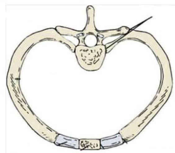
Identify the type of joint in the image provided.

Which type of collagen is most abundant in hyaline cartilage?
Antalgic hip gait is related to which of the following?
Lurching Gait is due to paralysis of which of the following?
While playing football, a 19-year-old college student receives a twisting injury to his knee when being tackled from the lateral side. Which of the following conditions most likely has occurred?
Lateral movement is produced by anterior translation of one condyle producing rotation about the
The axis of flexion and extension at the elbow joint passes through which of the following structures?
The mechanoreceptors in joints and ligaments are:
Which of the following is an intra-articular tendon?
Which of the following muscles is not in the pectoral region?
Explanation: ***Synovial joint*** - The image depicts a **costovertebral joint**, which connects a rib to a thoracic vertebra. These joints are **diarthrotic**, meaning they are freely movable, characteristic of synovial joints. - Synovial joints are characterized by the presence of a **synovial cavity**, articular cartilage, an articular capsule, and synovial fluid, allowing for a wide range of motion. *Syndesmosis* - A syndesmosis is a type of **fibrous joint** where two bones are joined by a ligament or a membrane, allowing for very limited movement, such as the distal tibiofibular joint. - This definition does not match the image, which shows a joint designed for movement between the rib and vertebra. *Synarthrosis* - Synarthrosis is a classification for **immovable joints**, such as sutures in the skull. - The costovertebral joints, as shown, allow for movement during respiration and are therefore not synarthrotic. *Symphysis* - A symphysis is a type of **cartilaginous joint** where bones are joined by **fibrocartilage**, allowing for slight movement. Examples include the pubic symphysis or intervertebral discs. - The costovertebral joint shown in the image is a synovial articulation, not a cartilaginous joint.
Explanation: ***Type II*** - **Type II collagen** is the predominant type found in **hyaline cartilage**, providing tensile strength and elasticity [1]. - It is crucial for the **structural integrity** and functionality of cartilage in articular surfaces [1]. *Type I* - Predominantly found in **bone**, tendons, and skin, contributing to tensile strength but not a major component of hyaline cartilage [2]. - It forms the structure of **fibrocartilage**, such as in the **intervertebral discs**. *Type IV* - Mainly located in **basement membranes** and plays a role in filtration and structural support of epithelial cells, not in hyaline cartilage. - It is critical in the formation of structures like **glomeruli** in kidneys, differing from cartilage's needs. *Type III* - Found in **reticular fibers** and supporting tissues throughout the body, important for organ structure but not prominent in hyaline cartilage. - Often associated with **vascular structures** and is not involved in the composition of cartilage.
Explanation: ***Painful hip gait*** - An **antalgic gait** is a deviation from a normal gait pattern caused by pain, most commonly experienced in the hip or knee. - The individual attempts to **minimize the time spent bearing weight** on the painful limb, resulting in a shortened stance phase on the affected side. *Waddling gait* - This gait is characterized by a **broad base** and a **swaying motion** from side to side, often due to weakness in the hip abductor muscles. - While sometimes seen in hip pathologies, it's not synonymous with an antalgic gait, which is specifically pain-driven. *Trendelenberg gait* - This gait occurs due to weakness of the **hip abductor muscles** (gluteus medius and minimus) on the stance leg, causing the pelvis to drop on the swing leg side. - It's a compensatory mechanism for muscle weakness, not directly caused by pain. *Short leg gait* - This gait arises from a **discrepancy in leg length**, leading to compensatory mechanisms like hip hiking or circumduction to clear the shorter limb during swing phase. - While it can lead to secondary pain, the primary cause is a structural difference, not acute pain influencing the weight-bearing phase.
Explanation: ***Gluteus medius*** * Paralysis of the **gluteus medius** leads to a **Trendelenburg gait** or **lurching gait**, where the pelvis drops on the unsupported side during walking. * This muscle is crucial for **stabilizing the pelvis** during the single-limb support phase of gait. *Adductor Magnus* * Paralysis of the adductor magnus would primarily affect **thigh adduction** and extension, not directly causing a lurching gait. * Problems with this muscle might impact the ability to bring the legs together or stabilize the leg during certain movements. *Hamstrings* * The hamstrings are responsible for **knee flexion** and **hip extension**. * Paralysis would result in difficulty bending the knee and limited hip extension, potentially leading to a stiff-knee gait, but not typically a lurching gait. *Quadriceps femoris* * The quadriceps femoris is essential for **knee extension** and is critical for activities like standing, walking, and climbing stairs. * Paralysis would cause the knee to buckle, leading to a **knee-hyperflexion gait** or difficulty with weight-bearing on that leg.
Explanation: **Tear of the medial meniscus** - A **twisting injury** to the knee, especially when tackled from the lateral side (which can force the leg into valgus stress), commonly causes a **tear of the medial meniscus**. - The **medial meniscus** is less mobile and more firmly attached than the lateral meniscus, making it more susceptible to injury during twisting forces. *Ruptured fibular collateral ligament* - A rupture of the **fibular collateral ligament (FCL)**, also known as the **lateral collateral ligament (LCL)**, typically results from a **varus stress** (a blow to the medial side of the knee), which is contrary to a tackle from the lateral side. - While twisting can contribute to knee injuries, isolate FCL tears from a lateral-side tackle are less likely than meniscal damage. *Tenderness on pressure along the fibular collateral ligament* - Tenderness along the **fibular collateral ligament** would indicate an injury to this structure, but a twisting injury from the lateral side is less likely to directly damage the FCL compared to the medial structures. - This symptom alone does not fully explain the mechanism of injury and the common resulting pathology in this scenario. *Injured posterior cruciate ligament* - The **posterior cruciate ligament (PCL)** is most commonly injured by a direct blow to the anterior tibia when the knee is flexed (a **dashboard injury**) or by a hyperflexion injury. - A twisting injury from the lateral side is a less common mechanism for isolated PCL injury.
Explanation: ***Center in the opposite condyle*** - **Lateral excursion** of the mandible involves the **working side condyle** rotating around a vertical axis, while the **non-working side condyle** translates anteriorly and medially (Bennett movement). - This anterior translation of the non-working condyle causes the entire mandible to pivot, with the center of rotation for the **lateral movement** being located roughly within the **condyle** on the **working (rotating)** side of the jaw. *Center in the opposite neck* - While the neck of the condyle is anatomically close to the condyle head, the **functional center of rotation** for lateral movement is typically described as being within the condyle itself, specifically its rotating component. - Positioning the center of rotation in the neck would imply a different biomechanical axis for the movement, which is not accurately reflected in standard mandibular kinematics. *Center in the opposite ramus* - The **ramus** is a broad part of the mandible, much larger than the condyle, and locating the center of rotation here would imply a much wider arc of movement, which is not consistent with the precise articulation of the **temporomandibular joint**. - The primary movements of the mandible during lateral excursion are centered on the condyle and its articular surfaces, not the entire ramus. *Center in the opposite angle* - The **angle of the mandible** is a distant anatomical landmark from the temporomandibular joint and is primarily involved in muscle attachments, not as a point of rotation for **lateral condylar movement**. - Placing the center of rotation at the angle would be biomechanically inaccurate for describing mandibular kinematics during lateral excursion.
Explanation: ***Trochlea*** - The **axis of flexion and extension at the elbow joint** (also called the carrying angle axis) passes through the **trochleo-capitellar region**, with the **trochlea forming the medial component** of this axis. - The **trochlea** articulates with the trochlear notch of the ulna and is the **primary structure** defining the medial aspect of the elbow's rotational axis. - This axis runs from the inferior aspect of the medial epicondyle, through the center of the trochlea and capitulum, to the inferior aspect of the lateral epicondyle. - In clinical and anatomical contexts, when asked about "the axis of the upper limb" at the elbow, **trochlea is the most appropriate answer** as it represents the dominant medial component. *Capitulum* - The **capitulum** forms the lateral part of the elbow axis and articulates with the head of the radius. - While the flexion-extension axis passes through the trochleo-capitellar region (including the capitulum), the **trochlea is considered the primary structure** as it provides the main hinge mechanism through its articulation with the ulna. *Olecranon* - The **olecranon** is the proximal end of the ulna, forming the prominent posterior bony point of the elbow. - It articulates with the **olecranon fossa** during extension and serves as the attachment for the triceps muscle. - The olecranon **rotates around the axis** but does not define the axis itself. *Radial styloid* - The **radial styloid process** is located at the distal end of the radius at the wrist. - It is involved in wrist articulation but is not related to the axis of the elbow joint.
Explanation: ***Slow adapting*** - **Mechanoreceptors** in joints and ligaments, such as **Ruffini endings** and **Golgi-type endings**, are primarily **slowly adapting**. - This characteristic allows them to provide continuous information about **joint position** and **pressure** over extended periods. *Adapt differentially for different stresses* - While different mechanoreceptors respond to different types of stimuli (e.g., pressure, stretch), this option describes varying responses rather than the fundamental **adaptation rate**. - The primary characteristic being asked for is how their firing rate changes over time in response to a constant stimulus. *Fast adapting* - **Fast-adapting mechanoreceptors**, like **Pacinian corpuscles** and **Meissner's corpuscles**, respond strongly at the onset and offset of a stimulus. - They are more involved in sensing **vibration** and **changes in pressure** rather than sustained joint position. *Non adapting* - All biological sensory receptors exhibit some degree of **adaptation** to a constant stimulus, meaning their firing rate changes over time. - A truly **non-adapting** receptor would fire at a constant rate indefinitely for a given stimulus, which is not characteristic of mechanoreceptors.
Explanation: ***Popliteus*** - The **popliteus tendon** originates within the knee capsule (intra-articular) before emerging to insert onto the posterior tibia. - It plays a crucial role in **unlocking the knee joint** from full extension and contributes to posterior stability. *Anconeus* - The **anconeus muscle** is located on the posterior aspect of the elbow, extending from the lateral epicondyle of the humerus to the ulna. - It is an **extra-articular muscle** that assists in elbow extension and stabilization. *Semitendinosus* - The **semitendinosus** is one of the hamstring muscles, located in the posterior thigh. - Its tendon contributes to the **pes anserinus**, inserting on the medial aspect of the tibia distal to the knee joint, making it an extra-articular tendon. *Sartorius* - The **sartorius** is the longest muscle in the body, running obliquely across the anterior aspect of the thigh. - Its tendon also contributes to the **pes anserinus**, inserting medially to the knee joint, and is considered extra-articular.
Explanation: ***Infraspinatus*** - The **infraspinatus** muscle is located in the **posterior scapular region**, specifically on the posterior aspect of the scapula, filling the infraspinous fossa. - Its primary function is **external rotation** of the humerus, and it is a key component of the **rotator cuff**. *Pectoralis major* - The **pectoralis major** is a large, superficial muscle located in the **anterior chest wall**, forming the bulk of the chest. [1] - It plays a significant role in **adduction**, **flexion**, and **medial rotation** of the humerus. *Pectoralis minor* - The **pectoralis minor** is a smaller, triangular muscle situated beneath the pectoralis major in the **anterior thoracic wall**. [1] - Its functions include **stabilizing the scapula** by pulling it inferiorly and anteriorly, and assisting in forced inspiration. [1] *Subclavius* - The **subclavius** is a small, triangular muscle located inferior to the clavicle in the **pectoral region**. - Its primary role is to **depress and stabilize the clavicle**, protecting the underlying neurovascular structures.
Get full access to all questions, explanations, and performance tracking.
Start For Free