
Embryology and Development — MCQs

On this page
All of the following are derivatives of the dorsal mesogastrium EXCEPT:
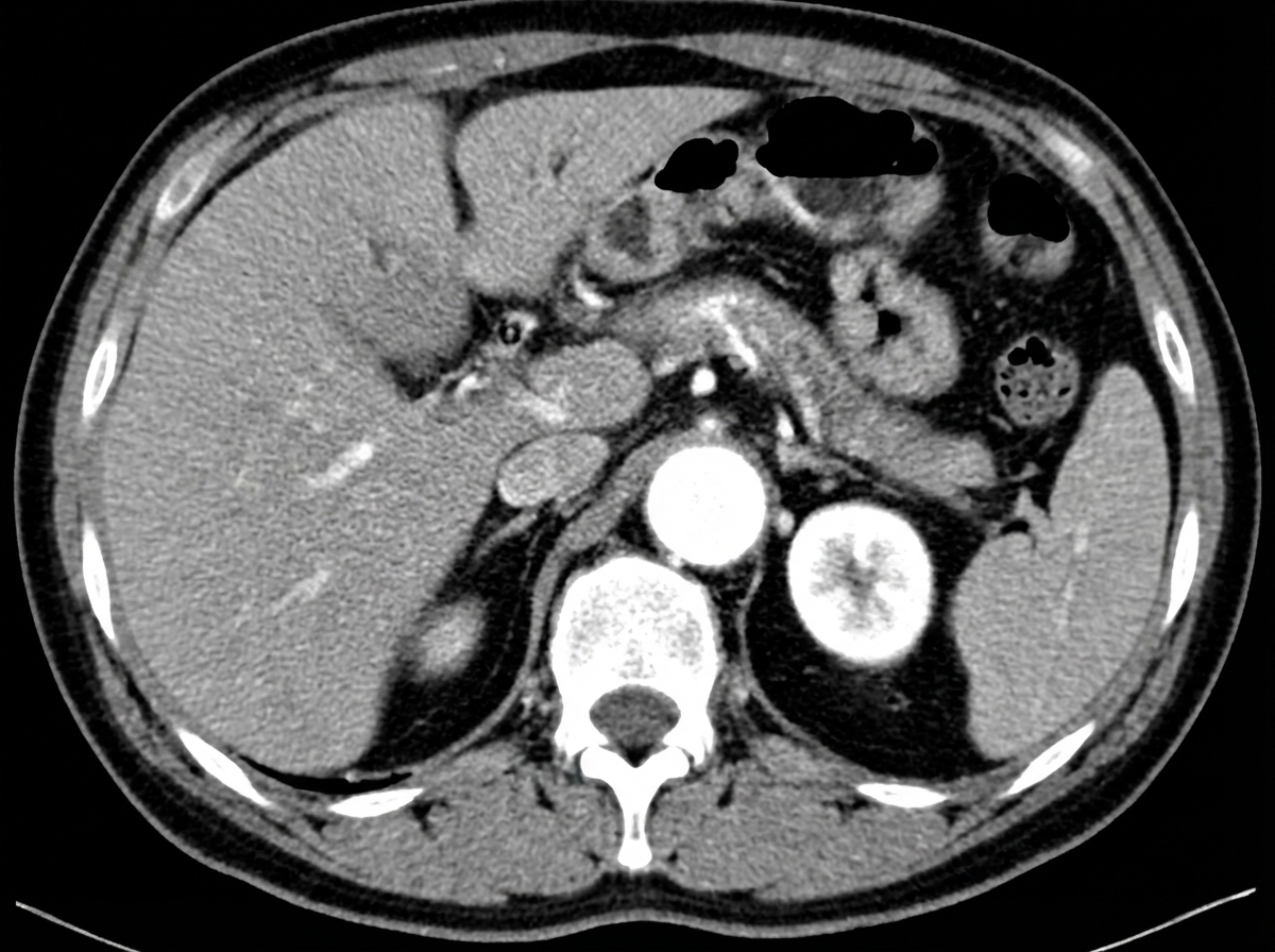
Complete regression of which of the following veins causes this anomaly in the development of the Inferior Vena Cava?
The excretory portion of the kidney is formed by which embryonic structure?
Merkel cells of the epidermis are derived from which embryonic structure?
Fusion of the caudal portions of the kidneys during embryonic development is most likely to result in which of the following congenital conditions?
At what gestational age are the ovaries and testes first distinguishable?
The fourth ventricle develops from which embryonic structure?
A baby is born with a large defect in the occipital bone through which the posterior portion of the brain has herniated. Which of the following terms best describes this lesion?
Bochdalek hernia is related to which of the following structures?
The head of a sperm is derived from which cellular organelle?
Practice by Chapter
Gametogenesis and Fertilization
Practice Questions
Early Embryonic Development
Practice Questions
Placentation
Practice Questions
Development of Nervous System
Practice Questions
Development of Cardiovascular System
Practice Questions
Development of Gastrointestinal System
Practice Questions
Development of Urogenital System
Practice Questions
Development of Musculoskeletal System
Practice Questions
Development of Head and Neck
Practice Questions
Congenital Anomalies
Practice Questions
Teratology
Practice Questions
Molecular Mechanisms in Development
Practice Questions
Want unlimited practice?
Get full access to all questions, explanations, and performance tracking.
Scan to download app