
Embryology and Development — MCQs

On this page
A newborn has multiple congenital defects owing to dysgenesis of the neural crest. Which of the following cells is most likely to be spared?
The bladder develops from which germ layer?
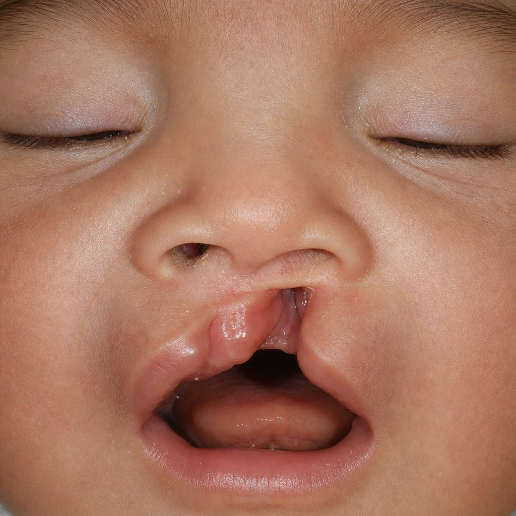
The given deformity is due to which of the following developmental anomalies?
Which of the following structures is derived from the surface ectoderm?
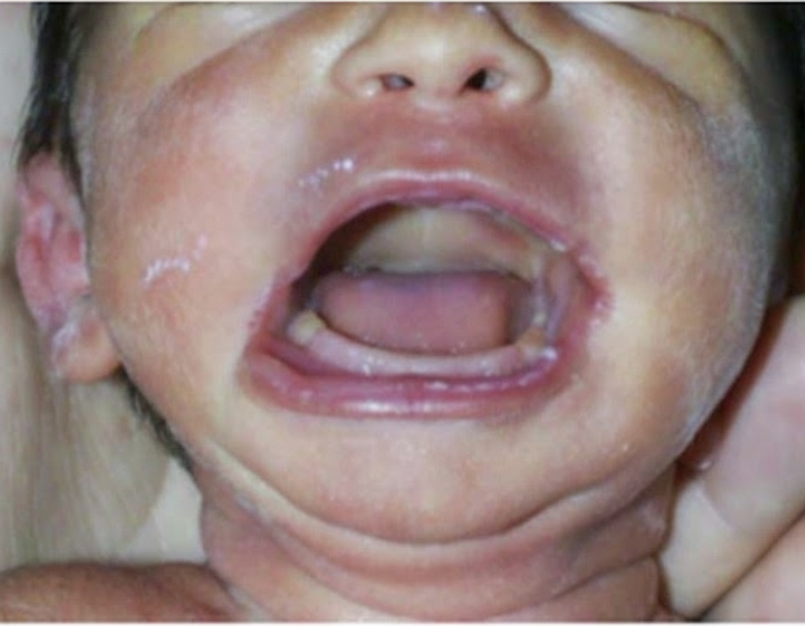
The given deformity is due to which of the following developmental anomalies?
Defect in any of the following may result in renal agenesis except?
The medial umbilical ligament is a remnant of which fetal structure?
What is the most common congenital anomaly of the upper renal tract?
The biceps brachii muscle develops from which of the following embryological structures?
Which gene is primarily responsible for eye morphogenesis?
Practice by Chapter
Gametogenesis and Fertilization
Practice Questions
Early Embryonic Development
Practice Questions
Placentation
Practice Questions
Development of Nervous System
Practice Questions
Development of Cardiovascular System
Practice Questions
Development of Gastrointestinal System
Practice Questions
Development of Urogenital System
Practice Questions
Development of Musculoskeletal System
Practice Questions
Development of Head and Neck
Practice Questions
Congenital Anomalies
Practice Questions
Teratology
Practice Questions
Molecular Mechanisms in Development
Practice Questions
Want unlimited practice?
Get full access to all questions, explanations, and performance tracking.
Scan to download app