
Embryology and Development — MCQs

On this page
In complete müllerian duct aplasia, all of the following are likely to be absent EXCEPT?
A 3-year-old male presents with a palpable mass in the right side of his scrotum. A preliminary diagnosis of congenital, indirect inguinal hernia is made. What is the most likely cause of an indirect inguinal hernia in this patient?
Where does the blastocyst normally implant?
Which of the following statements about fetal development is FALSE?
Which of the following structures in fetal life becomes the ligamentum teres in adult life?
What is the most common congenital defect of the face and jaws?
In the placenta, maternal blood comes in direct contact with which layer?
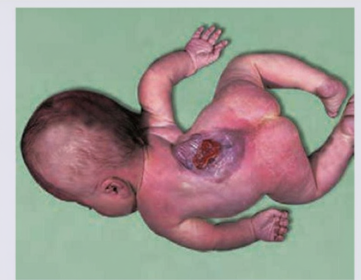
This defect most commonly involves which region of the spine?
The incus develops from which pharyngeal arch?
Extrophy of the bladder is associated with which of the following conditions?
Practice by Chapter
Gametogenesis and Fertilization
Practice Questions
Early Embryonic Development
Practice Questions
Placentation
Practice Questions
Development of Nervous System
Practice Questions
Development of Cardiovascular System
Practice Questions
Development of Gastrointestinal System
Practice Questions
Development of Urogenital System
Practice Questions
Development of Musculoskeletal System
Practice Questions
Development of Head and Neck
Practice Questions
Congenital Anomalies
Practice Questions
Teratology
Practice Questions
Molecular Mechanisms in Development
Practice Questions
Want unlimited practice?
Get full access to all questions, explanations, and performance tracking.
Scan to download app