
Embryology and Development — MCQs

On this page
To which group does the X chromosome belong?
All of the following are true about the notochord, EXCEPT:
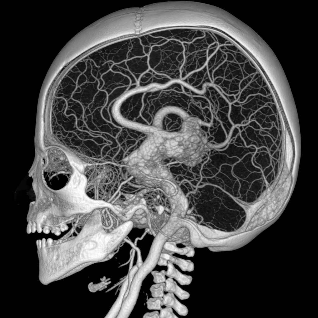
A neonate presents with features of heart failure. On examination, the anterior fontanelle was found to be bulging. On auscultation, a cranial bruit was heard. A CT angiography showed an abnormality. Which vessel is mainly involved in this abnormality?
Which of the following is the ovary-determining gene?
Permanent kidney in humans arise from which of the following embryological structures?
During the formation of the neural tube, in which direction do the neural folds fuse?
What is the remnant of the notochord in adults?
Spermatogonia divide by which type of cell division?
Which anatomical structure develops from the same embryonic precursor as the male genital swelling?
All of the following are features of Treacher Collins syndrome except:
Practice by Chapter
Gametogenesis and Fertilization
Practice Questions
Early Embryonic Development
Practice Questions
Placentation
Practice Questions
Development of Nervous System
Practice Questions
Development of Cardiovascular System
Practice Questions
Development of Gastrointestinal System
Practice Questions
Development of Urogenital System
Practice Questions
Development of Musculoskeletal System
Practice Questions
Development of Head and Neck
Practice Questions
Congenital Anomalies
Practice Questions
Teratology
Practice Questions
Molecular Mechanisms in Development
Practice Questions
Want unlimited practice?
Get full access to all questions, explanations, and performance tracking.
Scan to download app