Atavism is the resemblance of characteristics to which ancestral generation?
What is the most common site of fertilization?
How many vessels are typically seen in a cross-section of the umbilical cord?
In renal agenesis, where is the adrenal gland typically located?
The upper three-fourths of the vagina develops from which embryonic structure?
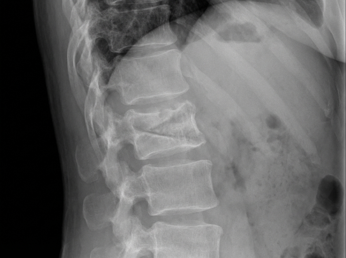
All of the following are components of the placental barrier, EXCEPT:
The stapes is a derivative of which embryonic pharyngeal arch?
What is true about Lyonization of the X chromosome?
Which of the following placental anomalies is characterized by an abnormal attachment of the umbilical cord?
A baby girl presents with bilateral inguinal masses, thought to be hernias but are found to be testes in the inguinal canals. Which karyotype would you expect to find in the child?
Explanation: **Explanation:** **Atavism** (also known as evolutionary throwback) refers to the reappearance of a biological trait in an organism after several generations of absence. In the context of medical genetics and embryology, it is defined as the resemblance of characteristics to **grandparents** or even more remote ancestors, rather than the immediate parents. 1. **Why Grandparents is correct:** Atavism occurs when genes for previously expressed ancestral traits—which were preserved in the DNA but "silenced" or suppressed during evolution—are suddenly reactivated due to genetic recombination or mutations. Since these traits skip the immediate generation (parents), the closest ancestral resemblance is noted in the grandparents or beyond. 2. **Why other options are wrong:** * **Parents:** Resemblance to parents is termed "heredity" or direct inheritance. * **Siblings:** Resemblance among siblings is due to shared parental alleles and is not considered an evolutionary throwback. **Clinical Pearls & High-Yield Facts for NEET-PG:** * **Examples of Atavism in Humans:** * **Atavistic Tail:** The presence of a true vestigial tail (containing muscle and blood vessels) in a newborn. * **Polythelia:** Presence of accessory nipples along the milk line. * **Persistent Lanugo:** Excessive body hair resembling an ancestral coat. * **Distinction:** Do not confuse Atavism with **Vestigial organs**. Vestigial organs (like the vermiform appendix) are present in all members of a species but have lost their original function, whereas Atavistic traits appear only in specific individuals. * **Mechanism:** It usually results from the loss of "suppressor genes" that normally keep ancestral developmental pathways dormant.
Explanation: **Explanation:** Fertilization is the process where the male and female gametes fuse to form a zygote. In humans, this occurs within the **Fallopian tube (uterine tube)** [1]. **Why Ampulla is the Correct Answer:** The **Ampulla** is the widest and longest part of the fallopian tube (approximately 5 cm long). Due to its spacious lumen and its location distal to the isthmus, it serves as the most favorable environment for the sperm to meet the oocyte. Statistically, it is the site of fertilization in over 90% of cases [1]. **Analysis of Incorrect Options:** * **Isthmus (A):** This is the narrow, thick-walled medial part of the tube. While sperm pass through it, its narrow lumen is not the primary site for fertilization. * **Infundibulum (C):** This is the funnel-shaped lateral end with fimbriae that "catch" the ovum from the ovary [1]. It is the entry point for the egg, not the site of fusion. * **Interstitial/Intramural (D):** This is the segment that pierces the uterine wall. It is the narrowest part of the tube and is not involved in fertilization. **NEET-PG High-Yield Pearls:** 1. **Ectopic Pregnancy:** The **Ampulla** is also the most common site for an ectopic pregnancy. 2. **Narrowest Part:** The **Interstitial (Intramural) part** is the narrowest portion of the fallopian tube (0.7 mm). 3. **Fertilization Timing:** Fertilization typically occurs within **12–24 hours** after ovulation. 4. **Implantation:** While fertilization occurs in the ampulla, implantation usually occurs in the **posterior wall of the body of the uterus** on approximately the 6th day after fertilization [1].
Explanation: **Explanation:** The umbilical cord typically contains **three vessels**: **two umbilical arteries** and **one umbilical vein** [1], all embedded in a gelatinous substance called **Wharton’s jelly**. 1. **Why Three is Correct:** During early development, there are initially four vessels (two arteries and two veins). However, the **right umbilical vein** undergoes regression (disappears) around the 8th week of gestation, leaving only the **left umbilical vein** and the two umbilical arteries. The vein carries oxygenated blood from the placenta to the fetus, while the arteries carry deoxygenated blood from the fetus back to the placenta [1]. 2. **Why Other Options are Incorrect:** * **One:** A single vessel is never normal and is incompatible with life. * **Two:** A **Single Umbilical Artery (SUA)** occurs in about 1% of pregnancies. While it can be an isolated finding, it is highly associated with congenital anomalies (renal or cardiac) and chromosomal trisomies, making it a significant clinical finding rather than the "typical
Explanation: The correct answer is **D. Present at the usual location.** **1. Why the correct answer is right:** The development of the adrenal gland and the kidney are embryologically independent processes. The **adrenal cortex** develops from the intermediate mesoderm (coelomic epithelium) between the root of the mesentery and the developing gonad, while the **adrenal medulla** is derived from neural crest cells. In contrast, the **permanent kidney (metanephros)** develops from the ureteric bud and metanephric blastema, which originate much lower in the pelvis and subsequently ascend to the lumbar region. Because the adrenal glands develop in situ in the upper abdomen and do not "ascend" like the kidneys, the absence of a kidney (renal agenesis) does not prevent the adrenal gland from forming or occupying its normal anatomical position [2]. **2. Why the incorrect options are wrong:** * **Option A:** The adrenal gland is not absent because its embryological origin is distinct from the metanephric system. * **Option B:** Adrenal development is bilateral and independent; the absence of one kidney does not cause the ipsilateral adrenal to migrate to the contralateral side. * **Option C:** The adrenal gland does not follow the kidney's migratory path [2]. Even in cases of pelvic kidneys (ectopy), the adrenal gland remains in its normal sub-diaphragmatic position [1]. **3. Clinical Pearls & High-Yield Facts for NEET-PG:** * **"Lying-down Adrenal" Sign:** On prenatal ultrasound or CT, in renal agenesis, the adrenal gland appears elongated and flattened (pancake-like) because it is not molded by the superior pole of the kidney. * **Potter Sequence:** Bilateral renal agenesis leads to oligohydramnios, which causes pulmonary hypoplasia, limb deformities, and characteristic facial features (Potter facies). * **Blood Supply:** The adrenal gland receives a rich supply from three sources (Superior, Middle, and Inferior suprarenal arteries), further highlighting its independent vascular and structural integrity.
Explanation: **Explanation:** The development of the female reproductive tract is a high-yield topic in embryology. The vagina has a **dual origin**, arising from two distinct embryonic structures: 1. **Mullerian (Paramesonephric) Ducts:** The caudal ends of the Mullerian ducts fuse to form the **uterovaginal canal**. This canal gives rise to the uterus, cervix, and the **upper 3/4th (upper portion)** of the vagina [1]. 2. **Urogenital Sinus (Sinovaginal Bulbs):** The lower part of the uterovaginal canal meets the urogenital sinus, inducing the formation of the **sinovaginal bulbs**. These bulbs proliferate and canalize to form the **lower 1/4th** (or 1/5th) of the vagina [1]. **Analysis of Options:** * **Option A (Correct):** As stated, the Mullerian ducts form the upper 3/4th of the vagina, along with the fallopian tubes and uterus [1]. * **Option B (Incorrect):** The **Wolffian (Mesonephric) ducts** regress in females due to the absence of testosterone. Remnants may persist as **Gartner’s cysts** in the lateral wall of the vagina. * **Option C (Incorrect):** The sinovaginal bulbs form the **lower 1/4th** of the vagina, not the upper portion [1]. * **Option D (Incorrect):** While the urogenital sinus is endodermal, the upper 3/4th of the vagina is derived from the Mullerian duct, which is **mesodermal** in origin [1]. **High-Yield Clinical Pearls for NEET-PG:** * **Mayer-Rokitansky-Küster-Hauser (MRKH) Syndrome:** Congenital absence of the uterus and the upper 2/3rd to 3/4th of the vagina due to Mullerian agenesis. * **Hymen:** Formed at the junction where the sinovaginal bulbs meet the urogenital sinus. * **Epithelium:** Initially, the entire vagina is lined by columnar epithelium (Mullerian), which is later replaced by **stratified squamous epithelium** from the urogenital sinus.
Explanation: The **placental barrier** (placental membrane) is a composite structure that separates the maternal blood in the intervillous spaces from the fetal blood within the villi [1]. Its primary function is to prevent the mixing of blood while allowing the exchange of nutrients, gases, and waste [3]. ### Why Amnion is the Correct Answer The **Amnion** is the innermost fetal membrane that encloses the amniotic cavity and the fetus [1]. While it is part of the fetal membranes, it **does not contribute to the structure of the chorionic villi** where gas and nutrient exchange occurs [1]. Therefore, it is not a component of the placental barrier. ### Explanation of Other Options The placental barrier is composed of four layers (from external to internal): * **Trophoblast (Option A):** Specifically the outer **Syncytiotrophoblast** (which is in direct contact with maternal blood) and the inner **Cytotrophoblast** [5]. * **Mesoderm (Option C):** The extraembryonic mesoderm forms the connective tissue core of the villus, providing structural support [4]. * **Fetal capillary endothelium (Option B):** The innermost layer that lines the fetal blood vessels within the villi [4]. ### High-Yield Facts for NEET-PG * **Thinning of the Barrier:** In early pregnancy, all four layers are present. In late pregnancy (after the 4th month), the barrier thins to facilitate faster exchange; the **cytotrophoblast** and **mesoderm** largely disappear, leaving only the syncytiotrophoblast and fetal endothelium. * **The "Barrier" is not absolute:** Most drugs with a molecular weight <600 Da, viruses (e.g., Rubella, CMV, HIV), and certain antibodies (IgG) can cross this barrier [3]. * **IgG Transfer:** IgG is the only immunoglobulin that crosses the placenta, providing passive immunity to the newborn [2].
Explanation: The pharyngeal (branchial) arches are fundamental to head and neck development. Each arch contains a central cartilaginous rod that serves as a precursor to specific skeletal structures [1]. **Correct Answer: B. Second arch (Reichert’s cartilage)** The second pharyngeal arch is responsible for forming the **stapes** (except for its vestibular part/footplate, which is partly derived from the neural crest and otic capsule). Other key skeletal derivatives of the second arch include the **styloid process** of the temporal bone, the **stylohyoid ligament**, and the **lesser cornu and upper part of the body of the hyoid bone**. **Explanation of Incorrect Options:** * **A. First arch (Meckel’s cartilage):** This arch gives rise to the **malleus** and **incus** (the other two middle ear ossicles), as well as the mandible, maxilla, and zygomatic bone. * **C. Third arch:** This arch forms the **greater cornu and lower part of the body of the hyoid bone**. * **D. Fourth arch:** This arch (along with the sixth) contributes to the **laryngeal cartilages** (thyroid, cricoid, arytenoid, etc.), excluding the epiglottis. **High-Yield Clinical Pearls for NEET-PG:** * **Nerve Supply:** Remember the "Rule of Nerves": 1st Arch = Trigeminal (V); 2nd Arch = **Facial (VII)**; 3rd Arch = Glossopharyngeal (IX); 4th/6th Arch = Vagus (X). Since the stapes is a 2nd arch derivative, it is moved by the **stapedius muscle**, which is supplied by the **Facial nerve** [1]. * **Treacher Collins Syndrome:** Results from the failure of neural crest cell migration into the **first and second arches**, leading to malformation of the ossicles (conductive hearing loss) and zygomatic hypoplasia. * **Mnemonic:** The **S**tructures of the **S**econd arch all start with **S**: **S**tapes, **S**tyloid process, **S**tylohyoid ligament, and **S**even (CN VII).
Explanation: ### Explanation: Lyonization of the X Chromosome **1. Understanding the Correct Answer (Option A)** Lyonization, or X-inactivation, is the process by which one of the two X chromosomes in female mammals is inactivated to ensure **dosage compensation** (equalizing gene expression between males and females) [1]. This process occurs **only in somatic cells** during early embryonic development (blastocyst stage). In individuals with multiple X chromosomes, only one remains active while the others are inactivated [1]. The inactivated X chromosome becomes heterochromatic and is visible as a **Barr body**. **2. Why Other Options are Incorrect** * **Options B & C:** Inactivation does **not** occur in germ cells. In fact, both X chromosomes must remain active in the germline to ensure successful oogenesis. If an X chromosome is inactivated in a germ cell, it is "reactivated" before meiosis begins. * **Option D:** The number of Barr bodies is calculated by the formula: **(Number of X chromosomes – 1)**. For example, a normal female (46,XX) has one Barr body, while a female with Turner syndrome (45,XO) has zero. **3. High-Yield Clinical Pearls for NEET-PG** * **Timing:** Lyonization occurs around the **3rd to 6th day** of gestation (late blastocyst stage). * **Randomness:** In humans, the choice of which X (maternal or paternal) is inactivated is random, making females **genetic mosaics** [1]. * **XIST Gene:** The *X-inactive specific transcript* (XIST) gene, located on the X-inactivation center (Xq13), is the master regulator that produces non-coding RNA to coat and silence the chromosome [1]. * **Clinical Correlation:** In **Klinefelter syndrome (47,XXY)**, there is one Barr body despite the patient being phenotypically male. In **Triple X syndrome (47,XXX)**, there are two Barr bodies.
Explanation: ### Explanation **Correct Answer: B. Battledore placenta (Marginal insertion)** The umbilical cord normally inserts into the center of the placental mass (eccentric or central insertion) [3]. In a **Battledore placenta**, the cord is attached at or within 2 cm of the **placental margin** [1]. The name is derived from its resemblance to a "battledore" (a historical badminton racket). While often clinically insignificant, it can be associated with preterm labor, fetal distress, or progress to a velamentous insertion. **Analysis of Incorrect Options:** * **A. Succenturiate lobe:** This is an anomaly of **placental morphology**, not cord attachment [2]. It involves one or more small accessory lobes of placental tissue situated at a distance from the main placental mass, connected by fetal vessels running through the membranes [2]. * **C. Horseshoe placenta:** This is a rare morphological variant where the placenta is shaped like a horseshoe, often associated with placenta previa. It does not specifically define an anomaly of cord attachment. **High-Yield Clinical Pearls for NEET-PG:** 1. **Velamentous Insertion:** A more severe attachment anomaly where the cord inserts into the **fetal membranes** (chorioamniotic membranes) rather than the placental mass itself [1]. The vessels run unprotected by Wharton’s jelly. 2. **Vasa Previa:** Often associated with velamentous insertion or succenturiate lobes; fetal vessels cross the internal os [1]. This is a surgical emergency due to the risk of fetal exsanguination upon rupture of membranes. 3. **Placenta Membranacea:** A rare condition where the entire chorionic sac is covered by functional villi, resulting in a thin, membranous placenta [2].
Explanation: ### Explanation **Correct Answer: B. 46,XY** **Concept:** The clinical presentation describes a classic case of **Androgen Insensitivity Syndrome (AIS)**, formerly known as Testicular Feminization Syndrome. In AIS, there is a mutation in the androgen receptor gene. Although the individual has a **46,XY karyotype** and functional testes that produce testosterone and Anti-Müllerian Hormone (AMH), the peripheral tissues cannot respond to androgens [1]. 1. **Testicular Development:** The presence of the SRY gene on the Y chromosome ensures the development of testes. 2. **Phenotype:** Because the body cannot respond to testosterone/DHT, external genitalia develop along female lines (default pathway). 3. **Clinical Clue:** The "inguinal hernias" in a phenotypic female infant are actually **undescended testes** located in the inguinal canal or labia majora. AMH causes regression of the uterus and fallopian tubes, leading to a blind-ending vaginal pouch. --- **Analysis of Incorrect Options:** * **A. 46,XX:** This is a normal female karyotype. While inguinal hernias can occur in females (containing ovaries), they would not contain histologically confirmed testes. * **C & D. 46,XXY / 47,XXY (Klinefelter Syndrome):** These individuals have a male phenotype (small testes, gynecomastia, tall stature) because they respond to androgens. They do not present as phenotypic females with inguinal masses in infancy. --- **High-Yield NEET-PG Pearls:** * **Most common cause of primary amenorrhea with absent uterus:** AIS (46,XY) or Müllerian Agenesis (46,XX). * **AIS vs. Müllerian Agenesis:** In AIS, testosterone levels are in the normal male range and pubic/axillary hair is absent or sparse. * **Management:** Testes are usually left in situ until after puberty to allow for natural estrogenization (via peripheral aromatization of testosterone), then removed to prevent **Gonadoblastoma/Dysgerminoma**.
Gametogenesis and Fertilization
Practice Questions
Early Embryonic Development
Practice Questions
Placentation
Practice Questions
Development of Nervous System
Practice Questions
Development of Cardiovascular System
Practice Questions
Development of Gastrointestinal System
Practice Questions
Development of Urogenital System
Practice Questions
Development of Musculoskeletal System
Practice Questions
Development of Head and Neck
Practice Questions
Congenital Anomalies
Practice Questions
Teratology
Practice Questions
Molecular Mechanisms in Development
Practice Questions
Get full access to all questions, explanations, and performance tracking.
Start For Free