Which gland is derived from the foramen caecum?
The outer cell mass of the blastocyst gives rise to which of the following?
Which substance is secreted by Sertoli cells of the fetal testis?
By which day does the caudal end of the neuropore close?
The palate is formed from which of the following embryonic structures?
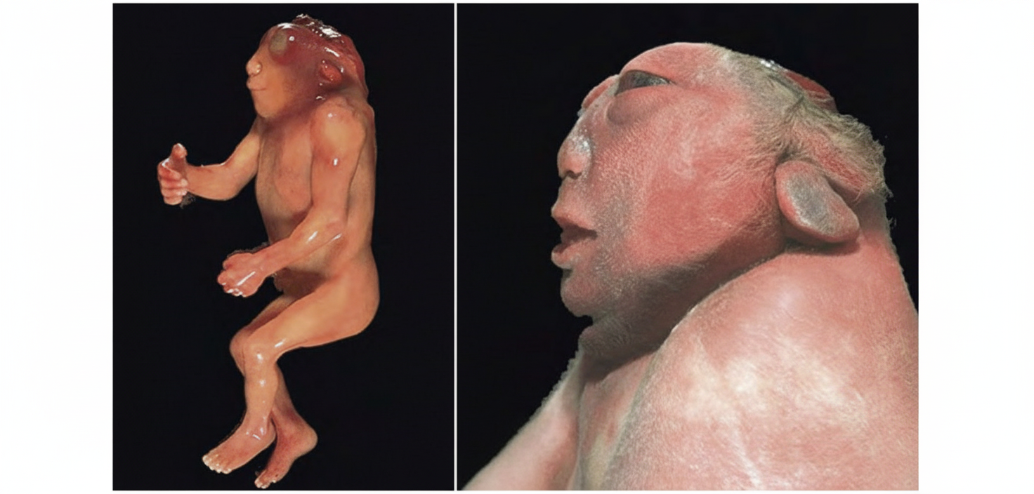
A fertilized ovum reaches the uterine cavity in how many days?
The hyoid bone is a derivative of which of the following?
Which of the following is NOT true about the primitive streak?
At what age does the fetal circulation first become intact and functional, separate from maternal circulation?
Ovaries develop from which of the following structures?
Explanation: ### Explanation **1. Why Thyroid is Correct:** The thyroid gland is the first endocrine gland to develop in the embryo (around the 24th day). It originates as a midline endodermal thickening in the floor of the primordial pharynx, just caudal to the tuberculum impar [1]. This site is marked by a small pit called the **foramen caecum**, located at the junction of the anterior two-thirds and posterior one-third of the tongue (the apex of the sulcus terminalis). The thyroid primordium descends as a diverticulum into the neck, remaining connected to the tongue via the **thyroglossal duct**, which normally disappears later [1]. **2. Why the Other Options are Incorrect:** * **Parathyroid Glands:** These do not arise from the foramen caecum. Instead, they develop from the **pharyngeal pouches**. * **Superior Parathyroid Glands:** Derived from the **4th pharyngeal pouch**. * **Inferior Parathyroid Glands:** Derived from the **3rd pharyngeal pouch** (along with the thymus). * *Note: Options B, C, and D all list the Parathyroid, which follows a different embryological pathway involving the branchial apparatus rather than the midline tongue base.* **3. Clinical Pearls for NEET-PG:** * **Thyroglossal Cyst:** The most common congenital midline neck swelling. It occurs due to a persistent remnant of the thyroglossal duct and characteristically **moves upward on protrusion of the tongue** [2]. * **Ectopic Thyroid:** The most common site is the **Lingual Thyroid**, occurring when the gland fails to descend from the foramen caecum [2]. * **Pyramidal Lobe:** A common anatomical variant representing a persistent distal end of the thyroglossal duct [1]. * **Parafollicular (C) cells:** Unlike the follicular cells (endoderm), these are derived from the **Ultimobranchial body** (Neural crest cells).
Explanation: ### Explanation At the **blastocyst stage** (approximately day 4–5 after fertilization), the cells of the morula undergo compaction and differentiation into two distinct populations based on their position [1]: 1. **Outer Cell Mass (Trophoblast):** These cells form the outer wall of the blastocyst. Their primary function is to provide nutrients to the embryo and facilitate implantation [1]. The trophoblast eventually differentiates into the **cytotrophoblast** and the **syncytiotrophoblast**, which contribute to the formation of the **placenta** [2]. 2. **Inner Cell Mass (Embryoblast):** These cells are situated at one pole of the blastocyst and give rise to the **embryo proper** and associated extraembryonic membranes (amnion, yolk sac) [1]. #### Analysis of Options: * **Option A (Embryo proper):** Incorrect. The embryo proper develops from the **Inner Cell Mass (Embryoblast)**, not the outer cell mass [1]. * **Option B (Trophoblast):** **Correct.** The outer cell mass is synonymous with the trophoblast [1]. * **Option C (Syncytiotrophoblast):** Incorrect. While the outer cell mass *eventually* gives rise to the syncytiotrophoblast, it does so by first forming the trophoblast [2]. In embryology questions, the immediate derivative is the preferred answer. The syncytiotrophoblast is a secondary differentiation that occurs during implantation (Day 7–8). #### High-Yield NEET-PG Pearls: * **Totipotency vs. Pluripotency:** The zygote and early blastomeres (up to the 8-cell stage) are **totipotent**. The Inner Cell Mass is **pluripotent** (can form all body tissues but not the placenta). * **Hatching:** The blastocyst must "hatch" from the **Zona Pellucida** before implantation can occur. * **Implantation Site:** The most common site for normal implantation is the **posterior wall of the body of the uterus** near the fundus. * **hCG Production:** The **Syncytiotrophoblast** (derived from the trophoblast) is responsible for secreting Human Chorionic Gonadotropin (hCG), which maintains the corpus luteum [2].
Explanation: **Explanation:** The differentiation of the male reproductive system is an active process driven by hormones secreted by the fetal testes. The correct answer is **Mullerian Inhibiting Substance (MIS)**, also known as Anti-Müllerian Hormone (AMH) [2]. 1. **Why it is correct:** In a male fetus (XY), the *SRY* gene on the Y chromosome triggers the differentiation of primitive gonads into testes. Within the testes, **Sertoli cells** secrete MIS [1]. The primary function of MIS is to cause the regression of the Paramesonephric (Müllerian) ducts, which would otherwise develop into the uterus, fallopian tubes, and upper vagina [2]. 2. **Why other options are incorrect:** * **hCG (Human Chorionic Gonadotropin):** Secreted by the syncytiotrophoblast of the placenta to maintain the corpus luteum. * **LH (Luteinizing Hormone):** Secreted by the anterior pituitary gland. * **Testosterone:** Secreted by the **Leydig cells** (interstitial cells) of the fetal testis, not Sertoli cells. Testosterone is responsible for the development of Mesonephric (Wolffian) ducts into the epididymis, vas deferens, and seminal vesicles [2]. **High-Yield Clinical Pearls for NEET-PG:** * **Sertoli Cells:** Produce MIS (inhibits Müllerian ducts) and Inhibin [1]. * **Leydig Cells:** Produce Testosterone (stimulates Wolffian ducts) and Dihydrotestosterone (DHT) via 5α-reductase (develops external genitalia) [2]. * **Persistent Müllerian Duct Syndrome:** Occurs due to a deficiency of MIS or its receptors, resulting in a male phenotype with a rudimentary uterus and tubes. * **Mnemonic:** **S**ertoli cells **S**top Müllerian ducts; **L**eydig cells **L**ead to Wolffian development.
Explanation: **Explanation:** The closure of the neural tube (neurulation) is a critical event in embryology that occurs during the 4th week of development. The neural tube does not close all at once; instead, it closes like a zipper, starting in the cervical region and proceeding cranially and caudally. This process leaves two openings called **neuropores**. 1. **Cranial (Anterior) Neuropore:** Closes first, typically on the **25th day** (or the 18-20 somite stage). 2. **Caudal (Posterior) Neuropore:** Closes approximately two days later, on the **27th day** (or the 25 somite stage). **Analysis of Options:** * **Option A (24th day):** This is too early for the caudal neuropore; it is closer to the timing of the cranial neuropore closure (Day 25). * **Option B (27th day):** **Correct.** This represents the standard embryological timeline for the completion of primary neurulation at the caudal end. * **Option C & D (32nd/40th day):** These are well beyond the window of primary neurulation. By this time, secondary neurulation (formation of the sacral and coccygeal segments) is underway. **High-Yield NEET-PG Pearls:** * **Clinical Correlation:** Failure of the cranial neuropore to close results in **Anencephaly**. Failure of the caudal neuropore to close results in **Spina Bifida** (most commonly Spina Bifida Cystica) [1]. * **Biomarker:** Neural tube defects (NTDs) are associated with elevated **Alpha-fetoprotein (AFP)** in maternal serum and amniotic fluid, along with the presence of **Acetylcholinesterase** [1]. * **Prevention:** Supplementation with **Folic Acid** (400 mcg/day) starting before conception significantly reduces the risk of NTDs.
Explanation: ### Explanation The development of the palate occurs between the **6th and 12th weeks** of gestation through the fusion of two distinct embryonic components: 1. **Primary Palate (Median Palatine Process):** Formed by the fusion of the two **medial nasal processes**. It gives rise to the premaxillary part of the maxilla, which holds the four upper incisor teeth. 2. **Secondary Palate (Lateral Palatine Processes):** These are two shelf-like projections (palatal shelves) that arise from the **maxillary processes**. Initially, they grow downward on either side of the tongue but later elevate and fuse horizontally in the midline. The final palate is formed when the primary palate fuses with the secondary palate at the **incisive foramen**. Therefore, both median and lateral processes are essential for complete formation. **Analysis of Incorrect Options:** * **Option A:** The median palatine process only forms the anterior (primary) palate. Relying solely on this would result in a massive posterior cleft. * **Option B:** The lateral palatine processes form the hard and soft palate posterior to the incisive foramen. Without the median process, the anterior alveolar ridge would be incomplete. * **Option D:** Development is a coordinated fusion of both structures; they are not alternatives to one another. **High-Yield Clinical Pearls for NEET-PG:** * **Critical Period:** Palatal fusion is completed by the 12th week. * **Cleft Lip:** Failure of fusion between the **maxillary process** and **medial nasal process**. * **Cleft Palate:** Failure of fusion of the **lateral palatine shelves** with each other or with the primary palate [1]. * **Incisive Foramen:** Serves as the anatomical landmark dividing the primary and secondary palate.
Explanation: **Explanation:** **1. Why Option A is Correct:** Fertilization typically occurs in the **ampulla** of the fallopian tube [2]. Following fertilization, the zygote undergoes a series of mitotic divisions called cleavage as it travels toward the uterus. It takes approximately **3 to 4 days** for the developing embryo (usually at the **morula stage**, consisting of 12–16 cells) to traverse the fallopian tube and enter the uterine cavity [1]. The movement is facilitated by ciliary action and rhythmic contractions of the tubal musculature [1]. **2. Analysis of Incorrect Options:** * **Option B (5-6 days):** This is the timeframe when the **blastocyst begins to implant** into the endometrial lining, not when it first reaches the cavity [4]. * **Option C (7-8 days):** By this stage, the blastocyst is already partially embedded in the endometrium (interstitial implantation) [4]. * **Option D (10 days):** By day 10, the blastocyst is almost completely buried in the decidua, and the primitive uteroplacental circulation begins to form [4]. **3. NEET-PG High-Yield Pearls:** * **Site of Fertilization:** Ampulla of the fallopian tube (most common) [2]. * **Stage of Entry:** The embryo enters the uterine cavity at the **Morula stage** (Day 4) [1]. * **Stage of Implantation:** Implantation occurs at the **Blastocyst stage** [3]. * **Implantation Window:** Begins on Day 6 and is completed by Day 10–12 [4]. * **Zona Pellucida:** It must disappear ("hatching") before implantation can occur; its presence prevents ectopic implantation within the tube [1].
Explanation: The hyoid bone is a unique structure in the neck that develops from the cartilaginous elements of the **second and third pharyngeal arches**. ### **Explanation of the Correct Answer** The hyoid bone is formed through the fusion of multiple components derived from different arches: * **Second Pharyngeal Arch (Reichert’s Cartilage):** Gives rise to the **lesser cornu (horn)** and the **upper part of the body** of the hyoid. * **Third Pharyngeal Arch:** Gives rise to the **greater cornu (horn)** and the **lower part of the body** of the hyoid. ### **Analysis of Incorrect Options** * **Option A (First Arch):** The first arch (Mandibular arch/Meckel’s cartilage) forms the malleus, incus, and the mandible (via membrane bone formation around the cartilage). It does not contribute to the hyoid. * **Option B (First and Second):** While the second arch contributes, the first arch does not. * **Option D (Fourth Arch):** The fourth and sixth arches contribute to the **laryngeal cartilages** (thyroid, cricoid, arytenoid, corniculate, and cuneiform), not the hyoid bone. ### **High-Yield Clinical Pearls for NEET-PG** * **Nerve Supply:** Because the hyoid is derived from the 2nd and 3rd arches, the muscles attaching to it are supplied by the **Facial nerve (CN VII)** and **Glossopharyngeal nerve (CN IX)** respectively. * **Stylohyoid Ligament:** This is also a derivative of the **second arch** and connects the styloid process to the lesser cornu of the hyoid. * **Fracture Significance:** A fractured hyoid bone is a classic forensic finding in cases of **strangulation or throttling**. * **Ectopic Tissue:** The **Thyroglossal duct** descends from the foramen caecum to the thyroid gland, passing either anterior to or through the hyoid bone. This is a common site for thyroglossal cysts.
Explanation: ### Explanation The **primitive streak** is a pivotal structure in embryology, marking the beginning of **gastrulation** (the process by which the bilaminar disc transforms into a trilaminar disc) [1]. **1. Why Option C is the correct answer (The False Statement):** The primitive streak appears at the **caudal (posterior) end** of the dorsal aspect of the epiblast, not the cranial end. It elongates cranially, and its appearance establishes the cranio-caudal axis, the medial-lateral axis, and the dorsal-ventral symmetry of the embryo. **2. Analysis of Incorrect Options:** * **Option A:** This is a true statement. The formation of the primitive streak on day 15 is the **first morphological sign of gastrulation**. * **Option B:** This is a true statement. The streak is formed by the proliferation and migration of **epiblast cells** toward the median plane of the embryonic disc [1]. These cells then undergo an epithelial-to-mesenchymal transition to form the intraembryonic mesoderm and endoderm. **3. NEET-PG High-Yield Facts & Clinical Pearls:** * **Primitive Node (Hensen’s Node):** The cephalic end of the streak expands to form this node, which acts as the "organizer." * **Fate of the Streak:** Under normal conditions, the primitive streak regresses and disappears by the end of the 4th week. * **Clinical Correlation (Sacrococcygeal Teratoma):** If the primitive streak fails to regress and remnants persist in the sacrococcygeal region, it can lead to a **Sacrococcygeal Teratoma**. This is the most common tumor in newborns and contains tissues from all three germ layers. * **Situs Inversus:** Abnormalities in the signaling molecules at the primitive streak/node (like *SHH* and *FGF8*) can lead to defects in left-right asymmetry.
Explanation: ### Explanation The development of the cardiovascular system is a critical milestone in embryology, as it is the first functional organ system to develop in the human embryo. **Why 21 Days is Correct:** By the beginning of the **third week** of development, the embryo can no longer satisfy its nutritional and oxygen requirements by simple diffusion alone. Vasculogenesis and angiogenesis begin in the extraembryonic mesoderm (yolk sac and stalk) and the intraembryonic mesoderm. By **Day 21**, the primitive heart tubes fuse and begin to beat. At this point, a continuous circuit is established between the heart, the intraembryonic vessels, and the extraembryonic vessels (umbilical and vitelline systems), making the fetal circulation **intact and functional** [1]. **Analysis of Incorrect Options:** * **A. 8 days:** This corresponds to the early implantation phase (blastocyst stage). The embryo is a bilaminar disc, and no vascular system exists. * **B. 12 days:** This is the stage of primary villi formation and the development of the extraembryonic mesoderm. Circulation has not yet begun. * **C. 17 days:** At this stage, blood islands begin to appear in the yolk sac and the primitive streak is active (Gastrulation), but the heart has not yet started pumping. **High-Yield NEET-PG Pearls:** * **First system to function:** Cardiovascular system (Week 3). * **Heartbeat begins:** Day 21–22. * **First site of hematopoiesis:** Yolk sac (extraembryonic mesoderm) starting around Day 17. * **Fetal Hemoglobin (HbF):** Has a higher affinity for oxygen than adult hemoglobin (HbA), facilitating oxygen transfer across the placenta [2]. * **Cardiac Progenitor Cells:** Derived from the **epiblast** (lateral plate mesoderm).
Explanation: **Explanation:** The development of the gonads (ovaries or testes) is a complex process involving three main sources: the mesothelium (lining the posterior abdominal wall), the underlying mesenchyme, and primordial germ cells. **Why Option A is Correct:** The **Genital Ridge** (or gonadal ridge) is a thickening of the intermediate mesoderm on the medial aspect of the mesonephros. During the 5th week of development, the proliferation of the surface epithelium and the condensation of the underlying mesenchyme form this ridge. In the presence of a female genotype (absence of the SRY gene), the primary sex cords of the genital ridge regress, and secondary cortical cords develop to form the **ovary**. **Why the other options are incorrect:** * **B. Mullerian duct (Paramesonephric duct):** These ducts give rise to the female internal genital tract, specifically the **fallopian tubes, uterus, and the upper 4/5th of the vagina** [1]. They do not form the ovaries. * **C. Endoderm of Urogenital sinus:** This structure gives rise to the **lower 1/5th of the vagina**, the urinary bladder, and the urethra [2]. * **D. Genital tubercle:** This is a component of the external genitalia. In females, it develops into the **clitoris**; in males, it forms the glans penis. **High-Yield NEET-PG Pearls:** * **Primordial Germ Cells:** These originate from the **epiblast**, migrate to the **yolk sac wall**, and eventually reach the genital ridge by the 6th week. If they fail to reach the ridge, the gonad does not develop (streak ovaries). * **Dual Origin of Vagina:** Remember the "4/5 and 1/5" rule—upper part from Mullerian ducts (mesoderm) and lower part from Urogenital sinus (endoderm) [2]. * **Ligaments:** The gubernaculum in females becomes the **ovarian ligament** and the **round ligament of the uterus**.
Gametogenesis and Fertilization
Practice Questions
Early Embryonic Development
Practice Questions
Placentation
Practice Questions
Development of Nervous System
Practice Questions
Development of Cardiovascular System
Practice Questions
Development of Gastrointestinal System
Practice Questions
Development of Urogenital System
Practice Questions
Development of Musculoskeletal System
Practice Questions
Development of Head and Neck
Practice Questions
Congenital Anomalies
Practice Questions
Teratology
Practice Questions
Molecular Mechanisms in Development
Practice Questions
Get full access to all questions, explanations, and performance tracking.
Start For Free