
Embryology and Development — MCQs

On this page
Which of the following statements about the development of the ear is true?
Holt-Oram syndrome is caused by a mutation of?
From which structure does the spleen develop?
Which of the following is NOT a recognized cause of renal agenesis?
Closure of the neural tube begins from which anatomical region?
Scaphocephaly is due to premature closure of which of the following sutures?
At 30 days of intrauterine life, what is the expected developmental milestone?
Which carpal bone is the first to ossify during fetal development?
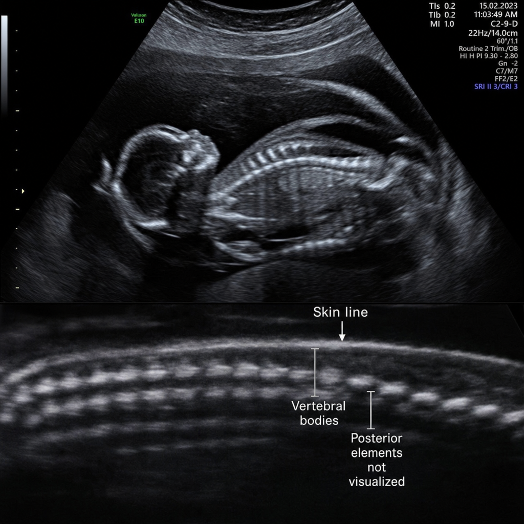
A 25-year-old pregnant female at 18 weeks' gestation presented to the Obstetrics OPD for a routine ultrasound. The USG of the fetus showed a posterior vertebral arch defect with incomplete closure of the neural tube. Which of the following embryological structures is primarily responsible for the formation of the structure affected in this abnormality?
What is the primary cause of omphalocele?
Practice by Chapter
Gametogenesis and Fertilization
Practice Questions
Early Embryonic Development
Practice Questions
Placentation
Practice Questions
Development of Nervous System
Practice Questions
Development of Cardiovascular System
Practice Questions
Development of Gastrointestinal System
Practice Questions
Development of Urogenital System
Practice Questions
Development of Musculoskeletal System
Practice Questions
Development of Head and Neck
Practice Questions
Congenital Anomalies
Practice Questions
Teratology
Practice Questions
Molecular Mechanisms in Development
Practice Questions
Want unlimited practice?
Get full access to all questions, explanations, and performance tracking.
Scan to download app