
Embryology and Development — MCQs

On this page
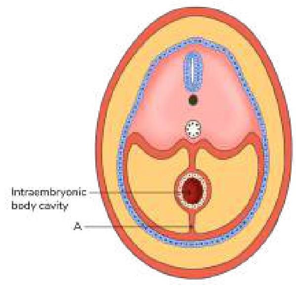
The structure marked A in the image below gives rise to which of the following structures?
Which of the following is a remnant of the Wolffian duct in females?
Which of the following statements about skin is false?
Most common site for fertilization?
Nerves of the Branchial arches are derived from:
Ureteric bud develops from:
Which of the following muscles is derived from the first pharyngeal arch?
Number of stem villi at term in human placenta is?
Which of the following statements about keratinocytes is true?
Embryo gets implanted at what stage of development?
Practice by Chapter
Gametogenesis and Fertilization
Practice Questions
Early Embryonic Development
Practice Questions
Placentation
Practice Questions
Development of Nervous System
Practice Questions
Development of Cardiovascular System
Practice Questions
Development of Gastrointestinal System
Practice Questions
Development of Urogenital System
Practice Questions
Development of Musculoskeletal System
Practice Questions
Development of Head and Neck
Practice Questions
Congenital Anomalies
Practice Questions
Teratology
Practice Questions
Molecular Mechanisms in Development
Practice Questions
Want unlimited practice?
Get full access to all questions, explanations, and performance tracking.
Scan to download app