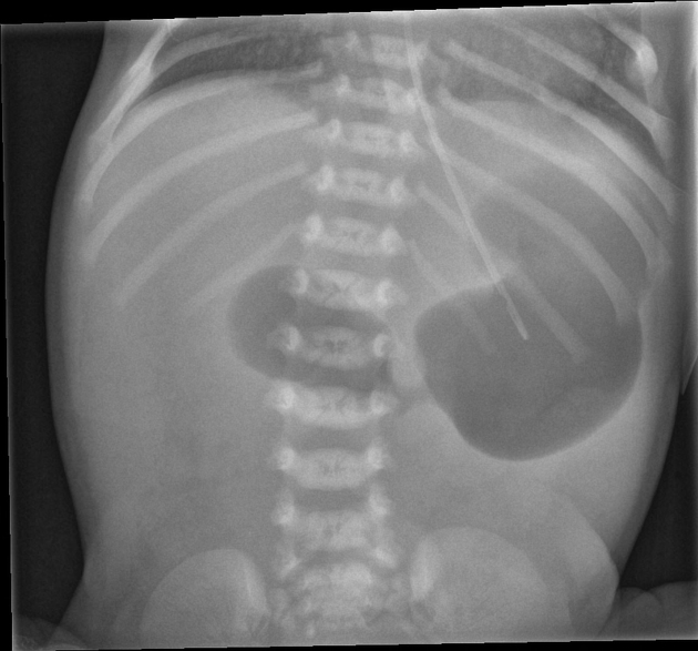
Double bubble sign is seen in -
Tetralogy of Fallot (TOF) includes all of the following components except:
Seal like limbs i.e. phocomelia is a specific side effect of -
The commonest type of congenital heart disease is –
Which of the following is the MOST accurate test for detecting neural tube defects?
Which of the following is a characteristic feature of Down syndrome?
All of the following are neural tube defects except:
What is the time period for the embryonic period?
The labia majora develop from which embryological structure?
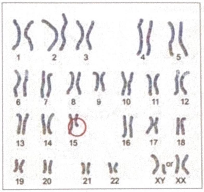
The diagrammatic representation of the karyotype of an individual indicates a specific genetic abnormality. What is the diagnosis?

Explanation: ***All of the options*** - The **"double bubble sign"** on an X-ray indicates **duodenal obstruction**, which can be caused by intrinsic factors like **duodenal atresia** or extrinsic compressions such as an **annular pancreas** or **Ladd's bands** associated with malrotation. - While differing in etiology, all these conditions lead to fluid and air accumulation in the stomach and proximal duodenum, creating the characteristic two dilated loops. *Duodenal atresia* - This is an **intrinsic congenital obstruction** of the duodenum, preventing the passage of gastric and duodenal contents. - On imaging, it shows **two distinct air-filled bubbles** (one for the stomach, one for the proximal duodenum) separated by the pylorus. *Ladd's band* - **Ladd's bands** are peritoneal fibrous bands that can compress the duodenum in cases of **intestinal malrotation**, leading to extrinsic obstruction. - The resulting proximal duodenal dilation, along with gastric distension, presents as the **double bubble sign**. *Annular pancreas* - An **annular pancreas** is a congenital anomaly where pancreatic tissue completely encircles and obstructs the second part of the duodenum. - This extrinsic compression causes significant dilation of the stomach and proximal duodenum, mimicking the appearance of the **double bubble sign**.
Explanation: ***ASD*** - While an **atrial septal defect (ASD)** can be present in some complex congenital heart diseases, it is **not considered a primary component of Tetralogy of Fallot** [1]. - The four classic components of **TOF** are **ventricular septal defect (VSD)**, **pulmonary stenosis**, **overriding aorta**, and **right ventricular hypertrophy (RVH)** [1]. *VSD* - A **ventricular septal defect (VSD)** is a **mandatory component** of Tetralogy of Fallot, allowing for mixing of oxygenated and deoxygenated blood [1]. - It's typically a **large, subaortic defect** that enables the overriding aorta to receive blood from both ventricles [1]. *Pulmonary stenosis* - **Pulmonary stenosis** (obstruction of blood flow from the right ventricle to the pulmonary artery) is a **key component** determining the severity of Tetralogy of Fallot [1]. - The degree of **pulmonary stenosis** dictates the amount of right-to-left shunting and the clinical manifestation of **cyanosis** [1]. *RVH* - **Right ventricular hypertrophy (RVH)** develops as a compensatory mechanism due to the increased workload on the right ventricle from the **pulmonary stenosis** and the **VSD** [1]. - It is a **consequence** of the increased pressure required to eject blood past the stenotic pulmonary valve and into the systemic circulation through the VSD [1].
Explanation: ***Thalidomide*** - **Phocomelia**, characterized by severely shortened or absent limbs resembling those of a seal, is a classic and well-documented **teratogenic effect** of thalidomide. - This drug, when taken during early pregnancy (especially between weeks 4 and 8), disrupts limb bud development. *Doxorubicin* - **Doxorubicin** is an **anthracycline antibiotic** used in cancer chemotherapy, known for its significant **cardiotoxicity**, leading to dilated cardiomyopathy. - While it has various side effects, **phocomelia** is not a reported teratogenic effect of doxorubicin. *Cyclophosphamide* - **Cyclophosphamide** is an **alkylating agent** used in chemotherapy and immunosuppression, with notable side effects including **hemorrhagic cystitis** and **myelosuppression**. - Although it is a teratogen and can cause various fetal malformations, it is not specifically associated with **phocomelia**. *Terazosin* - **Terazosin** is an **alpha-1 blocker** primarily used to treat hypertension and benign prostatic hyperplasia (BPH). - Its main side effects include **orthostatic hypotension** and dizziness; it is not known to be teratogenic or associated with **phocomelia**.
Explanation: ***VSD*** - **Ventricular Septal Defect (VSD)** is the most common type of **congenital heart disease**, accounting for approximately 25-30% of all congenital heart defects. - It involves a hole in the septum separating the **ventricles**, leading to a **left-to-right shunt** of blood. *ASD* - **Atrial Septal Defects (ASDs)** are common but less frequent than VSDs, typically accounting for about 10% of congenital heart defects. - ASDs involve a hole in the septum separating the **atria**, also causing a **left-to-right shunt**. *PDA* - **Patent Ductus Arteriosus (PDA)** is another common congenital heart defect, but it is less prevalent than VSD, accounting for around 5-10% of cases. - PDA is the persistence of the fetal **ductus arteriosus**, allowing blood to flow from the **aorta to the pulmonary artery**. *TOF* - **Tetralogy of Fallot (TOF)** is a complex cyanotic congenital heart defect, representing about 5-7% of all congenital heart diseases. - It is characterized by four distinct anomalies: **pulmonary stenosis**, **ventricular septal defect**, **overriding aorta**, and **right ventricular hypertrophy**.
Explanation: ***USG (Ultrasound)*** - **High-resolution ultrasound** is the **gold standard and most accurate imaging modality** for detecting **neural tube defects (NTDs)** due to its ability to directly visualize anatomical structures of the fetus. - **Diagnostic accuracy**: Detection rate >95% for anencephaly and 80-90% for open spina bifida with targeted anomaly scan at 18-20 weeks. - Can identify specific features such as **lemon sign** (frontal bone scalloping), **banana sign** (cerebellar compression), direct visualization of **spina bifida**, **anencephaly**, or **encephalocele**. - **Non-invasive, safe, and widely available**, making it the primary diagnostic tool in clinical practice. *Amniocentesis* - **Amniocentesis** measures **alpha-fetoprotein (AFP)** and **acetylcholinesterase (AChE)** in amniotic fluid, which are elevated in open NTDs. - While highly accurate as a **confirmatory test** (near 99% sensitivity with both markers), it is **invasive** with risk of miscarriage (0.1-0.3%). - Used primarily when ultrasound findings are **equivocal** or for **biochemical confirmation**, not as the first-line diagnostic test. - In modern practice, ultrasound has largely replaced amniocentesis for NTD diagnosis due to superior imaging technology. *Chromosomal analysis* - **Chromosomal analysis** (karyotyping) detects **chromosomal abnormalities** like trisomies (Down syndrome, Edwards syndrome). - NTDs are **structural malformations**, not chromosomal abnormalities, though some chromosomal disorders may have associated structural defects. - Does not directly diagnose NTDs. *Placentography* - **Placentography** is used to localize the **placenta** in cases of suspected **placenta previa** or for guiding invasive procedures. - Provides no information about **fetal anatomy** and is not used for detecting NTDs.
Explanation: ***Trisomy 21*** - **Down syndrome** is the most common autosomal chromosome abnormality and is characterized by the presence of an extra copy of chromosome 21 [1, 2]. - This extra genetic material leads to the characteristic physical features, intellectual disability, and medical conditions associated with the syndrome [1, 2]. *Trisomy 18* - **Trisomy 18**, also known as **Edwards syndrome**, is a serious chromosomal disorder distinct from Down syndrome [2]. - It is characterized by severe developmental problems, including **heart defects**, **kidney malformations**, and **severe intellectual disability**, with generally a much shorter life expectancy [2, 3]. *Robertsonian translocation involving chromosome 21* - A **Robertsonian translocation** involving chromosome 21 is a cause of Down syndrome, but it is not the characteristic feature itself; rather, it is a specific **chromosomal rearrangement** that can lead to an extra copy of chromosome 21 material [1, 2]. - This specific type of translocation accounts for only a small percentage (2-3%) of all Down syndrome cases, while **Trisomy 21** (nondisjunction) is the most common cause [1, 2]. *Trisomy 13* - **Trisomy 13**, also known as **Patau syndrome**, is a distinct chromosomal disorder characterized by the presence of an extra copy of chromosome 13 [2]. - It is associated with severe birth defects, including **cleft lip/palate**, **polydactyly**, and severe neurological problems, and is usually fatal within the first year of life [2]. **References:** [1] Kumar V, Abbas AK, et al.. Robbins and Cotran Pathologic Basis of Disease. 9th ed. Genetic Disorders, pp. 170-171. [2] Kumar V, Abbas AK, et al.. Robbins and Cotran Pathologic Basis of Disease. 9th ed. Genetic Disorders, pp. 171-172. [3] Kumar V, Abbas AK, et al.. Robbins and Cotran Pathologic Basis of Disease. 9th ed. Genetic Disorders, pp. 168-169.
Explanation: ***Holoprosencephaly*** - This condition results from the **failure of the prosencephalon (forebrain)** to properly divide into two hemispheres, often leading to facial abnormalities. - It is considered a **forebrain malformation**, distinct from neural tube closure defects. *Encephalocele* - An encephalocele is a **neural tube defect** characterized by the protrusion of brain tissue and meninges through an opening in the skull. - It results from the **incomplete closure of the neural tube** during embryonic development. *Myelomeningocele* - Myelomeningocele is a severe form of **spina bifida**, a neural tube defect where the spinal cord and meninges protrude through an opening in the spine [2]. - This defect arises from the **failure of the neural tube to close completely** in the caudal region [1]. *Anencephaly* - Anencephaly is a lethal **neural tube defect** characterized by the absence of a major portion of the brain, skull, and scalp [3]. - It occurs due to the **failure of the cranial end of the neural tube to close** [3].
Explanation: 3rd to 8th week of gestation - The embryonic period begins at the start of the 3rd week (around day 15-16, after gastrulation begins) and concludes at the end of the 8th week of gestation [2]. - During this critical period, all major organs and body systems are formed in a process called organogenesis [2]. - This is the most critical period of development when the embryo is most susceptible to teratogens [1]. 9 weeks to birth - This time frame describes the fetal period, which follows the embryonic period (starting at week 9) [2]. - The fetal period is primarily characterized by the growth and maturation of tissues and organs formed during the embryonic stage [1]. 22 weeks intrauterine to 7 days after birth - This period describes the perinatal period, which is a medical classification related to outcomes around birth. - It encompasses conditions affecting the fetus from viability until the first week postpartum, not the embryonic developmental stage. 0-14 days of gestation - This initial period is often referred to as the pre-embryonic stage or the period of the zygote and blastocyst [3]. - It involves fertilization, cleavage, blastulation, and implantation, leading to the formation of the bilaminar embryonic disc and early gastrulation [3].
Explanation: ***Labioscrotal swellings*** - The **labia majora** develop from the **labioscrotal swellings**, which are paired bilateral structures that appear around week 9-10 of development [1]. - These swellings arise lateral to the urogenital folds and do not fuse in females, forming the labia majora. - In males, these same structures fuse in the midline to form the scrotum. - This is a key example of **sexual differentiation** in embryological development [1]. *Urogenital folds* - The urogenital folds form the **labia minora** in females, not the labia majora. - In males, these folds fuse to form the ventral aspect of the penis and enclose the penile urethra. *Genital tubercle* - The genital tubercle forms the **clitoris** in females and the **glans penis** in males. - It does not contribute to the formation of the labia majora. *Müllerian ducts* - The Müllerian (paramesonephric) ducts form the **upper vagina, uterus, and fallopian tubes** in females. - They are internal structures and do not contribute to external genitalia like the labia majora.
Explanation: ***Prader-Willi syndrome*** - The karyotype shows an abnormality on **chromosome 15**, which is consistent with Prader-Willi syndrome caused by **deletion of 15q11-q13** inherited from the **paternal** chromosome or **maternal uniparental disomy**. - While PWS deletions are typically **submicroscopic**, larger deletions can occasionally be visible on standard karyotyping, particularly when they represent **class I deletions** that are more extensive and involve additional chromosomal material beyond the typical PWS critical region. *Angelman syndrome* - Although Angelman syndrome also involves **chromosome 15q11-q13 deletion**, it results from **maternal** deletion or **paternal uniparental disomy**, and presents with distinctly different clinical features. - Clinical presentation includes **severe intellectual disability**, **ataxia**, **seizures**, **absent speech**, and **inappropriate laughter** (happy demeanor), which differs significantly from the PWS phenotype. *DiGeorge syndrome* - DiGeorge syndrome is caused by **deletion of chromosome 22q11.2**, not chromosome 15 as shown in the karyotype. - Clinical features include **cardiac defects** (conotruncal abnormalities), **thymic hypoplasia**, **parathyroid hypoplasia** (hypocalcemia), **cleft palate**, and characteristic facial features (CATCH-22 syndrome). *Cri du Chat syndrome* - This syndrome results from **deletion of chromosome 5p** (short arm of chromosome 5), not chromosome 15 as indicated in the karyotype. - Characteristic features include **high-pitched cry** resembling a cat's meow in infancy, **intellectual disability**, **microcephaly**, and **distinctive facial features**.
Get full access to all questions, explanations, and performance tracking.
Start For Free