
Back — MCQs

On this page
Which of the following is NOT true about the iliolumbar ligament?
A 39-year-old male presents with severe neck pain after a whiplash injury, sustained when his car was struck from behind. Radiographic studies reveal trauma to the ligament lying on the anterior surface of the cervical vertebral bodies. Which ligament is this?
The triangle of auscultation is formed by which intercostal space?
The Grynfeltt triangle is not bounded by which of the following?
A 44-year-old woman presents with headache and backache. Examination reveals fluid accumulation in the spinal epidural space due to damage to blood vessels or meninges. Which of the following structures is most likely ruptured?
A 65-year-old male complains of severe back pain and inability to move his left lower limb. Radiographic studies demonstrate the compression of nerve elements at the intervertebral foramen between vertebrae L5 and S1. Which structure is most likely responsible for this space-occupying lesion?
Which of the following ligaments is derived from the thoracolumbar fascia?
All of the following muscles form the boundary of the suboccipital triangle, EXCEPT?
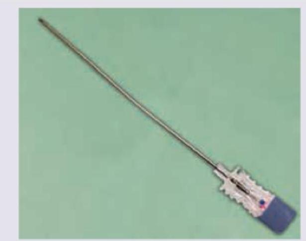
All of the following layers are pierced by the needle shown below except:
A patient undergoes spinal surgery at the L4-L5 level. During the procedure, which of the following ligaments must be divided first to access the spinal canal?
Practice by Chapter
Vertebral Column
Practice Questions
Spinal Cord and Meninges
Practice Questions
Back Muscles and Fasciae
Practice Questions
Vertebral Joints and Ligaments
Practice Questions
Vasculature of the Back
Practice Questions
Innervation of the Back
Practice Questions
Clinical Aspects of Back Disorders
Practice Questions
Applied Anatomy of the Back
Practice Questions
Surface Anatomy of the Back
Practice Questions
Development of the Vertebral Column
Practice Questions
Want unlimited practice?
Get full access to all questions, explanations, and performance tracking.
Scan to download app