
Back — MCQs

On this page
The posterior longitudinal ligament continues as which of the following structures?
A 26-year-old heavyweight boxer sustained a blow to the mandible, causing slight subluxation of the atlantoaxial joint. This injury resulted in a decreased range of motion at the joint. Which movement would be most affected?
Which cervical vertebra has a lateral mass?
Which structure forms the posterior one-third of the vertebral canal?
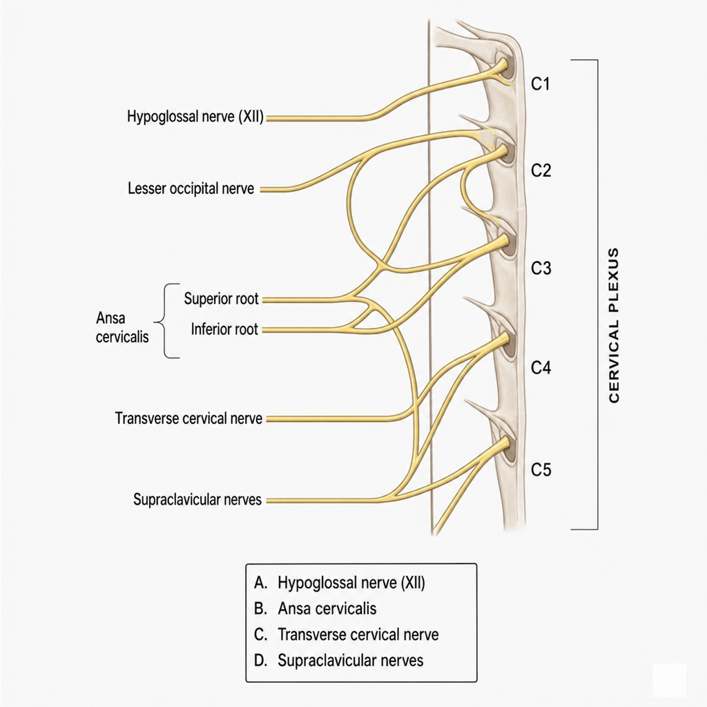
A lesion of the first cervical spinal nerve would cause functional impairment of which structure?
What is the primary vertebral curve?
Which ligament is primarily responsible for maintaining Atlantoaxial stability?
On which structure does the rhomboids major insert?
Which of the following vertebrae has the most prominent spine?
During an outbreak of meningitis, a 20-year-old student presents with headache, fever, chills, and stiff neck. A lumbar puncture is planned. Cerebrospinal fluid (CSF) is normally withdrawn from which of the following spaces?
Practice by Chapter
Vertebral Column
Practice Questions
Spinal Cord and Meninges
Practice Questions
Back Muscles and Fasciae
Practice Questions
Vertebral Joints and Ligaments
Practice Questions
Vasculature of the Back
Practice Questions
Innervation of the Back
Practice Questions
Clinical Aspects of Back Disorders
Practice Questions
Applied Anatomy of the Back
Practice Questions
Surface Anatomy of the Back
Practice Questions
Development of the Vertebral Column
Practice Questions
Want unlimited practice?
Get full access to all questions, explanations, and performance tracking.
Scan to download app