Where does the main part of vertebral venous plexus lie?
Epidural space lies between:
All are TRUE about intervertebral disc, EXCEPT:
Membrana tectoria is continuation of:
Which of the following parts of the vertebral canal shows the first secondary curve to develop?
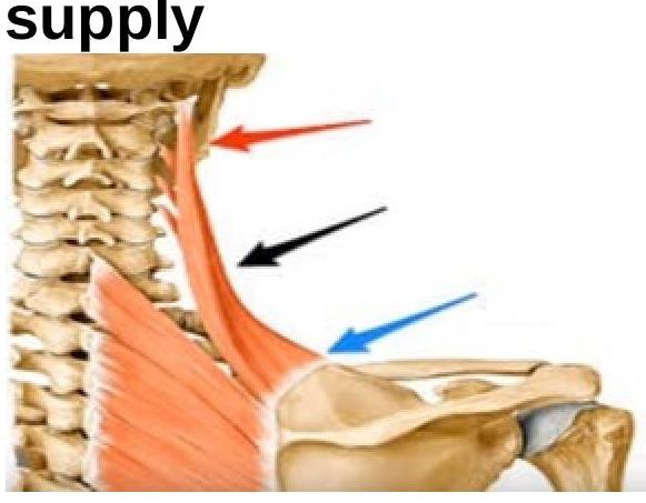
What is the nerve supply of the shown muscle?

All of the following contribute to the intervertebral disc EXCEPT:
In a lumbar puncture procedure, into which space is the needle inserted to access the cerebrospinal fluid?
Muscle lying between the anterior and middle layers of thoracolumbar fascia is?
Insertion of levator scapulae is?
Explanation: ***Epidural space*** - The **internal vertebral venous plexus** is located within the **epidural space**, surrounding the dura mater of the spinal cord. - This plexus is a valveless network of veins, which allows for bidirectional blood flow and provides an important pathway for the **spread of infection** and **metastasis**. *Subdural space* - The subdural space is a potential space between the **dura mater** and **arachnoid mater**, typically not containing significant venous plexuses. - While it can fill with blood in cases of **subdural hematoma**, it is not the primary location of the vertebral venous plexus. *External to the vertebral column* - The **external vertebral venous plexuses** are located outside the vertebral column, both anterior and posterior to the vertebrae. - Although these plexuses exist, the "main part" often refers to the internal plexus within the spinal canal due to its clinical significance and close association with the **spinal cord**. *Subarachnoid space* - The subarachnoid space lies between the **arachnoid mater** and **pia mater** and contains **cerebrospinal fluid (CSF)** and major arteries. - While small veins might traverse this space, it does not house the expansive network of the vertebral venous plexus.
Explanation: ***Dura and vertebral column*** - The **epidural space** is located between the dura mater and the surrounding vertebral column (specifically the **periosteum** lining the vertebral canal). - This space contains **fat, connective tissue**, and a **venous plexus**, acting as a protective cushion for the spinal cord. *Dura and arachnoid* - The space between the dura mater and the arachnoid mater is the **subdural space** [1]. - This is normally a **potential space** but can become a real space in pathological conditions like a subdural hematoma [1]. *Pia and arachnoid* - The space between the pia mater and the arachnoid mater is the **subarachnoid space** [1]. - This space normally contains **cerebrospinal fluid (CSF)** and blood vessels [1]. *Pia mater and grey matter* - The pia mater is directly apposed to the surface of the brain and spinal cord, including its **grey matter**. - There is no distinct "space" between the pia mater and the neural tissue it covers.
Explanation: ***Rich vascular supply*** - The intervertebral disc is largely **avascular** in adults, receiving nutrients primarily through diffusion from the vertebral bodies. - This lack of direct blood supply is why it has a limited capacity for self-repair and healing after injury. *Prolapse is most common in lumbosacral region* - The **lumbosacral region** (L4-L5 and L5-S1) bears the most weight and experiences the greatest biomechanical stress, making it the most common site for disc prolapse or herniation [1]. - This area is prone to injury due to the demands placed upon it during movement and lifting. *Nucleus pulposus is a remnant of notochord* - The **nucleus pulposus**, the gelatinous center of the intervertebral disc, is indeed a direct remnant of the embryonic **notochord**. - This embryological origin explains its high water content and elastic properties, which allow it to function as a shock absorber. *Annulus fibrosus is made up of fibrocartilage* - The **annulus fibrosus** is the tough outer layer of the intervertebral disc composed of concentric layers of **fibrocartilage**. - These collagen fibers are arranged in a crisscross pattern to provide strength and contain the nucleus pulposus.
Explanation: ***Posterior longitudinal ligament*** - The **membrana tectoria** is a broad, strong membrane that is the superior continuation of the **posterior longitudinal ligament**. - It extends from the body of the **axis (C2)** to the **basilar part of the occipital bone**, covering the dens and its associated ligaments. *Anterior atlanto-occipital membrane* - This membrane is a continuation of the **anterior longitudinal ligament** superiorly, connecting the anterior arch of the atlas to the anterior margin of the foramen magnum. - It is located **anterior** to the vertebral column, while the membrana tectoria is posterior. *Anterior longitudinal ligament* - This ligament runs along the **anterior surfaces of the vertebral bodies** and intervertebral discs from the atlas to the sacrum. - Its superior continuation is the **anterior atlanto-occipital membrane**, not the membrana tectoria. *Posterior atlanto-occipital membrane* - This membrane connects the **posterior arch of the atlas** to the posterior margin of the foramen magnum. - It is distinct from the membrana tectoria, which lies **deep** to it and is a continuation of the posterior longitudinal ligament.
Explanation: ***Cervical Vertebral Canal*** - The **cervical curve** is the **first secondary curve** to develop as an infant learns to hold their head up (around 3-4 months). - This **lordotic curve** is concave posteriorly and helps to balance the head on the vertebral column. - It develops before the lumbar curve, making it the earliest secondary curvature. *Lumbar Vertebral Canal* - The **lumbar curve** is also a **secondary curve** but develops later when an infant begins to stand and walk (around 12-18 months). - It is a **lordotic curve**, concave posteriorly, and helps maintain an upright posture. - This is the second secondary curve to develop. *Thoracic Vertebral Canal* - The **thoracic curve** is a **primary curve**, meaning it is present at birth. - This **kyphotic curve** is convex posteriorly and accommodates the thoracic organs. *Sacral Vertebral Canal* - The **sacral curve** is another **primary curve**, also present at birth. - It is a **kyphotic curve**, convex posteriorly, and contributes to the pelvic basin's shape.
Explanation: ***Dorsal scapular*** - The image points to the **levator scapulae muscle**, which elevates and rotates the scapula. - The **dorsal scapular nerve** (C5 root, with contributions from C3-C4) innervates the levator scapulae, as well as the rhomboid major and minor muscles. - This nerve arises from the C5 root of the brachial plexus and pierces through the middle scalene muscle. - Clinically, injury to the dorsal scapular nerve can cause **medial scapular winging** and difficulty elevating the shoulder. *Suprascapular* - The suprascapular nerve (C5-C6) primarily innervates the **supraspinatus and infraspinatus muscles**, which are involved in rotator cuff function. - It does not supply the levator scapulae muscle. - This nerve passes through the suprascapular notch beneath the superior transverse scapular ligament. *Dorsal rami of C1* - The **dorsal ramus of C1** (suboccipital nerve) primarily innervates the muscles of the suboccipital triangle: rectus capitis posterior major and minor, obliquus capitis superior and inferior. - These nerves are involved in fine head and neck movements but do not innervate the levator scapulae. - The levator scapulae receives segmental innervation from C3-C4 cervical nerves directly, in addition to the dorsal scapular nerve. *Subscapular* - The subscapular nerves (upper and lower, from C5-C6) innervate the **subscapularis muscle**, which is part of the rotator cuff. - They also innervate the **teres major muscle**, but not the levator scapulae. - These are branches from the posterior cord of the brachial plexus.
Explanation: ***Elastic cartilage*** - **Elastic cartilage** is characterized by the presence of **elastic fibers**, providing flexibility to structures like the ear and epiglottis. - It is **not found** within the intervertebral disc, which requires specific properties for weight-bearing and shock absorption. *Fibrocartilage* - **Fibrocartilage** is a primary component of the **annulus fibrosus** and plays a crucial role in providing tensile strength and resisting compressive forces [1]. - Its presence is essential for the structural integrity and function of the intervertebral disc. *Annulus fibrosus* - The **annulus fibrosus** is the **tough, outer fibrous ring** of the intervertebral disc, composed of concentric layers of fibrocartilage. - It encircles the nucleus pulposus, containing it and providing stability to the disc. *Nucleus pulposus* - The **nucleus pulposus** is the **gel-like core** of the intervertebral disc, rich in proteoglycans and water. - It acts as a shock absorber and allows for flexibility between vertebrae.
Explanation: ***Correct Answer: Subarachnoid space*** - The **subarachnoid space** is the primary target for a lumbar puncture to access **cerebrospinal fluid (CSF)**, as this fluid circulates within this space between the arachnoid mater and pia mater [1]. - The needle passes through skin, subcutaneous tissue, ligaments (supraspinous, interspinous, ligamentum flavum), and the **dura mater and arachnoid mater** before entering the subarachnoid space. - This procedure is typically performed between L3-L4 or L4-L5 vertebral levels, below the termination of the spinal cord (L1-L2), to minimize risk of cord injury. *Incorrect: Epidural space* - The **epidural space** is located outside the dura mater and contains fat, connective tissue, and blood vessels. - This space is targeted for **epidural anesthesia**, not for CSF collection. - Stopping the needle here would not yield CSF as the dura mater has not been penetrated. *Incorrect: Subdural space* - The **subdural space** is a potential space between the dura mater and the arachnoid mater. - It does not normally contain CSF and is not a target for lumbar puncture. - Entering this space would indicate improper needle placement and represents a complication. *Incorrect: Subcutaneous space* - The **subcutaneous space** is directly beneath the skin and consists of adipose tissue and connective tissue. - This is the most superficial layer and contains no CSF. - The needle must pass through this layer before reaching deeper anatomical structures.
Explanation: ***Quadratus Lumborum*** - The **quadratus lumborum** muscle is situated in the posterior abdominal wall, specifically within the compartment formed by the attachments of the **anterior and middle layers of the thoracolumbar fascia**. - This muscle extends from the iliac crest to the 12th rib and lumbar vertebrae, playing a key role in **lateral flexion of the trunk** and **stabilization of the lumbar spine**. *Psoas major* - The **psoas major** muscle lies anterior to the lumbar vertebrae and is covered by its own fascia, but it is not directly situated between the anterior and middle layers of the thoracolumbar fascia. - It runs in a more medial and anterior plane in the posterior abdominal wall, contributing to **hip flexion**. *Obturator internus* - The **obturator internus** is a muscle of the pelvic floor and hip, located deep within the pelvis. - It lies near the **obturator foramen** and is completely unrelated to the thoracolumbar fascia or the lumbar region. *External oblique* - The **external oblique** muscle is an anterolateral abdominal wall muscle, superficial to the transversus abdominis and internal oblique muscles [1]. - Its aponeurosis contributes to the rectus sheath but is not located between the layers of the **thoracolumbar fascia** in the lumbar region.
Explanation: ***Superior part of medial scapula border*** - The **levator scapulae muscle** originates from the **transverse processes of C1-C4 vertebrae** and **inserts** onto the superior part of the medial border of the scapula, between the **superior angle and the spine of the scapula**. - Its main actions are to **elevate** and **rotate** the scapula downward. *Lateral border of scapula* - The **lateral border of the scapula** primarily serves as the attachment site for muscles that move the **humerus**, such as the **teres major** and **teres minor**. - The levator scapulae has no direct insertion on the lateral border. *Suprolateral part of scapula* - This general description is vague and does not precisely identify the insertion point of the levator scapulae. - While it's located superiorly, the specific insertion is on the **medial border**, not broadly "suprolateral." *Inferior angle of scapula* - The **inferior angle of the scapula** is the insertion point for muscles like the **latissimus dorsi** (occasionally) and a key landmark for muscles involved in **scapular rotation**, such as the **serratus anterior**. - The levator scapulae is located much more superiorly and inserts onto the medial border at a higher level than the inferior angle.
Vertebral Column
Practice Questions
Spinal Cord and Meninges
Practice Questions
Back Muscles and Fasciae
Practice Questions
Vertebral Joints and Ligaments
Practice Questions
Vasculature of the Back
Practice Questions
Innervation of the Back
Practice Questions
Clinical Aspects of Back Disorders
Practice Questions
Applied Anatomy of the Back
Practice Questions
Surface Anatomy of the Back
Practice Questions
Development of the Vertebral Column
Practice Questions
Get full access to all questions, explanations, and performance tracking.
Start For Free