A 65-year-old male complains of severe back pain and inability to move his left lower limb. Radiographic studies demonstrate the compression of nerve elements at the intervertebral foramen between vertebrae L5 and S1. Which structure is most likely responsible for this space-occupying lesion?
Which of the following ligaments is derived from the thoracolumbar fascia?
All of the following muscles form the boundary of the suboccipital triangle, EXCEPT?
Which part of the vertebral canal shows secondary curves with concavity towards the back?
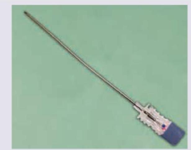
All of the following layers are pierced by the needle shown below except:

Regarding the epidural space, all are true except:
Which of the following structures provides the main nutritional supply to the intervertebral disc?
What constitutes a spinal motion segment?
All of the following are superficial muscles of the back, EXCEPT:
Where is the epidural venous plexus located?
Explanation: The clinical presentation describes a classic case of **Intervertebral Disc Herniation** (specifically a posterolateral protrusion) causing nerve root compression. [1] **Why Nucleus Pulposus is Correct:** The intervertebral disc consists of a peripheral fibrous ring, the **anulus fibrosus**, and a central gelatinous core, the **nucleus pulposus**. With age or trauma, the anulus fibrosus can develop tears. The nucleus pulposus, which is under pressure, then herniates through these tears. [1] Because the posterior longitudinal ligament is narrowest in the lumbar region, the nucleus typically protrudes **posterolaterally** into the intervertebral foramen, compressing the spinal nerve roots (in this case, the L5 or S1 roots). [1] Thus, the nucleus pulposus is the actual "space-occupying" material causing the compression. **Why Other Options are Incorrect:** * **Anulus fibrosus:** While this structure must rupture or bulge for herniation to occur, it is the extrusion of the *nucleus pulposus* that typically forms the symptomatic mass. [1] * **Posterior longitudinal ligament (PLL):** This ligament resists hyperflexion and sits behind the vertebral bodies. [2] While a disc herniation occurs *past* or through the weakened fibers of the PLL, the ligament itself is not the lesion; rather, it is often bypassed by the herniating nucleus. * **Anterior longitudinal ligament (ALL):** This ligament is located on the anterior aspect of the vertebral bodies and prevents hyperextension. [2] It is rarely involved in nerve compression syndromes of the spinal canal. **High-Yield Clinical Pearls for NEET-PG:** * **Most Common Site:** L4-L5 or L5-S1 are the most frequent sites for lumbar disc herniation. [1] * **Rule of Nerve Involvement:** In the lumbar spine, a posterolateral disc protrusion usually compresses the nerve root **below** the level of the disc (e.g., an L5-S1 protrusion typically affects the S1 nerve root). [1] * **Schmorl’s Nodes:** These are vertical herniations of the nucleus pulposus into the cartilaginous endplates of the vertebral bodies. [1] * **Nerve Exit:** Lumbar nerves exit the intervertebral foramen *below* their corresponding vertebrae.
Explanation: ### Explanation The **thoracolumbar fascia (TLF)** is a critical deep investing membrane in the back that consists of three layers (anterior, middle, and posterior). **Why the Lateral Arcuate Ligament is Correct:** The **lateral arcuate ligament** is a thickened arch of the **anterior layer of the thoracolumbar fascia** (which covers the quadratus lumborum muscle). It extends from the transverse process of the L1 vertebra to the 12th rib. It serves as one of the points of origin for the diaphragm. **Analysis of Incorrect Options:** * **A. Medial arcuate ligament:** This is a thickening of the **psoas fascia** (fascia covering the psoas major muscle), extending from the side of the L1/L2 vertebral bodies to the L1 transverse process. * **C. Lacunar ligament (Gimbernat’s ligament):** This is a triangular extension of the **inguinal ligament** (derived from the external oblique aponeurosis) that connects the inguinal ligament to the pectineal line of the pubis. * **D. Cruciate ligament:** This refers to the ligaments of the knee (ACL/PCL) or the cruciform ligament of the atlas, none of which are related to the thoracolumbar fascia. **High-Yield Clinical Pearls for NEET-PG:** * **Diaphragmatic Openings:** The medial and lateral arcuate ligaments form part of the "lumbocostal arches." * **Layers of TLF:** * **Anterior layer:** Covers Quadratus lumborum (gives rise to lateral arcuate ligament). * **Middle layer:** Lies between Quadratus lumborum and Erector spinae. * **Posterior layer:** Covers Erector spinae; it is the thickest layer and attaches to the spinous processes. * **The "V" Sign:** The internal oblique and transversus abdominis muscles originate from the fusion of the middle and posterior layers of the TLF [1].
Explanation: The **suboccipital triangle** is a high-yield anatomical region located deep to the semispinalis capitis muscle. It is bounded by three specific muscles, and identifying these boundaries is crucial for NEET-PG. ### **Explanation of the Correct Answer** **D. Rectus capitis posterior minor muscle:** This muscle is located **medial** to the suboccipital triangle. While it is part of the suboccipital group of muscles, it does not form a boundary of the triangle itself. It arises from the posterior tubercle of the atlas (C1) and inserts into the medial part of the inferior nuchal line. ### **Analysis of Incorrect Options (The True Boundaries)** * **A. Obliquus capitis superior:** Forms the **lateral (superolateral)** boundary. It runs from the transverse process of the atlas to the occipital bone. * **B. Obliquus capitis inferior:** Forms the **inferior (inferolateral)** boundary. It runs from the spine of the axis (C2) to the transverse process of the atlas. * **C. Rectus capitis posterior major:** Forms the **medial (superomedial)** boundary. It runs from the spine of the axis to the lateral part of the inferior nuchal line. ### **High-Yield NEET-PG Pearls** * **Contents of the Triangle:** The two most important contents are the **Vertebral Artery** (3rd part) and the **Suboccipital Nerve** (Dorsal ramus of C1). Note: The Greater Occipital Nerve (C2) is *not* a content; it crosses the triangle inferiorly. * **Roof:** Formed by the Semispinalis capitis and Longissimus capitis. * **Floor:** Formed by the posterior atlanto-occipital membrane and the posterior arch of the atlas. * **Mnemonic for Boundaries:** **"Major-Superior-Inferior"** (Rectus capitis posterior **Major**, Obliquus capitis **Superior**, and Obliquus capitis **Inferior**). Minor is always excluded.
Explanation: The vertebral column exhibits four physiological curvatures in the sagittal plane, categorized as primary or secondary. ### 1. Why the Correct Answer is Right **Cervical vertebral canal:** This is a **secondary (compensatory) curve**. Secondary curves are characterized by being **convex anteriorly** and **concave posteriorly** (concavity towards the back). * The cervical curve develops at approximately 3–4 months of age when the infant begins to lift their head. * The lumbar curve is the other secondary curve, developing when the child begins to sit and walk (12–18 months). ### 2. Why the Incorrect Options are Wrong * **Thoracic vertebral canal (Option B):** This is a **primary curve**, present at birth. Primary curves are **concave anteriorly** and **convex posteriorly**. They follow the original C-shape of the fetal vertebral column. * **Sacral vertebral canal (Option C):** Like the thoracic region, the sacrum is a primary curve. It maintains its anterior concavity to accommodate the pelvic viscera. * **Coccyx vertebral canal (Option D):** The coccygeal region is part of the primary sacrococcygeal curve, showing anterior concavity. ### 3. Clinical Pearls & High-Yield Facts * **Kyphosis:** An exaggeration of the primary (thoracic) curve. * **Lordosis:** An exaggeration of the secondary (lumbar) curve, often seen in late pregnancy or obesity. * **Scoliosis:** An abnormal lateral curvature of the spine (most common in the thoracic region). * **Mnemonic:** **S**econdary curves are **S**haped like a "C" facing backward (Concave posteriorly). * **Developmental Milestone:** Remember: **C**ervical = **C**ontrol of head; **L**umbar = **L**ocomotion (walking).
Explanation: ***Ligamentum denticulatum*** - The **ligamentum denticulatum** is an internal structure of the spinal cord (specifically, pia mater extensions) and is not pierced during an LP, which targets the subarachnoid space. - It functions to suspend the spinal cord within the dura mater and is not in the pathway of the needle during a lumbar puncture. *Supraspinous ligament* - This ligament is the **first major ligament** encountered posterior to the spinous processes in the midline and must be pierced by the needle during a lumbar puncture. - It runs along the tips of the spinous processes. *Inter-spinous ligament* - Located between the spinous processes, this ligament is encountered **after the supraspinous ligament** and must be pierced during a lumbar puncture to reach deeper structures. - It connects adjacent spinous processes. *Ligamentum flavum* - This ligament is rich in elastic fibers and is pierced **before entering the epidural space** on the way to the subarachnoid space. - Its piercing is often felt as a distinct "pop" or "give" by the clinician performing the lumbar puncture.
Explanation: ***Continues through foramen magnum into the skull*** - The **epidural space** in the spinal column ends superiorly at the **foramen magnum** and does **not continue into the skull** as a defined space. - Within the cranial vault, the dura mater is fused with the periosteum of the skull, meaning there is no true epidural space like that found in the spine. *Lies outside of the dura mater* - The epidural space is indeed located **outside the dura mater**, which is the outermost layer of the meninges in the spinal cord. - This space contains **fat**, **loose connective tissue**, and a **venous plexus**. *Is an open space* - The epidural space is considered an **open or potential space**, meaning it is not normally filled with fluid but can be expanded by injections (e.g., epidural anesthesia) or pathology (e.g., hematoma). - Its contents allow for flexibility and cushioning of the spinal cord within the vertebral canal. *Ends at the sacrococcygeal membrane* - Inferiorly, the spinal epidural space terminates at the **sacrococcygeal membrane**, covering the sacral hiatus. - This anatomical landmark is important for procedures like **caudal epidural blocks**.
Explanation: ***Vertebral endplates*** - The **vertebral endplates** are thin layers of cartilage and bone that cover the superior and inferior surfaces of the vertebral bodies. - They are crucial for nutrient diffusion from the vertebral body capillaries into the avascular intervertebral disc. *Anterior longitudinal ligament* - The **anterior longitudinal ligament** is a strong, fibrous band that runs along the anterior surfaces of the vertebral bodies and intervertebral discs. - Its primary role is to prevent hyperextension of the spine and it does not directly supply nutrients to the disc. *Posterior longitudinal ligament* - The **posterior longitudinal ligament** is located on the posterior aspect of the vertebral bodies, inside the vertebral canal. - Its main function is to restrict hyperflexion and posterior disc herniation, without providing significant nutritional support. *Ligamentum flavum* - The **ligamentum flavum** connects the laminae of adjacent vertebrae, primarily composed of elastic tissue. - It helps maintain the upright posture and provides flexibility to the vertebral column but is not involved in disc nutrition.
Explanation: ***A disc and the vertebrae above and below, including their interlocking facet joints.*** - A **spinal motion segment** or **functional spinal unit** is defined as two adjacent vertebrae and the intervertebral disc between them. - This unit includes all the associated **ligaments**, **capsules**, and especially the **facet joints**, which together allow for complex movements. *A disc and the facet joints at that level.* - This definition is incomplete as it misses the crucial component of the **vertebral bodies** themselves. - The vertebral bodies provide the main structural support and articulation points for the disc and facet joints. *A vertebral body and the disc above.* - This partial definition describes only a fraction of the components required for a functional segment. - It omits the **inferior vertebral body** and the critical **facet joints** that enable motion. *A section of the spine involved in a physiological curve with the similar function (i.e. thoracic kyphosis).* - This option describes a broader **region** of the spine rather than a single, functional motion unit. - A physiological curve involves multiple motion segments working in concert, not a single segment.
Explanation: ***Correct: Erector spinae*** - The **erector spinae** muscles (iliocostalis, longissimus, spinalis) are part of the **intermediate layer** of back muscles, lying deep to the superficial extrinsic muscles. - They primarily function in **postural support** and **extension of the vertebral column**. - This is the correct answer because erector spinae is NOT a superficial muscle. *Incorrect: Levator scapulae* - The **levator scapulae** is a **superficial extrinsic back muscle** that elevates the scapula. - It connects the cervical vertebrae to the superior angle of the scapula. *Incorrect: Trapezius* - The **trapezius** is a large, **superficial extrinsic back muscle** involved in moving the scapula and supporting the arm. - It covers a significant portion of the upper back and neck. *Incorrect: Latissimus dorsi* - The **latissimus dorsi** is another large, **superficial extrinsic back muscle** responsible for extending, adducting, and internally rotating the arm. - It is located in the lower and middle back.
Explanation: ***In vertebral canal above dura mater*** - The **epidural venous plexus** (also known as **Batson's plexus**) is located in the **epidural space**, which is external to the dura mater. - In the spinal canal, this space is found between the **vertebral canal** (specifically, the periosteum lining the canal) and the **dura mater**. *At the junction of middle and posterior cranial fossa* - This location relates more to specific cranial nerves or arterial structures at the skull base and is not where the epidural venous plexus of the spine is primarily found. - The epidural space in the cranium is a potential space, and the venous plexuses associated with it are typically referred to as **dural venous sinuses**. *Basal ganglia adjacent to pons* - The **basal ganglia** are deep brain structures involved in motor control, and the pons is part of the brainstem, both located within the cranium. - This location is entirely within the brain parenchyma and not in the epidural space of the vertebral canal. *In vertebral canal below dura mater* - Below the dura mater is the **subdural space** (a potential space) and then the **subarachnoid space**, which contains cerebrospinal fluid and is where the spinal cord and nerve roots are located. - The epidural venous plexus is explicitly located *above* (external to) the dura mater, not below it.
Vertebral Column
Practice Questions
Spinal Cord and Meninges
Practice Questions
Back Muscles and Fasciae
Practice Questions
Vertebral Joints and Ligaments
Practice Questions
Vasculature of the Back
Practice Questions
Innervation of the Back
Practice Questions
Clinical Aspects of Back Disorders
Practice Questions
Applied Anatomy of the Back
Practice Questions
Surface Anatomy of the Back
Practice Questions
Development of the Vertebral Column
Practice Questions
Get full access to all questions, explanations, and performance tracking.
Start For Free