A tooth characterized by a single conical cusp and a single root is classified as:
What differences can be seen in skulls of male and female before puberty?
Which is best used for sex differentiation?
Which of the following is an aberrant epiphysis?
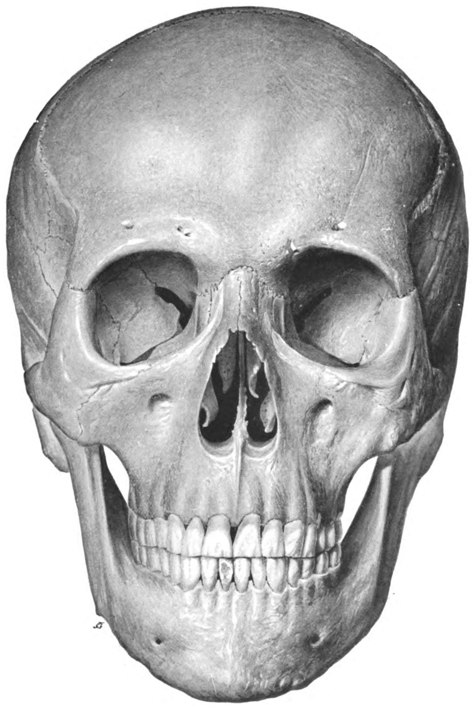
True statement about the skull shown below:

If both the alae of the sacrum are absent, it is called:
Which tendon is frequently absent in the upper limb?
A 45-year-old woman presents with severe headaches and is found to have a berry aneurysm. Which anatomical structure is most likely involved?
A surgeon notes increased bleeding during a splenectomy and suspects an anomaly in the vascular supply. Which variant of arterial supply could be responsible for this unexpected complication?
A patient presents with acute appendicitis. Upon reviewing the CT scan, the radiologist notes an abnormally positioned appendix. What anatomical variation could explain a hidden appendix that was not detected during a physical examination?
Explanation: ***Haplodont*** - **Haplodont** teeth are characterized by a **single conical cusp** and a **single root**, representing a simple, primitive tooth morphology. - This term describes the **tooth structure itself** - simple, cone-shaped teeth found in some reptiles and primitive mammals. - While not common terminology for modern primate teeth, it describes the basic morphological form being asked about. *Heterodont* - **Heterodont** refers to having teeth of **different types** specialized for different functions (incisors, canines, premolars, molars). - This describes the **dentition pattern** of an organism, not individual tooth morphology. - Primates are heterodont animals, but this doesn't describe a single tooth's shape. *Acrodont* - **Acrodont** refers to a **tooth attachment method** where teeth are fused to the top of the jawbone without sockets. - Seen in some reptiles (e.g., lizards and some snakes). - This is about **how teeth attach**, not their morphological structure. *Thecodont* - **Thecodont** describes teeth that are **set in sockets** within the jawbone. - Characteristic of mammals including primates, crocodilians, and some extinct reptiles. - This describes **tooth attachment**, not the cusp or root morphology.
Explanation: ***None of the options*** - Before **puberty**, the **skulls** of males and females are largely indistinguishable in terms of differentiating characteristics. - **Sex-specific differences** in skull morphology, such as pronounced brow ridges or larger mastoid processes, primarily develop during and after puberty due to hormonal influences. *Difference in capacity* - While adult males typically have slightly larger cranial capacities than adult females, this difference is not significant or reliably identifiable **before puberty**. - **Cranial capacity** continues to develop throughout childhood, and pre-pubertal variations are more reflective of individual growth rather than sex. *Difference in size* - **Skull size** differences between sexes become noticeable mostly **after puberty** due to the impact of sex hormones on bone growth. - In children, skull size varies greatly among individuals, with no consistent or significant difference based on sex that allows for reliable differentiation. *Difference in weight* - **Skull weight** is directly correlated with its size and bone density. - Similar to size and capacity, significant and consistent differences in skull weight between males and females appear **post-puberty**, not before.
Explanation: ***Pelvis*** - The **pelvis** exhibits the most significant and consistent **morphological differences** between sexes due to its role in childbirth in females, making it the most reliable osteological indicator for sex determination [1]. - Key features include the **subpubic angle**, **greater sciatic notch**, and the overall **pelvic inlet shape**, which are distinctly different between males and females [1]. *Femur* - While the **femur** can show some sexual dimorphism, such as differences in **head diameter** and **robusticity**, these tend to overlap significantly between sexes, making it less reliable than the pelvis. - Its use in sex differentiation is often secondary and relies on **metric analyses** rather than distinct morphological features. *Skull* - The **skull** presents several features that can aid in sex differentiation, including traits like the **mastoid process size**, **supraorbital ridge prominence**, and **nuchal crest development**. - However, these features are often influenced by **population variability** and can be less definitive than pelvic characteristics, especially in cases of ambiguous or juvenile remains. *Humerus* - The **humerus**, like other long bones, exhibits some sexual dimorphism, primarily in **overall size** and **robusticity**. - Measurements like the **humeral head diameter** can be used, but similar to the femur, there is considerable overlap between sexes, making it a less accurate indicator compared to the pelvis.
Explanation: ***Base of 1st metacarpal*** - The **base of the 1st metacarpal** is considered an **aberrant epiphysis** because its epiphysial plate is located at the base, similar to a phalanx, rather than at the head, like other metacarpals. - This anatomical variation is important for understanding bone growth and potential injury patterns in the **thumb**. *Coracoid process* - The coracoid process is a **secondary ossification center** with its own epiphyseal plate, but it is not typically classified as an aberrant epiphysis in the same context as the 1st metacarpal. - It forms part of the **scapula** and provides attachment for several muscles and ligaments. *Greater tubercle of humerus* - The greater tubercle of the humerus is a prominent feature that develops from a **secondary ossification center** and fuses with the shaft of the humerus. - It is an important site for the attachment of **rotator cuff muscles**, but it's not defined as an aberrant epiphysis. *Base of 2nd metacarpal* - The 2nd through 5th metacarpals typically have their **epiphysial plates at their heads** (distal ends), making the base a part of the diaphysis. - This is the standard pattern for metacarpals, in contrast to the unique arrangement of the 1st metacarpal.
Explanation: ***Prominent supraorbital ridges, prominent glabella, square chin, acute fronto-nasal angle - Male*** - The image displays features typical of a **male skull**, including a pronounced **supraorbital ridge** (brow ridge), a prominent **glabella** (area between the eyebrows), a more **square and robust chin**, and an **acute fronto-nasal angle**. - These characteristics reflect the usually heavier and denser bone structure in males, adapted for stronger muscle attachments. *Round shaped orbit, prominent frontal eminences - Female* - **Female skulls generally have rounder orbits** and more prominent frontal eminences, giving the forehead a fuller, more rounded appearance. - The skull in the image does not exhibit these characteristics; the orbits are squarer, and the frontal eminences are not particularly prominent. *Prominent supraorbital ridges, prominent glabella, square chin, acute fronto-nasal angle - Female* - This statement incorrectly attributes features typically found in **male skulls** to a female skull. - Female skulls are generally smoother and less rugged, lacking the prominently developed ridges and angles seen in the image. *Round shaped orbit, prominent frontal eminences - Male* - This statement incorrectly attributes features typical of **female skulls** to a male skull. - Male skulls usually have squarer orbits and less prominent frontal eminences, with a more sloping forehead compared to females.
Explanation: Robert's pelvis - This condition is characterized by the **absence of both sacral alae** (wings), leading to a narrow and contracted pelvic inlet. - It is a rare, severe congenital malformation that can significantly impact the birthing process due to the **severely constricted birth canal**. *Triradiate pelvis* - A **triradiate pelvis** is typically associated with **osteomalacia** [1] or rickets, where the softened bones are pushed inwards, giving the pelvic inlet a three-pronged appearance. - Unlike Robert's pelvis, it does not involve the absence of sacral alae but rather a deformation due to inadequate mineralization. *Rachitic pelvis* - This term refers to a pelvis deformed by **rickets**, a condition caused by **vitamin D deficiency** leading to soft, weakened bones. - The classic finding is a **flattened anteroposterior diameter** with an increased transverse diameter, often resulting in a kidney-shaped inlet rather than absent alae. *Naegele's pelvis* - **Naegele's pelvis** is a rare, obliquely contracted pelvis caused by the **absence of one sacral ala** [1] and usually **ankylosis of the sacroiliac joint** on the affected side. - This differs from Robert's pelvis, where **both sacral alae are absent**, resulting in symmetrical but severe contraction.
Explanation: ***Palmaris longus*** - The **palmaris longus** tendon is frequently absent in the upper limb, with reported prevalence rates of absence varying between **10% and 15%** in various populations. - Its absence is generally considered a **normal anatomical variation** and does not typically result in significant functional deficit. - Clinically used as a donor tendon for reconstructive surgeries due to its dispensable nature. *Flexor carpi ulnaris* - The **flexor carpi ulnaris** is a critical muscle for **wrist adduction** and **flexion**, making its absence extremely rare and usually associated with severe developmental anomalies. - Its presence is crucial for the stability and function of the ulnar side of the wrist. *Extensor carpi radialis brevis* - The **extensor carpi radialis brevis** is a primary muscle involved in **wrist extension** and **radial deviation**. - It is consistently present due to its significant role in wrist mechanics, and its absence would severely compromise hand function. *Flexor digitorum profundus* - The **flexor digitorum profundus** is essential for **flexion of the distal interphalangeal joints** of the fingers. - Its absence would result in a profound inability to grasp and manipulate objects, making it an anatomically consistent and vital structure.
Explanation: ***Anterior communicating artery*** - The **anterior communicating artery** is the most common site for **berry aneurysms** due to its complex hemodynamics and congenital deficiencies in its muscular wall [2, 3]. - Rupture of an aneurysm at this location typically leads to **subarachnoid hemorrhage**, causing severe headaches [1, 2]. *Internal carotid artery* - While aneurysms can occur on the **internal carotid artery**, particularly at its bifurcation or ophthalmic segment, they are less common than those at the anterior communicating artery [2]. - Aneurysms on the ICA may present with **visual symptoms** if they compress the optic nerve or chiasm. *Middle cerebral artery* - Aneurysms on the **middle cerebral artery (MCA)** are frequently found at its bifurcation or trifurcation, and are the second most common site for berry aneurysms [2]. - Ruptured MCA aneurysms often present with **intracerebral hemorrhage** in addition to subarachnoid hemorrhage [3]. *Basilar artery* - Aneurysms of the **basilar artery** are less common than those in the anterior circulation and are often found at its tip or along its course [2]. - Rupture of a basilar artery aneurysm can lead to severe **brainstem dysfunction** and high mortality due to its critical location.
Explanation: ***Supernumerary splenic artery*** - The presence of a **supernumerary splenic artery** means there are additional, unexpected arterial branches supplying the spleen, leading to increased bleeding if not identified and ligated during splenectomy. - These **anomalous vessels** can originate from various arteries, including the aorta, renal, or superior mesenteric arteries, making their presence unpredictable and a potential source of significant hemorrhage. *Accessory left gastric artery* - An accessory left gastric artery would supply the **stomach**, specifically the lesser curvature, and would not directly contribute to splenic bleeding during a splenectomy. - While it is a vascular variation in the upper abdomen, its anastomosis with the splenic circulation is typically not robust enough to cause significant intrasplenic hemorrhage post-ligation of the main splenic artery. *Aberrant hepatic artery* - An aberrant hepatic artery typically supplies the **liver** and is not directly involved in the vascular supply of the spleen. - While an aberrant hepatic artery might complicate other upper abdominal surgeries, it would not be a direct cause of unexpected bleeding during splenectomy unless injured by proximity. *Left gastroepiploic artery* - The left gastroepiploic artery is a **normal anatomical branch** that typically arises from the splenic artery and supplies the greater curvature of the stomach and greater omentum [1]. - It is an **expected structure**, not a vascular variant or anomaly, and would be routinely identified and ligated during splenectomy; therefore, it would not cause unexpected bleeding complications [1].
Explanation: ***Retrocecal appendix*** - A **retrocecal appendix** lies behind the cecum, making it less accessible to palpation and often masking typical signs of appendicitis on physical examination [1]. - This position can lead to a delayed diagnosis due to the absence of classic **right lower quadrant tenderness**, sometimes presenting instead with flank or back pain [1]. *Pelvic appendix* - A **pelvic appendix** extends into the pelvis, which can cause symptoms mimicking gynecological or urological conditions, such as bladder irritability or rectal tenesmus [1]. - While it may also be difficult to palpate directly, it typically presents with **suprapubic** or pelvic pain rather than being "hidden" in the right lower quadrant [1]. *Subhepatic appendix* - A **subhepatic appendix** is located below the liver, usually resulting from a **non-descended cecum** during fetal development. - Pain in this position would be in the **right upper quadrant**, making it easily detectable but atypical for classic appendicitis presentation. *Paracecal appendix* - A **paracecal appendix** lies adjacent to the cecum, either to the left or right, within the peritoneal cavity. - This position generally leads to more **typical right lower quadrant pain** and tenderness, making it more readily detectable on physical examination.
Principles of Anatomical Variations
Practice Questions
Variations in Vascular Anatomy
Practice Questions
Variations in Musculoskeletal System
Practice Questions
Variations in Nervous System
Practice Questions
Variations in Visceral Anatomy
Practice Questions
Clinically Significant Anatomical Variations
Practice Questions
Congenital Malformations
Practice Questions
Genetic Basis of Anatomical Variations
Practice Questions
Surgical Implications of Variations
Practice Questions
Imaging Aspects of Anatomical Variations
Practice Questions
Get full access to all questions, explanations, and performance tracking.
Start For Free