Which of the following are congenital abnormalities of the gall bladder? I. The phrygian cap II. Floating gall bladder III. Absence of gall bladder IV. Spigelian gall bladder Select the correct answer using the code given below :
The most common site of urethral opening in cases of hypospadias is :
Dysphagia lusoria is a condition which results from
All of the following are congenital sinuses except:
Which of the following are probable sites for Ectopic pancreas? 1. Submucosa of the stomach and duodenum 2. Liver 3. Small bowel mesentery 4. Splenic hilum
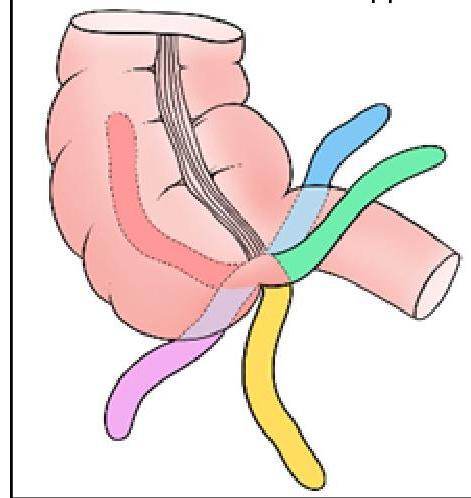
Identify the position of the appendix marked in BLACK in the given image:

In pediatric assessment, a cephalic index of 75-80 is classified as:
A skull is classified as dolichocephalic when the cephalic index is
Yoyo reflex is seen in:
Which of the following is an atavistic epiphysis?
Explanation: **I, II and III** - **Phrygian cap**, **floating gallbladder**, and **absence of gallbladder** are all recognized congenital anomalies of the gallbladder. [1] - These conditions arise from variations in gallbladder development during the **embryonic period**. *I, II and IV* - This option incorrectly includes **Spigelian gallbladder** (IV) as a congenital anomaly, while excluding **absence of gallbladder** (III). - The Spigelian lobe is a part of the liver, and **Spigelian hernia** is a distinct condition, not a congenital anomaly of the gallbladder itself. *II, III and IV* - This option incorrectly includes **Spigelian gallbladder** (IV) as a congenital anomaly and excludes the **Phrygian cap** (I), which is a common and benign congenital variation. - The Phrygian cap represents a folding of the gallbladder fundus and is universally recognized as congenital. *I, III and IV* - This option incorrectly includes **Spigelian gallbladder** (IV) as a congenital anomaly and excludes the **floating gallbladder** (II). - A floating gallbladder, due to a long mesentery, is a known congenital anatomical variation that can predispose to torsion. [1]
Explanation: ***Just proximal to the glans*** - The most frequent location for the urethral opening in **hypospadias** is the **subcoronal** or glanular region, which is just proximal to the glans. - This accounts for approximately 50-70% of all hypospadias cases, making it the **mildest and most common form**. *On the penile shaft* - While hypospadias can manifest with an opening on the **penile shaft** (midshaft or proximal shaft), these are less common than glanular or subcoronal types. - Penile shaft hypospadias usually indicates a more severe form compared to distal types and often presents with more significant **chordee**. *On the perineum* - An opening on the **perineum** represents the most severe form of hypospadias, often classified as **perineal hypospadias**. - This severe anomaly is associated with other urogenital defects and often presents with a **bifid scrotum** and ambiguous genitalia, which are rare compared to distal forms. *At the junction of penile shaft and scrotum* - This location, called **penoscrotal hypospadias**, is another severe form but is still less common than glanular or subcoronal types. - **Penoscrotal hypospadias** is characterized by a high degree of **chordee** and usually requires more complex surgical correction.
Explanation: ***aberrant right subclavian artery*** - Dysphagia lusoria is specifically caused by compression of the **esophagus** by an **aberrant right subclavian artery** [2]. - This congenital anomaly involves the right subclavian artery arising from the distal aortic arch and passing behind the esophagus [2]. *oesophageal atresia* - **Oesophageal atresia** is a congenital condition where the esophagus ends in a blind pouch, preventing food from reaching the stomach [1]. - It usually presents in neonates with **vomiting** and **choking** during feeds, not progressive dysphagia in an adult [1]. *oesophageal web* - An **oesophageal web** is a thin, eccentric protrusion of normal esophageal tissue into the lumen, typically causing intermittent dysphagia to solids. - While it causes dysphagia, it is a structural abnormality within the esophageal lumen, not external compression. *corrosive stricture* - A **corrosive stricture** results from chemical burns to the esophagus, leading to scarring and narrowing of the lumen. - This is an acquired condition with a clear history of corrosive ingestion, unlike the congenital anomaly seen in dysphagia lusoria.
Explanation: ***Pilonidal*** - A **pilonidal sinus** is an acquired condition, usually developing after puberty, linked to **hair follicles** and repeated trauma or pressure in the **sacrococcygeal region**. - It is not present at birth but rather develops over time due to **local trauma**, infection, and foreign body reaction to hair. *Coccygeal* - A **coccygeal dimple** or sinus is a **congenital pit** or tract located over the coccyx. - While sometimes referred to as coccygeal sinuses, these are developmental defects present **from birth**. *Urachal sinus* - An **urachal sinus** is a **congenital anomaly** resulting from incomplete closure of the **urachus**, a fetal remnant connecting the bladder to the umbilicus. - This condition is present at birth, often manifesting with **umbilical discharge**. *Pre auricular* - A **preauricular sinus** is a **congenital malformation** of the outer ear, typically appearing as a small pit or opening near the front of the ear. - It arises from incomplete fusion of the **auricular hillocks** during embryonic development.
Explanation: Correct: 1, 2 and 3 - The **submucosa of the stomach and duodenum** is the most common site for ectopic pancreatic tissue (25-40% of cases). - Ectopic pancreas can also be found in the **liver** and **small bowel mesentery**, though less frequently than in the upper gastrointestinal tract. - These three locations represent the well-recognized probable sites for ectopic pancreatic tissue. *Incorrect: 1, 2 and 4* - This option incorrectly includes the **splenic hilum**, which is not a recognized typical site for ectopic pancreas. - While ectopic pancreatic tissue can occur in various abdominal locations, the splenic hilum is not among the probable sites. *Incorrect: 2, 3 and 4* - This option omits the **submucosa of the stomach and duodenum**, which is the most frequent site for ectopic pancreas. - This makes the option incomplete as it misses the primary location for this condition. *Incorrect: 1, 3 and 4* - This option omits the **liver** as a probable site for ectopic pancreatic tissue. - While the liver is less common than the stomach or duodenum, it is a recognized location for ectopic pancreas.
Explanation: ***Retrocecal*** - The **retrocecal** position (represented by the black color in the image) indicates the appendix is located behind the cecum, often a common variant. - This position can make diagnosis of appendicitis challenging as it may cause atypical pain patterns. *Pelvic* - The **pelvic** appendix descends into the true pelvis, which can mimic gynecological or urological conditions. - It usually causes pain that is more generalized in the lower abdomen or suprapubic region. *Subcecal* - The **subcecal** appendix is located directly below the cecum and is a relatively rare position. - While somewhat straightforward in presentation, it is less common than retrocecal or pelvic positions. *Preileal* - The **preileal** position indicates the appendix lies in front of the terminal ileum. - This is a less common anatomical variation, often associated with specific clinical presentations related to its anterior location.
Explanation: ***Mesocephalic head shape*** - A cephalic index between **75 and 80** indicates a **mesocephalic** head shape, which means the head has a medium or normal width-to-length ratio. - This is considered the **average** or typical head shape in most populations. *Dolichocephalic head shape* - A **dolichocephalic** head shape is characterized by a cephalic index typically **below 75**, meaning the head is relatively **long and narrow**. - This head shape is often seen in individuals with certain genetic backgrounds or conditions that affect skull development. *Brachycephalic head shape* - A **brachycephalic** head shape is characterized by a cephalic index typically **above 80**, meaning the head is relatively **short and wide**. - This can be naturally occurring or a result of conditions like **craniosynostosis** [1] or positional molding. *Scaphocephalic head shape* - **Scaphocephaly** is a specific type of **dolichocephaly** resulting from the premature fusion of the **sagittal suture** [1]. - This condition leads to a very long, narrow, and keeled head shape that would fall into the severe dolichocephalic range (cephalic index well below 75).
Explanation: ***Below 75*** - A **cephalic index below 75** signifies a **dolichocephalic skull**, which is characterized by a relatively **long and narrow head**. - This classification is derived from the ratio of the maximum breadth to the maximum length of the skull (breadth/length × 100). - Dolichocephalic individuals have a **cranial index < 75**, indicating the skull is longer than it is wide. *75-80* - A cephalic index between 75 and 80 signifies a skull classification of **mesocephalic**, meaning a head of **medium breadth and length**. - This is considered the average or intermediate head shape. - This range represents the transition between dolichocephalic and brachycephalic skull types. *Above 80* - A cephalic index above 80 indicates a **brachycephalic skull**, characterized by a relatively **short and broad head**. - This is on the opposite end of the spectrum from dolichocephalic skulls. - Brachycephalic skulls are wider relative to their length. *None of the options* - This option is incorrect because one of the provided options correctly defines a dolichocephalic skull. - The cephalic index below 75 is the accepted criterion for dolichocephaly in anatomical classification.
Explanation: ***Duplication of ureter*** - The **yoyo reflex (yo-yo phenomenon)** describes the bidirectional flow of urine between the upper and lower moieties of a duplicated collecting system. - This occurs in **duplex kidney/ureter** when there is communication between the two ureters, allowing urine to reflux back and forth between the upper and lower pole segments [1]. - The phenomenon results from **pressure gradients** between the two systems, particularly when one moiety has an anomalous insertion, obstruction, or functional impairment. - The term "yo-yo" aptly describes the to-and-fro movement of urine within the duplicated system. *Megaureter* - A **megaureter** is characterized by an abnormally dilated ureter (>7mm), often due to obstruction or primary muscular abnormality. - It does not typically involve the bidirectional flow between two separate ureteral systems described as the yoyo reflex. *Vesicoureteral reflux* - **Vesicoureteral reflux (VUR)** involves the retrograde flow of urine from the bladder into the ureters and kidneys. - While it involves reflux, VUR is a **bladder-to-ureter reflux**, distinct from the yoyo reflex which is an **inter-ureteral phenomenon** within a duplicated system. *Medullary sponge kidney* - **Medullary sponge kidney** is a congenital disorder characterized by cystic dilation of the renal collecting tubules. - It presents with kidney stones and infections but is not associated with ureteral reflexes or the yoyo phenomenon.
Explanation: ***Coracoid process*** - An **atavistic epiphysis** is a phylogenetically older bone or part of a bone that ossifies separately and may fuse later, representing a remnant from ancestral forms. The **coracoid process** of the scapula is considered an atavistic epiphysis in humans, as it corresponds to the complete coracoid bone found in lower vertebrates. - In humans, it forms from a separate center of ossification and fuses with the scapula, reflecting its evolutionary origin. *Condyles of femur* - The condyles of the femur are typical **pressure epiphyses**, designed to withstand significant compressive forces at joints. - They ossify in a manner consistent with their role in forming the knee joint, not as a remnant of a separate ancestral bone. *Lower end of radius* - The lower end of the radius is also a **pressure epiphysis**, forming part of the wrist joint and bearing substantial weight and force. - Its development and function are primarily related to articulation and load-bearing, rather than being a phylogenetic remnant. [1] *Tubercle of humerus* - The tubercles of the humerus (greater and lesser tubercles) are **traction epiphyses**, where muscles and ligaments attach and exert pulling forces. - Their development is directly linked to the attachment of muscles of the rotator cuff, which is distinct from the concept of an atavistic epiphysis.
Principles of Anatomical Variations
Practice Questions
Variations in Vascular Anatomy
Practice Questions
Variations in Musculoskeletal System
Practice Questions
Variations in Nervous System
Practice Questions
Variations in Visceral Anatomy
Practice Questions
Clinically Significant Anatomical Variations
Practice Questions
Congenital Malformations
Practice Questions
Genetic Basis of Anatomical Variations
Practice Questions
Surgical Implications of Variations
Practice Questions
Imaging Aspects of Anatomical Variations
Practice Questions
Get full access to all questions, explanations, and performance tracking.
Start For Free