The accessory obturator artery most commonly arises from which of the following arteries?
Where is the fabella typically present?
Which of the following statements about anodontia is false?
Dysphagia lusoria means dysphagia:
What is the term for a tongue anomaly?
A dens in dente is usually caused by:
Which of the following is the most common renal vascular anomaly?
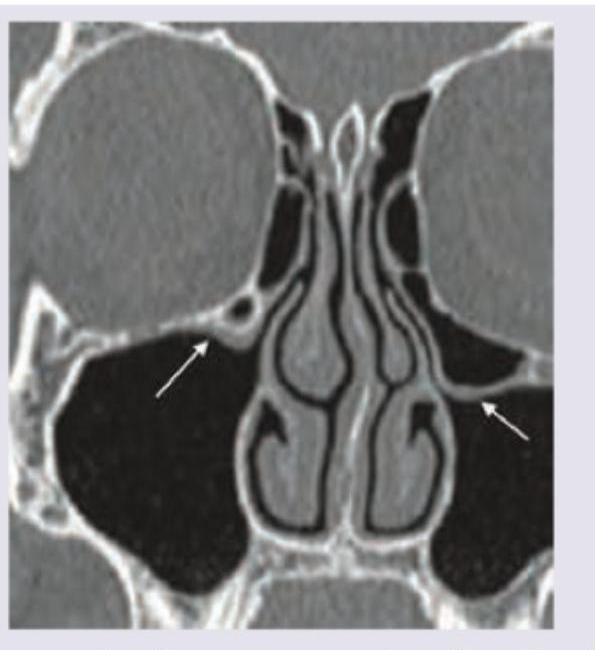
The CT scan of paranasal sinuses shows:

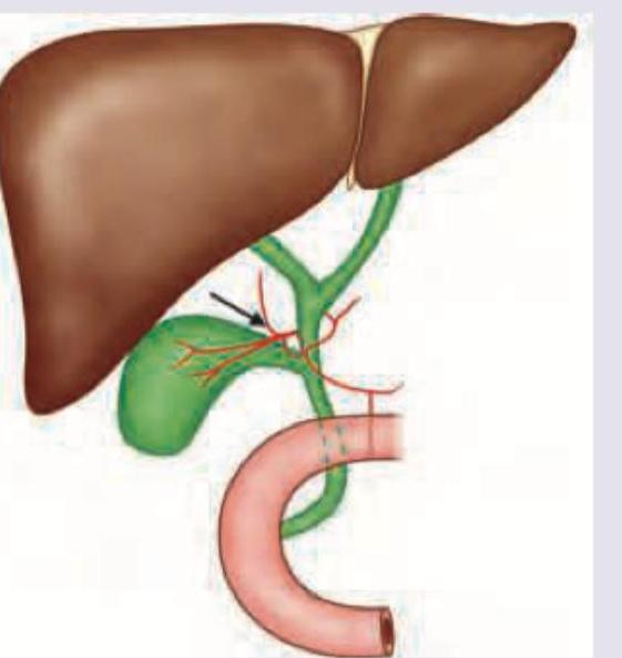
The following arrow marked vessel can cause torrential hemorrhage during cholecystectomy. Which of the following is the correct description?

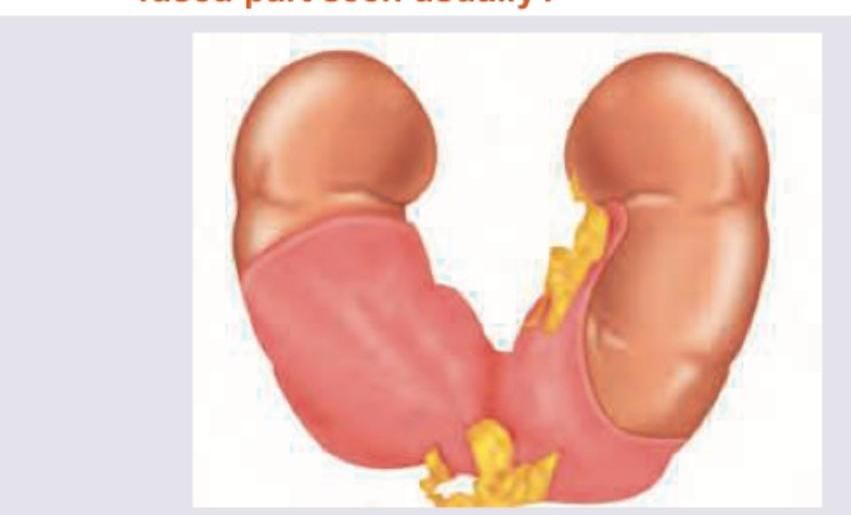
A 28 -year-old male was found to have a condition shown in the image below. At which level is the fused part seen usually?

Explanation: **Explanation:** The **accessory (or abnormal) obturator artery** is a common vascular variation where the arterial supply to the obturator canal originates from the external iliac system rather than the internal iliac system. **1. Why the Inferior Epigastric Artery is Correct:** Normally, the obturator artery is a branch of the **internal iliac artery**. However, in approximately **20–30% of individuals**, a large pubic branch of the **inferior epigastric artery** (a branch of the external iliac) [1] replaces or supplements the normal obturator artery. This vessel travels across the superior pubic ramus to reach the obturator foramen, earning the name "accessory obturator artery." **2. Analysis of Incorrect Options:** * **Femoral Artery:** This is the continuation of the external iliac artery distal to the inguinal ligament. While it supplies the lower limb, it does not typically give rise to the accessory obturator artery. * **Obturator Artery:** This is the "normal" source. The question asks for the origin of the *accessory* version, which by definition is an anomalous origin from a different source. * **Profunda Femoris Artery:** This is a deep branch of the femoral artery. While it has circumflex branches that anastomose around the hip, it is not the primary source of an accessory obturator artery. **3. Clinical Pearls for NEET-PG:** * **Corona Mortis (Crown of Death):** This is the clinical term for the vascular anastomosis between the obturator artery and the inferior epigastric artery. It is located behind the lacunar ligament. * **Surgical Significance:** It is high-yield for **femoral hernia repairs** and pelvic fractures. Accidental injury to this vessel during surgery can lead to massive, difficult-to-control hemorrhage, hence the name "Crown of Death." * **Location:** It typically runs over the **Cooper’s (pectineal) ligament**. [1] PRE-FORMATTED CITATION: "Townsend. Sabiston Textbook Of Surgery. 20E ed. Abdominal Wall, Umbilicus, Peritoneum, Mesenteries, Omentum, Retroperitoneum, pp. 1092-1093."
Explanation: ### Explanation **Correct Answer: B. Lateral head of gastrocnemius** The **fabella** is a small sesamoid bone (a bone embedded within a tendon) typically located in the **lateral head of the gastrocnemius muscle**. It is a common anatomical variation found in approximately 10% to 30% of the population. It articulates with the posterior surface of the lateral femoral condyle and is often bilateral. **Why the other options are incorrect:** * **Medial head of gastrocnemius:** While sesamoid bones can rarely occur here, the fabella is classically and most frequently associated with the lateral head. * **Adductor magnus & Adductor longus:** These are muscles of the medial compartment of the thigh. They do not typically contain sesamoid bones near the knee joint. The adductor magnus inserts into the adductor tubercle, but no "fabella" is associated with its tendon. **High-Yield Clinical Pearls for NEET-PG:** * **Radiological Significance:** On a lateral X-ray of the knee, the fabella can be mistaken for a **loose body** (joint mouse) or an avulsion fracture. Its smooth, rounded borders and specific location help distinguish it. * **Fabella Syndrome:** This is a clinical condition where the fabella causes posterolateral knee pain due to compression of the common peroneal nerve or friction against the femoral condyle. * **Evolutionary Note:** It is more common in Asian populations compared to Caucasians and is considered a "vestigial" structure that is actually increasing in prevalence globally over the last century. * **Other Sesamoids to Remember:** The **Patella** (largest sesamoid in the body, in the quadriceps tendon) and the **Pisiform** (in the flexor carpi ulnaris tendon).
Explanation: **Explanation:** **Anodontia** is a congenital anomaly characterized by the absence of teeth. Understanding the terminology is crucial for NEET-PG: 1. **Why Option B is False (The Correct Answer):** **False anodontia** (also known as pseudoanodontia) occurs when teeth are present in the jaw but fail to erupt into the oral cavity. This is typically due to impaction or systemic conditions like Cleidocranial Dysplasia. The statement in Option B is misleading because while the tooth germ exists, the clinical presentation is the absence of teeth. In contrast, **True anodontia** involves the complete failure of the tooth germ to develop (Agenesis). 2. **Analysis of Other Options:** * **Option A:** Anodontia can indeed affect both dentitions. If the primary (deciduous) tooth germ is absent, the permanent successor will also be absent. * **Option C:** Anodontia is a spectrum. **Hypodontia** refers to the absence of 1–5 teeth (most common: 3rd molars, followed by maxillary lateral incisors and mandibular 2nd premolars). **Oligodontia** is the absence of 6 or more teeth. * **Option D:** **Total anodontia** is the rare, complete absence of all teeth, often associated with hereditary **Ectodermal Dysplasia**. **Clinical Pearls for NEET-PG:** * **Most common missing tooth:** 3rd Molar (Wisdom tooth). * **Most common missing tooth (excluding 3rd molars):** Maxillary lateral incisor. * **Ectodermal Dysplasia:** Look for the triad of anodontia/hypodontia, hypotrichosis (sparse hair), and anhidrosis (lack of sweat glands). * **Hyperdontia:** The presence of extra teeth (e.g., **Mesiodens** – a supernumerary tooth between maxillary central incisors).
Explanation: **Explanation:** **Dysphagia lusoria** (derived from the Latin *lusus naturae*, meaning "freak of nature") is a clinical condition where swallowing is impaired by the extrinsic compression of the esophagus by an **aberrant right subclavian artery (ARSA)** [1]. 1. **Why the correct answer is right:** In normal anatomy, the right subclavian artery arises from the brachiocephalic trunk. In this anomaly, the brachiocephalic trunk is absent, and the right subclavian artery arises as the last branch of the aortic arch (distal to the left subclavian) [1]. To reach the right side, it must cross the midline, usually passing **posterior to the esophagus** (80% of cases). This creates a vascular indentation on the esophagus, leading to difficulty in swallowing. 2. **Why incorrect options are wrong:** * **Option A:** Dysphagia for cold items is typically associated with esophageal motility disorders like **Diffuse Esophageal Spasm (DES)**. * **Option C:** **Esophageal atresia** is a congenital failure of the esophagus to lumenize, presenting immediately at birth with drooling and inability to feed, rather than mechanical compression [2]. * **Option D:** **Benign strictures** are usually secondary to chronic GERD (Peptic stricture) or corrosive ingestion, involving intrinsic narrowing rather than extrinsic vascular compression. **High-Yield NEET-PG Pearls:** * **Barium Swallow Finding:** Shows a characteristic **oblique/spiral indentation** on the posterior aspect of the esophagus at the level of the T3-T4 vertebrae. * **Kommerell’s Diverticulum:** A dilated origin of the aberrant right subclavian artery, which can further exacerbate the compression. * **Surgical Note:** If the aberrant artery also compresses the trachea, it is part of a "vascular ring" configuration [1].
Explanation: The correct answer is **Bifid tongue (Option B)**. This condition, also known as a cleft tongue, is a rare developmental anomaly resulting from the **failure of the two lateral lingual swellings to fuse** in the midline during the fourth week of intrauterine life. These swellings normally merge over the tuberculum impar to form the anterior two-thirds of the tongue. **Analysis of Options:** * **Bifid Tongue (Correct):** It is a true structural anomaly of development. It is frequently associated with **Orofacial Digital Syndrome Type 1**. * **Fissured Tongue (Incorrect):** Also known as "scrotal tongue," this is characterized by deep grooves on the dorsal surface. While it can be congenital, it is often considered a **variation of normal anatomy** or an acquired condition rather than a primary developmental anomaly of tongue formation. It is a key feature of **Melkersson-Rosenthal syndrome**. * **Macroglossia (Incorrect):** This refers to an enlarged tongue. It is a **clinical sign** rather than a specific developmental anomaly of the tongue's structural formation. It is commonly associated with Down syndrome, hypothyroidism (cretinism), and amyloidosis. **High-Yield Clinical Pearls for NEET-PG:** * **Embryology:** The anterior 2/3 of the tongue develops from the **1st pharyngeal arch** (lateral lingual swellings and tuberculum impar), while the posterior 1/3 develops from the **3rd and 4th arches** (hypobranchial eminence). * **Ankyloglossia (Tongue-tie):** Another common anomaly where the lingual frenulum is abnormally short, tethering the tongue to the floor of the mouth. * **Thyroglossal Duct Cyst:** The most common developmental anomaly related to the tongue's descent, usually presenting as a midline neck swelling that moves with protrusion of the tongue.
Explanation: ### Explanation **Dens in dente** (also known as *Dens invaginatus*) is a developmental malformation resulting from a **deep invagination of the enamel organ** into the dental papilla before calcification occurs. #### Why the Correct Answer is Right: During the morphodifferentiation stage of tooth development, a portion of the enamel organ (specifically the internal enamel epithelium) proliferates and invaginates into the underlying dental papilla. This creates a "tooth within a tooth" appearance on radiographs, where a pocket of enamel and sometimes dentin is lined within the pulp cavity. It most commonly affects the **maxillary lateral incisors**. #### Why Other Options are Wrong: * **Option A:** Abnormal proliferation of pulp tissue does not lead to the formation of enamel structures inside the tooth; pulp proliferation is typically a reactive process (e.g., chronic hyperplastic pulpitis). * **Option B:** **Denticles** (pulp stones) are calcified masses found within the pulp chamber, but they are separate entities and do not involve the invagination of the enamel organ. * **Option C:** A supernumerary tooth bud enclaved within another is a description of **fusion** or **gemination** anomalies, not the invagination characteristic of dens in dente. #### High-Yield Clinical Pearls for NEET-PG: * **Most Common Site:** Maxillary permanent lateral incisors (followed by central incisors). * **Radiographic Appearance:** A pear-shaped radiopacity (enamel) within the radiolucency of the pulp canal. * **Clinical Significance:** The invagination often communicates with the oral cavity via a small pit, making the tooth highly susceptible to **early dental caries, pulpitis, and periapical lesions**, even without visible decay. * **Classification:** The **Oehlers’ Classification** is used to categorize the severity based on how far the invagination extends toward the root apex.
Explanation: **Explanation:** The correct answer is **Supernumerary renal arteries**. This is the most common vascular anomaly of the renal system, occurring in approximately **25–30%** of the population. **1. Why Supernumerary Renal Arteries are Correct:** During embryonic development, the kidneys "ascend" from the pelvis to the lumbar region. As they move upward, they receive blood supply from successive arterial sprouts arising from the aorta at higher levels. Normally, the lower vessels degenerate as new ones form. If these transient embryonic vessels persist, they result in **supernumerary (accessory) renal arteries**. These are typically end-arteries, meaning their ligation can lead to segmental renal ischemia. **2. Why the Other Options are Incorrect:** * **Supernumerary renal veins (B & D):** These are significantly less common than arterial variations (occurring in ~10–15% of cases). This is because the venous system undergoes a more complex process of fusion and regression (involving the supracardinal, subcardinal, and postcardinal veins), which typically results in a single definitive vein. * **Double renal arteries (C):** While "double" is a type of supernumerary artery, the term "supernumerary" is the broader, more accurate anatomical classification used in exams to include all variations (accessory, polar, or multiple arteries). **Clinical Pearls for NEET-PG:** * **Origin:** Most accessory renal arteries arise from the abdominal aorta, but they can also arise from the common iliac or mesenteric arteries. * **Lower Polar Artery:** An accessory artery to the lower pole can cross the ureteropelvic junction (UPJ), causing extrinsic compression and resulting in **Hydronephrosis**. * **Surgical Significance:** Knowledge of these variations is critical during renal transplantation, abdominal aortic aneurysm (AAA) repair, and nephrectomy. * **Left vs. Right:** Supernumerary veins are more common on the right side, whereas supernumerary arteries occur with equal frequency on both sides.
Explanation: ***Haller cells*** - The image shows **pneumatized ethmoid air cells** located inferior to the orbit and lateral to the maxillary sinus ostium (arrows), which are characteristic of Haller cells. - These ethmoid cells, also known as **infraorbital ethmoid cells**, can narrow the maxillary sinus ostium, potentially contributing to **recurrent sinusitis**. *Agger nasi cells* - **Agger nasi cells** are the most anterior ethmoid cells, located in the lacrimal bone, anterior and lateral to the frontal recess. - They are typically situated at the level of the frontal sinus ostium, not inferior to the orbit. *Onodi cells* - **Onodi cells** are sphenoethmoidal air cells that extend posteriorly and laterally over the sphenoid sinus, in close proximity to the optic nerve. - Their location is **posterior and superior** to the structures shown in the image, making them incorrectly identified here. *Frontoethmoid cells* - **Frontoethmoid cells** can be a broad category, but specifically refer to ethmoid cells that extend into the frontal sinus or drain into the frontal recess. - While they are ethmoid cells, the specific location shown in the image (inferior to the orbit and maxillary sinus ostium) is more precisely described as Haller cells.
Explanation: ***Moynihan's caterpillar hump due to bend of right hepatic artery*** - The image shows an anatomical variation where the **right hepatic artery** forms a tortuous bend near the cystic duct, resembling a "caterpillar hump." - This anatomical anomaly, known as **Moynihan's hump**, places the right hepatic artery in close proximity to the operative field during cholecystectomy, making it vulnerable to accidental injury and potentially causing torrential hemorrhage. *Moynihan's caterpillar hump due to bend of left hepatic artery* - The left hepatic artery originates from the common hepatic artery and supplies the left lobe of the liver, typically staying well away from the area of concern during routine cholecystectomy. - A bend in the **left hepatic artery** would not be located in the position shown or pose the same risk during gallbladder removal. *Moynihan's caterpillar hump due to bend of cystic artery* - The cystic artery typically arises from the right hepatic artery and is ligated during cholecystectomy to devascularize the gallbladder. - While it supplies the gallbladder, the described "caterpillar hump" refers specifically to a tortuous **right hepatic artery**, not the cystic artery itself. *Moynihan's caterpillar hump due to bend of common hepatic artery* - The common hepatic artery branches into the proper hepatic artery and gastroduodenal artery, located more proximally to the area depicted. - A bend in the **common hepatic artery** would not be found in such close proximity to the cystic duct and would not be described as Moynihan's caterpillar hump in this context.
Explanation: ***L4-L5 vertebral level (due to inferior mesenteric artery)*** - The image depicts a **horseshoe kidney**, a congenital condition where the lower poles of the kidneys are fused. - The fused portion, or **isthmus**, is typically located at the **L4-L5 vertebral level** because its ascent is blocked by the **inferior mesenteric artery**. *L1 - L2 vertebral level* - This level is usually where the **renal arteries** originate and corresponds to the normal position of the majority of the kidney mass, not typically the fused inferior pole. - The kidneys normally ascend during development to this level, but in a horseshoe kidney, the fusion prevents this full ascent. *T12 - L1 vertebral level* - This vertebral level is too high for the fused portion of a horseshoe kidney. - The kidneys initially develop in the pelvis and ascend; T12-L1 would be the upper extent of normal kidney position. *S1 - S2 vertebral level* - This level is too low for the fused portion of a horseshoe kidney, which typically is found in the lumbar region. - S1-S2 is more associated with pelvic structures, indicating the kidneys would have barely ascended at all.
Principles of Anatomical Variations
Practice Questions
Variations in Vascular Anatomy
Practice Questions
Variations in Musculoskeletal System
Practice Questions
Variations in Nervous System
Practice Questions
Variations in Visceral Anatomy
Practice Questions
Clinically Significant Anatomical Variations
Practice Questions
Congenital Malformations
Practice Questions
Genetic Basis of Anatomical Variations
Practice Questions
Surgical Implications of Variations
Practice Questions
Imaging Aspects of Anatomical Variations
Practice Questions
Get full access to all questions, explanations, and performance tracking.
Start For Free