Which of the following disorders is most commonly associated with multifactorial inheritance?
In which condition is the presence of an extra pair of ribs sometimes observed?
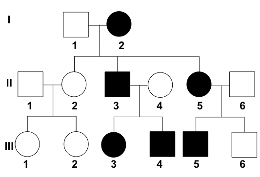
What is the interpretation of the given pedigree chart?

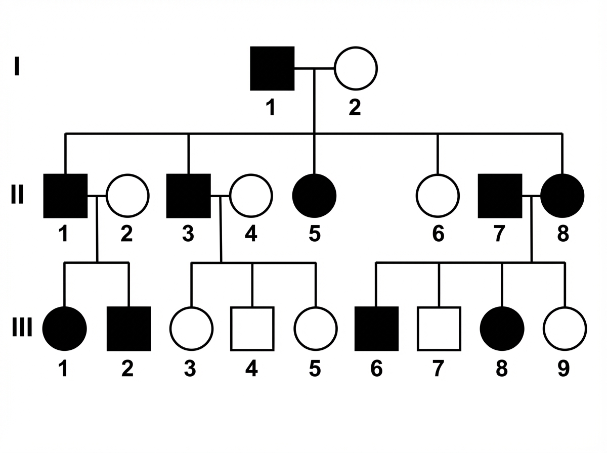
Which disease will show the mode of inheritance depicted in this pedigree?

Mutations are due to changes in:
Which investigation is the gold standard for diagnosing Edwards syndrome?
Which malformation is associated with mutations in the HOX gene?
Which of the following statements about polymorphism is true?
Which testis is typically positioned higher?
Which of the following represents a common variation in the arteries arising from the arch of the aorta?
Explanation: ***Cleft lip*** - **Cleft lip** is a classic example of a **multifactorial disorder**, resulting from the interaction of multiple genes and environmental factors. - Its recurrence risk is typically observed to be around 2-5% in affected families, consistent with **multifactorial inheritance**. *Achondroplasia* - Achondroplasia is an **autosomal dominant disorder**, caused by a single gene mutation in the **FGFR3 gene**. - It does not primarily involve the complex interplay of multiple genes and environmental factors characteristic of multifactorial inheritance. *Lysosomal storage disease* - Lysosomal storage diseases are a group of **autosomal recessive disorders**, each caused by a defect in a specific lysosomal enzyme. - They follow classic **Mendelian inheritance patterns** rather than multifactorial models. *Huntington disease* - Huntington disease is an **autosomal dominant neurodegenerative disorder**, caused by a trinucleotide repeat expansion in the **HTT gene**. - It exhibits a clear dominant inheritance pattern and does not involve multiple genes or significant environmental contributions in its etiology.
Explanation: ***Turner syndrome*** - **Turner syndrome** (45,X) is often associated with skeletal abnormalities, including an extra pair of ribs (cervical ribs) in some cases. - Other common skeletal features include **short stature**, a **shield chest**, and **cubitus valgus**. *Down syndrome* - **Down syndrome** (trisomy 21) is characterized by specific facial features, intellectual disability, and congenital heart defects. - While it can manifest with various skeletal anomalies, an extra pair of ribs is not a characteristic feature. *Klinefelter syndrome* - **Klinefelter syndrome** (47,XXY) is characterized by hypogonadism, tall stature, gynecomastia, and learning difficulties. - Skeletal features may include long limbs and decreased bone density, but cervical ribs are not typically associated with this condition. *Holt-Oram syndrome* - **Holt-Oram syndrome** is a genetic disorder affecting heart and limb development, specifically the upper limbs (thumb abnormalities, phocomelia). - It does not typically involve the presence of an extra pair of ribs. *Fibrous dysplasia* - **Fibrous dysplasia** is a bone disorder where normal bone is replaced by fibrous tissue, leading to weakened bone and fractures. - It is a localized bone condition and does not involve the presence of supernumerary ribs.
Explanation: ***Autosomal dominant*** - The trait appears in **every generation**, indicated by affected individuals in the first, second, and third generations [1]. - Both males and females are affected relatively equally, and affected individuals transmit the trait to approximately half of their offspring, consistent with **vertical transmission** [1]. - **Male-to-male transmission** is present, which definitively rules out X-linked inheritance patterns [1]. *Incomplete penetrance* - This pattern would typically show some individuals who carry the disease-causing genotype but **do not express the phenotype** (unaffected). - In this pedigree, all individuals who are expected to express the trait based on the clear dominant pattern are indeed affected. *Autosomal recessive* - This mode of inheritance would typically show **skipped generations**, meaning affected individuals would often have unaffected parents who are carriers [1]. - In this chart, every affected individual has at least one affected parent, ruling out an autosomal recessive pattern [1]. X-linked dominant - In X-linked dominant inheritance, **no male-to-male transmission** would be observed, as fathers pass their X chromosome only to daughters. - The presence of affected males transmitting the trait to male offspring rules out this inheritance pattern. **References:** [1] Kumar V, Abbas AK, et al.. Robbins and Cotran Pathologic Basis of Disease. 9th ed. Genetic Disorders, pp. 148-151.
Explanation: ***Achondroplasia*** - This pedigree shows an **autosomal dominant** inheritance pattern, characterized by affected individuals in every generation, affected offspring with at least one affected parent, and direct transmission from father to son (which **rules out X-linked**). Achondroplasia is an autosomal dominant disorder. - The presence of **affected individuals (shaded squares/circles)** in each successive generation and the 1:1 ratio of affected to unaffected offspring from affected parents mating with unaffected individuals supports autosomal dominant inheritance. *Prader-Willi syndrome* - This syndrome is caused by **genomic imprinting** or a chromosomal deletion on chromosome 15, typically inherited from the father. It does not follow a simple Mendelian dominant or recessive inheritance pattern. - While it has a genetic basis, its inheritance pattern is **complex** and involves specific parental origin of the genetic defect, unlike the clear autosomal dominant pattern shown. *Wilson disease* - Wilson disease is an **autosomal recessive** disorder, meaning affected individuals inherit two copies of the mutated gene (one from each parent). - This pedigree does not show skipped generations or unaffected parents having affected offspring, which would be characteristic of **autosomal recessive** inheritance. *Wiskott-Aldrich syndrome* - Wiskott-Aldrich syndrome is an **X-linked recessive** disorder. This means it primarily affects males, and affected fathers cannot pass the trait to their sons. - The pedigree shows **affected females** (shaded circles) and **father-to-son transmission** (e.g., father in second generation to son in third generation, assuming the leftmost branch is the paternal line), which rules out X-linked inheritance. *Hemophilia A* - Hemophilia A is an **X-linked recessive** disorder affecting the Factor VIII gene, predominantly affecting males. - Similar to Wiskott-Aldrich syndrome, the presence of **father-to-son transmission** in the pedigree rules out X-linked inheritance patterns, as affected fathers cannot pass X-linked traits to their sons.
Explanation: ***DNA nucleotide sequence*** - **Mutations** are defined as changes in the **genetic material**, which is primarily composed of **DNA**. - These changes in the **nucleotide sequence** of DNA can alter the genetic code, leading to changes in **protein structure and function**. *RNA nucleotide sequence* - While RNA can have its nucleotide sequence altered, these changes are generally not considered true **mutations** in the heritable sense for most organisms. - RNA is typically a temporary molecule, and changes to its sequence are usually not passed down to subsequent generations. *Amino acid sequence of ribonuclease* - An altered **amino acid sequence** in a protein like ribonuclease is a consequence of a **mutation in the DNA**, not the mutation itself. - **Ribonucleases** are enzymes that catalyze the degradation of RNA, and their structure is determined by the **DNA sequence**. *Cell membrane* - The cell membrane is a **lipid bilayer** with embedded proteins that regulates cellular transport and communication. - While its components can be affected by genetic mutations, alterations in the cell membrane itself do not constitute the primary definition of a **mutation**.
Explanation: ***Karyotype*** - A **karyotype** is considered the **gold standard** for diagnosing chromosomal abnormalities like **Edwards syndrome** (Trisomy 18) because it allows for the visualization and analysis of all 46 chromosomes [1]. - It can detect changes in chromosome number (aneuploidy) and large structural rearrangements, directly confirming the presence of an extra chromosome 18 [2]. *MLPA (Multiplex Ligation-dependent Probe Amplification)* - **MLPA** is a molecular technique used to detect **copy number variations** for specific targeted regions but does not provide a comprehensive view of the entire karyotype. - While it can detect trisomies, it is generally used for specific gene deletions or duplications and is not the first-line diagnostic method for whole chromosome aneuploidies like Edwards syndrome. *Microarray (Chromosomal Microarray Analysis)* - **Microarray** is a high-resolution method that can detect smaller deletions and duplications (microdeletions/microduplications) unidentifiable by traditional karyotyping. - However, for whole chromosome trisomies, a **karyotype** remains the gold standard as it directly visualizes the extra chromosome rather than inferring its presence through copy number changes of multiple probes [3]. *FISH (Fluorescence In Situ Hybridization)* - **FISH** is a targeted technique that uses fluorescent probes to detect specific chromosomal regions or whole chromosomes [3]. - While useful for rapid detection of common aneuploidies or specific translocations, it requires prior suspicion of a particular chromosomal abnormality and does not provide a comprehensive global view of all chromosomes like a traditional **karyotype** [3]. **References:** [1] Kumar V, Abbas AK, et al.. Robbins and Cotran Pathologic Basis of Disease. 9th ed. Genetic Disorders, pp. 167-168. [2] Cross SS. Underwood's Pathology: A Clinical Approach. 6th ed. (Basic Pathology) introduces the student to key general principles of pathology, both as a medical science and as a clinical activity with a vital role in patient care. Part 2 (Disease Mechanisms) provides fundamental knowledge about the cellular and molecular processes involved in diseases, providing the rationale for their treatment. Part 3 (Systematic Pathology) deals in detail with specific diseases, with emphasis on the clinically important aspects., pp. 54-55. [3] Kumar V, Abbas AK, et al.. Robbins and Cotran Pathologic Basis of Disease. 9th ed. Genetic Disorders, pp. 186-187.
Explanation: ***Polysyndactyly*** - The **HOX gene** plays a critical role in limb development and is associated with the malformation of **polysyndactyly**, which is characterized by extra fingers or toes [1]. - This condition is due to the disruption of the normal **patterning** during limb formation, directly involving the action of HOX genes [1]. *Gorlin syndrome* - Gorlin syndrome is primarily caused by mutations in the **PTCH1 gene**, linked to **basal cell carcinoma** and other abnormalities. - It does not involve HOX gene mutations, hence is **not** related to limb malformations. *Holoprosencephaly* - Holoprosencephaly is a developmental condition often linked to **chromosomal anomalies** and abnormal embryonic development, **not specifically** HOX gene mutations. - It refers to the incomplete separation of the forebrain, distinct from the **limb malformations** associated with HOX genes. *Mayer Rokitansky syndrome* - Mayer-Rokitansky syndrome involves **agenesis** or **hypoplasia** of the uterus and upper two-thirds of the vagina, which is due to other genetic factors. - This condition is not related to the functions of the **HOX genes** in limb or skeletal development. **References:** [1] Kumar V, Abbas AK, et al.. Robbins and Cotran Pathologic Basis of Disease. 9th ed. Bones, Joints, and Soft Tissue Tumors, p. 1186.
Explanation: ***Single locus with multiple normal alleles.*** - **Polymorphism** refers to the existence of multiple alleles at a **single genetic locus** within a population. - For a variant to be considered a polymorphism, the most common allele must have a frequency of **less than 99%**, meaning at least two alleles are common. *Single locus with multiple abnormal alleles, not linked to a specific phenotype.* - While polymorphism involves multiple alleles at a single locus, classifying them as "abnormal" is misleading, as polymorphism often refers to **variations that are common** in the population and not necessarily disease-causing or abnormal. - The definition emphasizes the presence of multiple alleles, not their clinical implications, and many polymorphisms have **no overt phenotypic effect**. *Single phenotype linked to a single locus with both normal and abnormal alleles.* - Polymorphism primarily describes **genetic variation (alleles)**, not direct links to a single phenotype. A single locus can influence **multiple phenotypes**, and a single phenotype can be influenced by multiple loci. - Grouping alleles as "normal" and "abnormal" oversimplifies the concept; **many polymorphisms are neutral** or beneficial, and some "normal" alleles can become "abnormal" in certain contexts. *Single phenotype linked to a single locus with multiple abnormal alleles.* - This option incorrectly narrows the definition by focusing on a **single phenotype** and exclusively "abnormal" alleles. Polymorphism encompasses any common variation, regardless of its phenotypic effect or whether the alleles are considered abnormal. - Many polymorphic variations are **silent mutations** or variations that do not result in overt phenotypic changes or disease.
Explanation: ***Right testis*** - The **right testis** is commonly positioned slightly higher than the left testis in most males [1]. - This anatomical variation is due to the **left spermatic cord** being inherently longer, which allows the left testis to hang lower. *Left testis* - The **left testis** is typically positioned lower than the right testis. - Its lower position is attributed to the generally **longer left spermatic cord**. *It varies between individuals* - While minor individual variations exist, a consistent pattern of the **right testis** being higher is observed in the majority of males. - The differences in cord length lead to a general trend, not complete randomness in height. *Both are at the same level* - It is uncommon for both testes to be at precisely the **same level**. - The **asymmetrical length** of the spermatic cords makes equal positioning rare.
Explanation: ***Left common carotid artery arising from brachiocephalic trunk*** - Normally, the **brachiocephalic trunk** gives rise to the right subclavian and right common carotid arteries, while the left common carotid and left subclavian arteries arise directly from the aortic arch. - However, in this common variation (sometimes called a **bovine arch**), the left common carotid artery originates from the brachiocephalic trunk, reducing the number of direct branches from the arch to two. *Absence of brachiocephalic trunk* - The **brachiocephalic trunk** is one of the three major vessels normally arising from the aortic arch [1]. Its absence is a very rare and significant anomaly, not a common variation. - This would imply direct origins for the right subclavian and right common carotid arteries from the aortic arch, which is not typical. *Left vertebral artery arising from the arch* - The **left vertebral artery** typically arises from the first part of the **left subclavian artery**. - Its direct origin from the aortic arch is a known anatomical variant, but it is less common than the left common carotid artery arising from the brachiocephalic trunk. *Presence of retroesophageal subclavian artery* - A **retroesophageal subclavian artery** (usually the right subclavian artery) is a congenital anomaly where the artery takes an abnormal course behind the esophagus [1]. - While it is a recognized variant, it is considered less common than the "bovine arch" configuration.
Get full access to all questions, explanations, and performance tracking.
Start For Free