NEET-PG 2025 — Anatomy
9 Previous Year Questions with Answers & Explanations
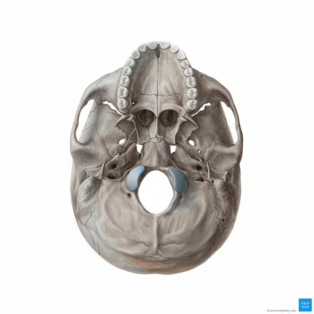
The image below highlights the jugular foramen. Which of the following does NOT pass through this foramen?
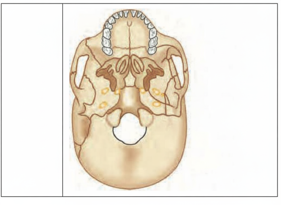
The image below highlights the jugular foramen. Which of the following does NOT pass through this foramen?
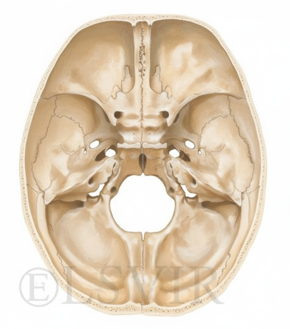
The image below highlights the jugular foramen. Which of the following does NOT pass through this foramen?
The image shows a congenital cardiac defect. Abnormal development of which branch of aortic arch leads to this defect?
During a neck dissection, a nerve was identified and marked that is most likely the vagus nerve (CN X). Which of the following is NOT a functional component of the vagus nerve?
What is the correct nerve supply to the muscles labelled as A and B ?
A patient presents with meralgia paresthetica. Based on the diagram, identify the nerve involved in this condition.
Which of the following organs does this epithelium most likely belong to?
During hyperextension, the long head of triceps gets detached from which site?
NEET-PG 2025 - Anatomy NEET-PG Practice Questions and MCQs
Question 1: The image below highlights the jugular foramen. Which of the following does NOT pass through this foramen?
- A. Hypoglossal nerve (Correct Answer)
- B. Accessory nerve
- C. Glossopharyngeal nerve
- D. Vagus nerve
Explanation: ***Hypoglossal nerve*** - The **Hypoglossal nerve (CN XII)** does not pass through the jugular foramen; instead, it utilizes a distinct opening called the **Hypoglossal canal**. - This canal is found in the **occipital bone**, separate from the structures passing through the jugular foramen. *Accessory nerve* - The **Accessory nerve (CN XI)** is one of the three cranial nerves (IX, X, XI) that pass through the jugular foramen. - It provides motor supply to the **sternocleidomastoid** and **trapezius muscles**. *Glossopharyngeal nerve* - The **Glossopharyngeal nerve (CN IX)** exits the skull through the anterior part of the jugular foramen. - It is known for innervating the **stylopharyngeus muscle** and carrying sensation and taste from the posterior one-third of the tongue. *Vagus nerve* - The **Vagus nerve (CN X)** passes through the jugular foramen, primarily through its middle compartment. - It is the major parasympathetic nerve supplying the **thorax and abdomen**, as well as motor supply to the **larynx** and pharynx.
Question 2: The image below highlights the jugular foramen. Which of the following does NOT pass through this foramen?
- A. Hypoglossal nerve (Correct Answer)
- B. Accessory nerve
- C. Glossopharyngeal nerve
- D. Vagus nerve
Explanation: ***Hypoglossal nerve*** - The **Hypoglossal nerve (CN XII)** does not pass through the jugular foramen; instead, it utilizes a distinct opening called the **Hypoglossal canal**. - This canal is found in the **occipital bone**, separate from the structures passing through the jugular foramen. *Accessory nerve* - The **Accessory nerve (CN XI)** is one of the three cranial nerves (IX, X, XI) that pass through the jugular foramen. - It provides motor supply to the **sternocleidomastoid** and **trapezius muscles**. *Glossopharyngeal nerve* - The **Glossopharyngeal nerve (CN IX)** exits the skull through the anterior part of the jugular foramen. - It is known for innervating the **stylopharyngeus muscle** and carrying sensation and taste from the posterior one-third of the tongue. *Vagus nerve* - The **Vagus nerve (CN X)** passes through the jugular foramen, primarily through its middle compartment. - It is the major parasympathetic nerve supplying the **thorax and abdomen**, as well as motor supply to the **larynx** and pharynx.
Question 3: The image below highlights the jugular foramen. Which of the following does NOT pass through this foramen?
- A. Hypoglossal nerve (Correct Answer)
- B. Accessory nerve
- C. Glossopharyngeal nerve
- D. Vagus nerve
Explanation: ***Hypoglossal nerve*** - The **Hypoglossal nerve (CN XII)** does not pass through the jugular foramen; instead, it utilizes a distinct opening called the **Hypoglossal canal**. - This canal is found in the **occipital bone**, separate from the structures passing through the jugular foramen. *Accessory nerve* - The **Accessory nerve (CN XI)** is one of the three cranial nerves (IX, X, XI) that pass through the jugular foramen. - It provides motor supply to the **sternocleidomastoid** and **trapezius muscles**. *Glossopharyngeal nerve* - The **Glossopharyngeal nerve (CN IX)** exits the skull through the anterior part of the jugular foramen. - It is known for innervating the **stylopharyngeus muscle** and carrying sensation and taste from the posterior one-third of the tongue. *Vagus nerve* - The **Vagus nerve (CN X)** passes through the jugular foramen, primarily through its middle compartment. - It is the major parasympathetic nerve supplying the **thorax and abdomen**, as well as motor supply to the **larynx** and pharynx.
Question 4: The image shows a congenital cardiac defect. Abnormal development of which branch of aortic arch leads to this defect?
- A. Right 6th aortic arch
- B. Left 6th aortic arch (Correct Answer)
- C. Left 4th aortic arch
- D. Right 4th aortic arch
Explanation: ***Left 6th aortic arch*** - The **Ductus Arteriosus**, which shunts blood from the pulmonary artery to the aorta in fetal life, is embryologically derived from the distal portion of the **Left 6th Aortic Arch**. - **Patent Ductus Arteriosus (PDA)** is the failure of this fetal connection to close after birth. *Right 4th aortic arch* - The **Right 4th Aortic Arch** contributes to the formation of the proximal segment of the **Right Subclavian Artery**. - Defects in the right 4th arch are typically associated with vascular ring anomalies, such as an aberrant right subclavian artery. *Right 6th aortic arch* - The **Right 6th Aortic Arch** forms the proximal segment of the **Right Pulmonary Artery**. - The distal part of the right 6th arch normally involutes and disappears completely, unlike the persistence seen on the left side (PDA). *Left 4th aortic arch* - The **Left 4th Aortic Arch** forms the segment of the **Arch of the Aorta** located between the left common carotid and the left subclavian arteries. - This arch is primarily involved in forming the main aortic arch structure.
Question 5: During a neck dissection, a nerve was identified and marked that is most likely the vagus nerve (CN X). Which of the following is NOT a functional component of the vagus nerve?
- A. General visceral afferent
- B. General somatic efferent (Correct Answer)
- C. General visceral efferent
- D. General somatic afferent
Explanation: ***General somatic efferent*** - **GSE** fibers innervate muscles derived from **somites**, typically cranial nerves that control the extraocular muscles (CN III, IV, VI) or the tongue muscles (CN XII). - The vagus nerve (CN X) does not carry GSE fibers; its motor components are **Special Visceral Efferent (SVE)** for pharyngeal/laryngeal muscles, and **General Visceral Efferent (GVE)** for parasympathetic supply. *General visceral afferent* - **GVA** fibers are a major functional component of the vagus nerve, providing **visceral sensation** from the respiratory, cardiovascular, and gastrointestinal systems. - These fibers monitor stretch receptors in the lungs, **baroreceptors** in the aortic arch, and sensation from the abdominal viscera, crucial for reflex regulation. *General visceral efferent* - **GVE** fibers represent the **parasympathetic outflow** of the vagus nerve below the neck, innervating smooth muscle, cardiac muscle, and glands. - This component is responsible for decreasing **heart rate**, promoting **bronchoconstriction**, and increasing gastrointestinal motility and secretion. *General somatic afferent* - **GSA** fibers carry general sensory information (pain, temperature, touch) from parts of the head and are present in the vagus nerve. - CN X GSA fibers provide sensation from a small area of the external auditory meatus and the external surface of the **tympanic membrane**.
Question 6: What is the correct nerve supply to the muscles labelled as A and B ?
- A. A - Facial nerve, B - Spinal accessory nerve
- B. A - Spinal accessory nerve, B - Mandibular nerve
- C. A - Facial nerve, B - Nerve to mylohyoid
- D. A - Mandibular nerve, B - Facial nerve (Correct Answer)
Explanation: ***A - Mandibular nerve, B - Facial nerve*** - Label A points to the **Masseter** muscle, which is supplied by the **Mandibular nerve (V3)** via the masseteric nerve. - Label B points to the **Platysma** muscle, which is a muscle of facial expression supplied by the **Facial nerve (VII)** via the cervical branch. *A - Facial nerve, B - Nerve to mylohyoid* - The **Masseter** (A) is a muscle of mastication supplied by the **Mandibular nerve (V3)**, not the facial nerve. - The **Nerve to mylohyoid** supplies the mylohyoid muscle and anterior belly of digastric, whereas B is the **Platysma** muscle supplied by the facial nerve. *A - Facial nerve, B - Spinal accessory nerve* - The **Facial nerve (VII)** supplies muscles of facial expression, not muscles of mastication like the **Masseter** (A). - The **Spinal accessory nerve (XI)** supplies the sternocleidomastoid and trapezius muscles; it does not supply the **Platysma** (B). *A - Spinal accessory nerve, B - Mandibular nerve* - The **Masseter** (A) is supplied by the **Mandibular nerve (V3)**, not the spinal accessory nerve (XI). - The **Platysma** (B) is supplied by the **Facial nerve (VII)**, not the mandibular nerve (V3).
Question 7: A patient presents with meralgia paresthetica. Based on the diagram, identify the nerve involved in this condition.
- A. A
- B. B
- C. C (Correct Answer)
- D. D
Explanation: ***C (Lateral Femoral Cutaneous Nerve)*** - Meralgia paresthetica is an entrapment neuropathy caused by compression of the **Lateral Femoral Cutaneous Nerve (LFCN)**, which corresponds to C in the diagram and arises from **L2 and L3** roots. - Compression usually occurs as the nerve passes under the **inguinal ligament**, resulting in pain, numbness, and tingling over the **anterolateral thigh**. *A (Ilioinguinal/Iliohypogastric Nerve)* - Nerve A, usually the Ilioinguinal or Iliohypogastric nerve (T12, L1), innervates the **inguinal region** and lower abdominal wall. - Entrapment of these nerves results in pain radiating towards the **groin** or superior thigh, not the characteristic distribution of meralgia paresthetica. *B (Genitofemoral Nerve)* - Nerve B is the **Genitofemoral nerve** (L1, L2), which supplies sensation to the superior medial thigh and genitalia. - Injury results in loss of the **cremasteric reflex** and sensory changes in the scrotal/labial and proximal anterior thigh area. *D (Femoral Nerve)* - Nerve D is the large **Femoral Nerve** (L2-L4), responsible for motor supply to the **quadriceps** and sensation to the anterior thigh and medial leg. - Compression typically causes prominent **quadriceps weakness** (difficulty extending the knee) in addition to sensory loss, unlike the purely sensory presentation of meralgia paresthetica.
Question 8: Which of the following organs does this epithelium most likely belong to?
- A. Trachea
- B. Gallbladder
- C. Intestine
- D. Ureter (Correct Answer)
Explanation: ***Ureter*** - The image displays **transitional epithelium** (urothelium), which is characterized by multiple cell layers and large, dome-shaped (**umbrella**) cells on the apical surface. - This specialized stratified epithelium is highly stretchable and is exclusively found lining the urinary tract, including the renal pelvis, ureter, and bladder. *Gallbladder* - The gallbladder is lined by **simple columnar epithelium**, which is specialized for water reabsorption and concentration of bile, lacking the stratified layers seen here. - It has a single layer of cells with microvilli but does not possess the unique apical **umbrella cells** of the urothelium. *Trachea* - The trachea is lined by **pseudostratified ciliated columnar epithelium**, which includes numerous **goblet cells** and distinct cilia on the apical surface. - This tissue type appears stratified but all cells contact the basement membrane, unlike the true stratification seen in the image. *Intestine* - The small and large intestines are lined by **simple columnar epithelium** with prominent **microvilli** (forming a striated border) and numerous **goblet cells**. - This is a single-layered epithelium primarily designed for absorption and secretion, which is structurally distinct from the urinary epithelium shown.
Question 9: During hyperextension, the long head of triceps gets detached from which site?
- A. Supraglenoid tubercle
- B. Shaft of humerus
- C. Olecranon process
- D. Infraglenoid tubercle (Correct Answer)
Explanation: ***Infraglenoid tubercle*** - The **long head of the triceps brachii** muscle originates from the **infraglenoid tubercle** of the scapula. - Violent or sudden hyperextension of the shoulder places maximum tensile stress on this specific origin site, predisposing it to avulsion or detachment. *Supraglenoid tubercle* - This tubercle is the origin point for the **long head of the biceps brachii** muscle, which is not the muscle relevant to this injury scenario. - Injuries involving the supraglenoid tubercle are classically associated with **SLAP lesions** and glenohumeral instability. *Shaft of humerus* - The **medial and lateral heads** of the triceps originate along the posterior surface of the shaft of the humerus. - These muscular origins are less prone to acute avulsion during hyperextension compared to the tendinous long head origin on the scapula. *Olecranon process* - The olecranon process of the ulna is the **insertion** point for all three heads of the triceps, not the origin. - Detachment at this site typically occurs due to direct impact or strong eccentric contraction during forced elbow flexion, often resulting in an **olecranon fracture**.