All (378)Anatomy (22)Anatomy (5)Anesthesiology (9)Behavioral Science (1)Biochemistry (3)Biochemistry (21)Biostatistics (4)Community Medicine (20)Dermatology (13)Diagnosis (3)ENT (11)Forensic Medicine (13)General Medicine (1)Internal Medicine (19)Internal Medicine (26)Microbiology (20)OB/GYN (16)Obstetrics and Gynecology (16)Ophthalmology (10)Orthopaedics (14)Pathology (25)Pediatrics (18)Pediatrics (2)Pharmacology (37)Physiology (10)Psychiatry (6)Radiology (12)Surgery (21)
Q221
What is the sequence of rigor mortis in the human body?
Q222
From a medico-legal perspective, in cases of sexual assault involving a female victim, what type of court proceeding is typically used to record medical evidence and testimony to protect the victim's privacy?
Q223
Police brought a person from a railway track with features of dry dilated pupils, dry skin, slurred speech, and altered sensorium. What is the most likely cause of poisoning?
Q224
A child was born 8 months after the father's death. The grandparents filed a case claiming that the baby is not their son's, but DNA testing confirmed paternity. What is the child called?
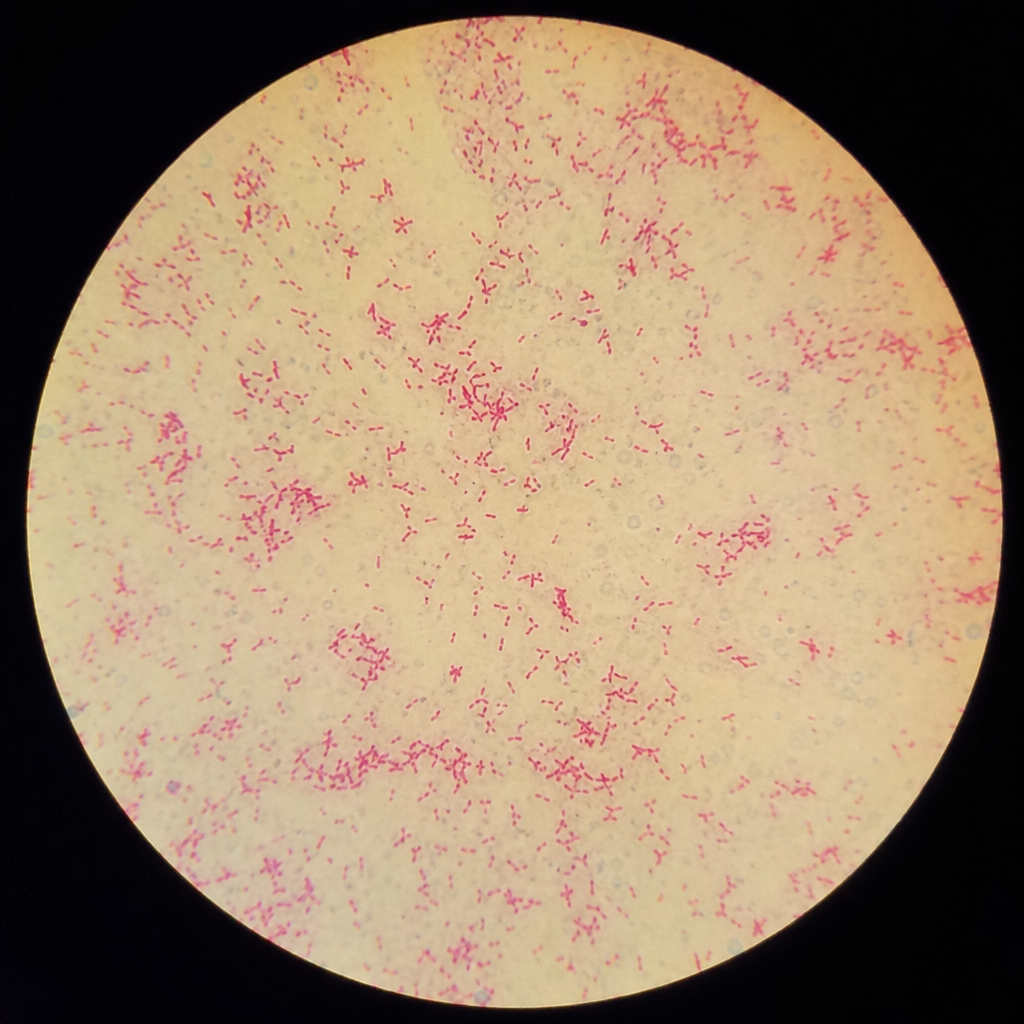
Q225
Examination of a suspicious stain shows the following finding. Which test is being depicted? 
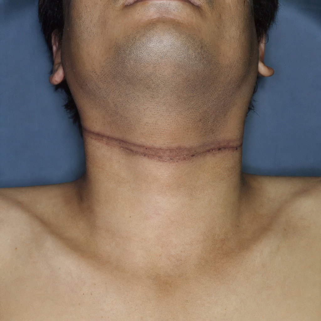
Q226
The dead body shows the following finding as seen in the image. What is the most likely cause of death?
Q227
In a medicolegal examination, an 18-year-old male claims he is 16 years old. Which joint X-ray should be done to estimate his age?
Q228
A 14-year-old victim of sexual assault with 22 weeks gestation has been brought for Medical Termination of Pregnancy (MTP). Which of the following statements is true?