All SubjectsAnatomy (22)Anatomy (5)Anesthesiology (9)Behavioral Science (1)Biochemistry (3)Biochemistry (21)Biostatistics (4)Community Medicine (20)Dermatology (13)Diagnosis (3)ENT (11)Forensic Medicine (13)General Medicine (1)Internal Medicine (19)Internal Medicine (26)Microbiology (20)OB/GYN (16)Obstetrics and Gynecology (16)Ophthalmology (10)Orthopaedics (14)Pathology (25)Pediatrics (18)Pediatrics (2)Pharmacology (37)Physiology (10)Psychiatry (6)Radiology (12)Surgery (21)
Q11
A pregnant female at 37 weeks of gestation with a history of prosthetic heart valves is currently taking warfarin. She comes for a routine antenatal check-up. What is the appropriate management advice?
Q12
A pregnant lady delivers a healthy baby via normal delivery. What is the earliest time at which an intrauterine contraceptive device (IUCD) can be inserted?
Q13
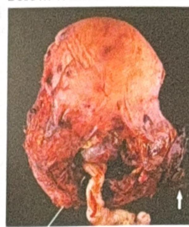
A G2P1L1 woman with a history of previous cesarean section presents with complications related to the placenta. The image below shows the gross appearance of the uterus. What is the most likely diagnosis?
Q14
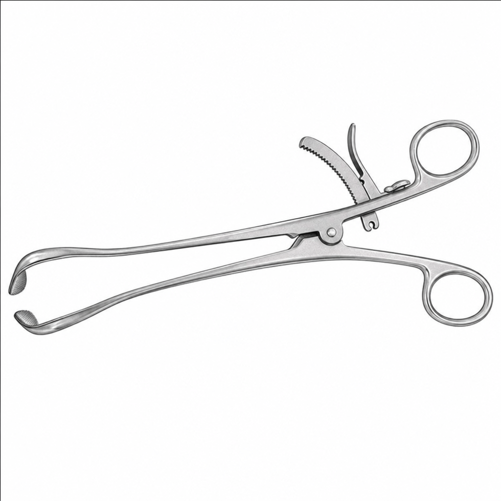
Identify the instrument shown in the image below: 
Q15
Based on the educational diagram showing different degrees of perineal tears, which degree involves only the perineal skin and vaginal mucosa without affecting the underlying muscle?
Q16
Identify the maneuver shown in the image: