All SubjectsAnatomy (22)Anatomy (5)Anesthesiology (9)Behavioral Science (1)Biochemistry (3)Biochemistry (21)Biostatistics (4)Community Medicine (20)Dermatology (13)Diagnosis (3)ENT (11)Forensic Medicine (13)General Medicine (1)Internal Medicine (19)Internal Medicine (26)Microbiology (20)OB/GYN (16)Obstetrics and Gynecology (16)Ophthalmology (10)Orthopaedics (14)Pathology (25)Pediatrics (18)Pediatrics (2)Pharmacology (37)Physiology (10)Psychiatry (6)Radiology (12)Surgery (21)
Q11
A man presents with dysuria and urethral discharge after a history of unprotected sex. The Gram stain of his discharge is shown. What is the best culture medium for isolating the organism responsible?

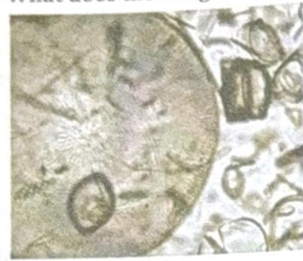
Q12
A young boy who used to wash his contact lenses in tap water or with unhygienic lens fluid developed keratitis. Microscopy revealed an organism with spiked or star-shaped structures. Identify the correct organism responsible.
Q13
A patient in the ICU with a central venous catheter (CVC) develops an infection. Microscopy reveals ovoid budding yeast cells. What is the most likely organism?
Q14
A farmer presents with a subcutaneous wound on his foot with discharge. Microscopy of a white granule from the wound shows Gram-positive filamentous rods. What is the most likely organism?
Q15
The image shows microscopic organisms. What is the most appropriate description of these organisms?
Q16
A patient is a known case of thalassemia. Which of the following viruses would be responsible for attacking progenitor cells and causing aplastic anemia?
Q17
A 27-year-old patient presents with motheaten alopecia, moist perianal lesions, and an asymptomatic macular rash. What is the most likely causative organism?
Q18
A patient presents with sinus tracts on the foot, and a smear reveals filamentous organisms.
Q19
A female presents with dysuria and vaginal discharge. Wet mount examination shows pear-shaped organisms. What is the most likely diagnosis?
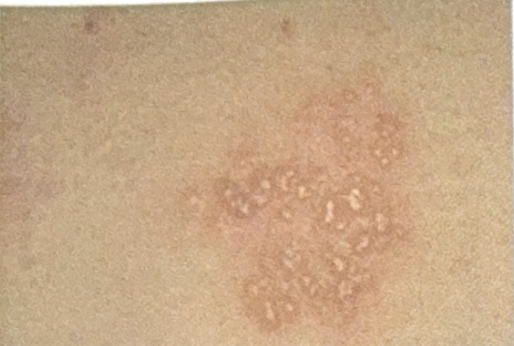
Q20
A patient presents with genital grouped vesicles, as shown in the image. What is the most likely causative organism?