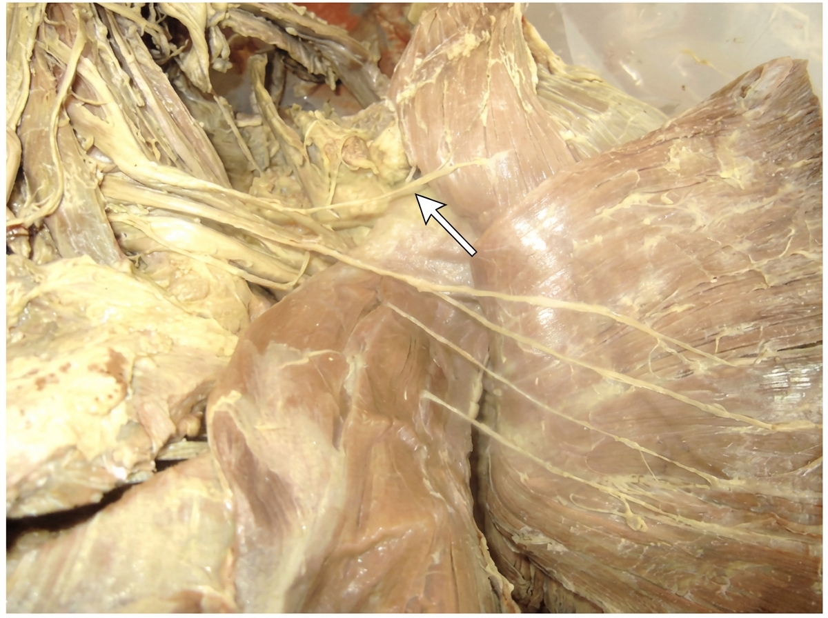
Identify the arrow marked nerve
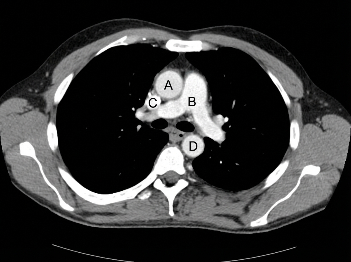
Identify the labeled structures correctly in the axial CT image of the thorax
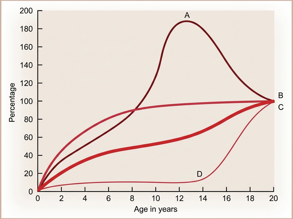
The Image shows the growth curve of different organs with age. Identify A in the graph.
NEET-PG 2024 - Anatomy NEET-PG Practice Questions and MCQs
Question 21: Identify the arrow marked nerve
- A. Medial pectoral nerve
- B. Lateral pectoral nerve (Correct Answer)
- C. Long thoracic nerve
- D. Thoracodorsal nerve
- E. Nerve to subclavius
Explanation: ***Lateral pectoral nerve*** - The arrow points to a nerve originating from the **lateral cord of the brachial plexus**, traveling laterally to innervate the **pectoralis major muscle**. - Its position, lateral to the medial pectoral nerve and supplying the pectoralis major, confirms it as the lateral pectoral nerve. *Medial pectoral nerve* - The medial pectoral nerve typically arises from the **medial cord of the brachial plexus** and passes through both **pectoralis major** and **pectoralis minor**. - It lies more medially and generally pierces the pectoralis minor, unlike the nerve indicated. *Long thoracic nerve* - The long thoracic nerve innervates the **serratus anterior muscle** and runs along the lateral aspect of the chest wall. - Its course is distal and distinct from the nerve shown, which is clearly positioned in the pectoral region. *Thoracodorsal nerve* - The thoracodorsal nerve innervates the **latissimus dorsi muscle** and descends on the posterior axillary wall. - It is not located in the shown pectoral region and has a different trajectory. *Nerve to subclavius* - The nerve to subclavius arises from the **upper trunk of the brachial plexus** (C5-C6) and descends to innervate the **subclavius muscle**. - It has a more superior course compared to the lateral pectoral nerve and is not visible in the position indicated by the arrow.
Question 22: Identify the labeled structures correctly in the axial CT image of the thorax
- A. A - Pulmonary trunk, B - Ascending aorta, C - Superior vena cava, D - Descending aorta
- B. A - Superior vena cava, B - Pulmonary trunk, C - Ascending aorta, D - Descending aorta
- C. A - Ascending aorta, B - Pulmonary trunk, C - Superior vena cava, D - Descending aorta (Correct Answer)
- D. A - Ascending aorta, B - Superior vena cava, C - Pulmonary trunk, D - Descending aorta
- E. A - Pulmonary trunk, B - Superior vena cava, C - Ascending aorta, D - Descending aorta
Explanation: ***A - Ascending aorta, B - Pulmonary trunk, C - Superior vena cava, D - Descending aorta*** - **A** points to the **ascending aorta**, which is the large artery arising from the left ventricle and supplying oxygenated blood to the systemic circulation. On this axial view, it is typically located anterior and to the right of the pulmonary artery. - **B** points to the **pulmonary trunk**, which emerges from the right ventricle and bifurcates into the pulmonary arteries to carry deoxygenated blood to the lungs. It is positioned anterior and to the left of the ascending aorta at this level. - **C** points to the **superior vena cava**, a large vein that collects deoxygenated blood from the upper half of the body and drains into the right atrium. It is typically located to the right and slightly posterior to the ascending aorta at this level. - **D** points to the **descending aorta**, which continues from the aortic arch downwards through the chest and abdomen to supply blood to the lower body. It is visible posteriorly and to the left of the vertebral body on this axial CT image. *A - Pulmonary trunk, B - Ascending aorta, C - Superior vena cava, D - Descending aorta* - This option incorrectly identifies A as the pulmonary trunk and B as the ascending aorta; the **ascending aorta** is typically positioned more anteriorly and to the right compared to the **pulmonary trunk** at this level. - The relative positions of the pulmonary trunk and ascending aorta are swapped, leading to an incorrect labeling. *A - Superior vena cava, B - Pulmonary trunk, C - Ascending aorta, D - Descending aorta* - This option incorrectly identifies A as the superior vena cava and C as the ascending aorta. The **superior vena cava** is typically located to the right of the ascending aorta, not anterior-central. - The **ascending aorta** is usually the most anterior and central great vessel in the mediastinum at this level, which does not correspond to C. *A - Ascending aorta, B - Superior vena cava, C - Pulmonary trunk, D - Descending aorta* - This option incorrectly identifies B as the superior vena cava and C as the pulmonary trunk. **Superior vena cava** is a venous structure and is not typically located in the position of B, which is an arterial structure (pulmonary trunk). - The **pulmonary trunk** is usually more anterior and central than the position of C, which correctly identifies the superior vena cava in other options.
Question 23: The Image shows the growth curve of different organs with age. Identify A in the graph.
- A. Brain Growth
- B. Somatic Growth
- C. Lymphoid Growth (Correct Answer)
- D. Gonadal Growth
- E. Reproductive Growth
Explanation: ***Lymphoid Growth*** - Curve 'A' shows a rapid increase in size during **childhood**, peaking around **10-12 years of age**, and then declining to adult levels. - This pattern is characteristic of **lymphoid tissues** (e.g., thymus, lymph nodes, tonsils), which are larger relative to body size in childhood and undergo involution post-puberty. *Brain Growth* - **Neural growth** (like the brain) typically shows very rapid growth in early childhood, reaching close to adult size by about 6-7 years of age, and then leveling off. - Curve 'A' continues to grow rapidly much longer than expected for brain development and then shows a distinct decline. *Somatic Growth* - **General somatic growth** (e.g., body as a whole) shows a sigmoid curve, with rapid growth in infancy and adolescence, and a plateau in adulthood. - Curve 'A' peaks significantly above the 100% mark and then declines, which is not characteristic of overall somatic growth. *Gonadal Growth* - **Genital (gonadal) growth** remains relatively flat until puberty, after which it experiences a rapid increase. - Curve 'A' shows significant growth in early childhood and a peak before puberty, which is inconsistent with typical gonadal development. *Reproductive Growth* - **Reproductive growth** follows the same pattern as gonadal growth, remaining minimal until puberty with subsequent rapid increase. - Curve 'A' demonstrates early childhood growth and pre-pubertal peak, which does not match the reproductive growth pattern.