NEET-PG 2022 — Radiology
4 Previous Year Questions with Answers & Explanations
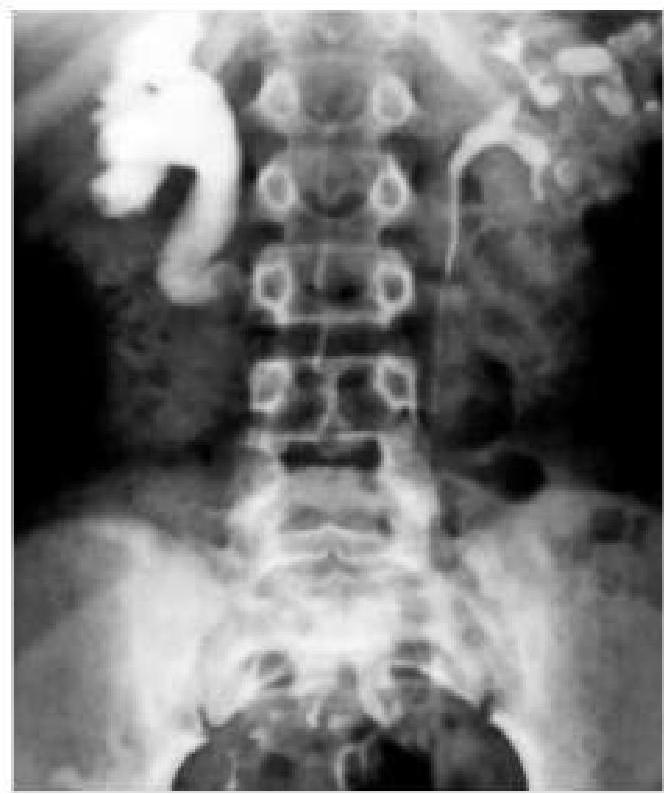
A delayed intravenous urogram of a patient is given below. What is the likely diagnosis?
A 60-year-old male with a history of smoking presents with severe abdominal pain and a pulsatile abdominal mass. What is the most appropriate next step in managing this patient?
Most sensitive investigation for abdominal trauma in a hemodynamically stable patient is-
What is the role of fixer?
NEET-PG 2022 - Radiology NEET-PG Practice Questions and MCQs
Question 1: A delayed intravenous urogram of a patient is given below. What is the likely diagnosis?
- A. Pelviureteric junction obstruction (Correct Answer)
- B. Putty kidney
- C. Staghorn calculus
- D. Cystic kidney
Explanation: ***Pelviureteric junction obstruction*** - The image shows marked **dilatation of the renal pelvis and calyces** on the right side, with a relatively abrupt narrowing at the junction of the pelvis and ureter. - The delayed nature of the urogram suggests **impaired drainage** of contrast from the renal pelvis, accumulating proximal to the obstruction. *Putty kidney* - A "putty kidney" (or **autonecrotic kidney**) refers to a chronic, severely diseased kidney, often seen in end-stage **renal tuberculosis**, that has become calcified and non-functional. - This image demonstrates active contrast excretion and pelvicalyceal dilatation, not a calcified, non-functional organ. *Staghorn calculus* - A staghorn calculus is a **large, branched kidney stone** that occupies a significant portion of the renal collecting system. - While it can cause hydronephrosis, the image does not show a dense, radiopaque calculus filling the collecting system. *Cystic kidney* - **Cystic kidneys**, such as in polycystic kidney disease, are characterized by multiple fluid-filled sacs within the kidney parenchyma. - The image depicts dilatation of the collecting system, not diffuse cystic changes throughout the renal parenchyma.
Question 2: A 60-year-old male with a history of smoking presents with severe abdominal pain and a pulsatile abdominal mass. What is the most appropriate next step in managing this patient?
- A. Immediate surgery
- B. CT angiography (Correct Answer)
- C. Ultrasound of the abdomen
- D. Observation
Explanation: ***CT angiography*** - **CT angiography** is the most appropriate next step for a **hemodynamically stable** patient with suspected **abdominal aortic aneurysm (AAA)**, as suggested by severe abdominal pain and a pulsatile abdominal mass in a smoker. - **CT angiography** is the gold standard for delineating the size, extent, anatomical relationships, and most importantly, the **rupture status** of an AAA, providing critical information for surgical planning. - This imaging is essential for determining the appropriate surgical approach (open repair vs. endovascular repair/EVAR) and identifying contained ruptures that may not be immediately life-threatening but require urgent intervention. - The patient presentation suggests a **symptomatic or contained rupture**, and assuming hemodynamic stability, imaging should precede surgery. *Immediate surgery* - Immediate surgery **without imaging** is indicated only when the patient is **hemodynamically unstable** (hypotension, shock) or in frank rupture with peritoneal signs, where delays for imaging would be fatal. - In a **stable** patient, proceeding directly to surgery without CT angiography increases operative risks due to lack of precise anatomical information about aneurysm size, location, proximal/distal extent, and involvement of renal or iliac arteries. - The question scenario, while concerning, does not explicitly indicate hemodynamic instability, making imaging the preferred next step. *Ultrasound of the abdomen* - **Ultrasound** is excellent for screening and confirming the presence of AAA, measuring aortic diameter, but it has significant limitations in acute settings. - **Ultrasound cannot reliably detect rupture** or provide the detailed anatomical information necessary for surgical planning (proximal/distal extent, branch vessel involvement). - In this acute presentation with suspected rupture, ultrasound would be insufficient and would delay definitive diagnosis, making **CT angiography** superior. *Observation* - **Observation** is absolutely contraindicated in a patient with severe abdominal pain and a pulsatile abdominal mass, as this presentation strongly suggests **symptomatic or ruptured AAA**. - AAA rupture carries mortality rates of 50-80% even with treatment, and any delay in diagnosis and intervention significantly increases mortality. - The combination of symptoms (severe pain) with a pulsatile mass in a high-risk patient (elderly male smoker) mandates immediate diagnostic workup, not observation.
Question 3: Most sensitive investigation for abdominal trauma in a hemodynamically stable patient is-
- A. Ultrasonography (FAST)
- B. Diagnostic peritoneal lavage (DPL)
- C. MRI (Magnetic Resonance Imaging)
- D. CT Scan (Computed Tomography) (Correct Answer)
Explanation: ***CT Scan (Computed Tomography)*** - **CT scans** offer superior anatomical detail and can accurately detect organ damage, hemorrhage, and other injuries in **hemodynamically stable** patients with abdominal trauma. - It is considered the **most sensitive** and specific imaging modality for evaluating blunt and penetrating abdominal trauma when the patient can tolerate the study. *Ultrasonography (FAST)* - While effective for detecting **free fluid** (blood) in specific abdominal areas, **Focused Assessment with Sonography for Trauma (FAST)** has lower sensitivity for solid organ injuries or bowel perforations. - Its primary role is rapid assessment for **hemoperitoneum** to guide immediate management in unstable patients, not detailed injury characterization. *Diagnostic peritoneal lavage (DPL)* - **DPL** is an invasive procedure with high sensitivity for detecting **intraperitoneal bleeding**, but it does not identify specific organ injuries or retroperitoneal hemorrhage. - It is rarely used in hemodynamically stable patients due to its invasiveness and the availability of more detailed imaging techniques. *MRI (Magnetic Resonance Imaging)* - **MRI** provides excellent soft tissue contrast but is typically too **time-consuming** and less accessible in urgent trauma settings compared to CT. - It's generally not the first-line investigation for acute abdominal trauma due to motion artifacts and limited utility in detecting air or bone injuries.
Question 4: What is the role of fixer?
- A. It binds developer to film.
- B. It removes the extra silver halides which are unfixed. (Correct Answer)
- C. It takes away extra developer solution.
- D. It strengthens/fixes the silver halides on to X-ray film.
Explanation: ***It removes the extra silver halides which are unfixed.*** - The fixer solution plays a crucial role in creating a permanent radiographic image by **dissolving and removing all unexposed and undeveloped silver halide crystals** from the film emulsion. - This process prevents the film from darkening over time and ensures that only the areas exposed to radiation, forming the latent image, remain visible. *It binds developer to film.* - The developer's role is to **convert exposed silver halide crystals into metallic silver**, creating the visible image, but it does not bind to the film permanently. - The fixer step follows development to remove unexposed crystals, not to bind the developer. *It takes away extra developer solution.* - While the fixer follows the developer bath, its primary role is not simply to remove residual developer solution; that function is more closely associated with the **rinse step** between development and fixing. - The main action of the fixer involves chemical removal of silver halides. *It strengthens/fixes the silver halides on to X-ray film.* - The developer is responsible for converting exposed silver halides into visible silver, but the fixer actually **removes the *unfixed*** silver halides, rather than strengthening or "fixing" them onto the film. - This removal is essential for a stable and clear image, as any remaining unfixed halides would eventually darken.