All (140)Anatomy (8)Anesthesiology (2)Biochemistry (10)Community Medicine (10)Dental (3)Dermatology (5)ENT (3)Forensic Medicine (8)Internal Medicine (22)Microbiology (10)Obstetrics and Gynecology (17)Ophthalmology (2)Orthopaedics (3)Pathology (5)Pediatrics (7)Pharmacology (8)Physiology (3)Psychiatry (1)Psychiatry (2)Radiology (2)Surgery (9)
Q91
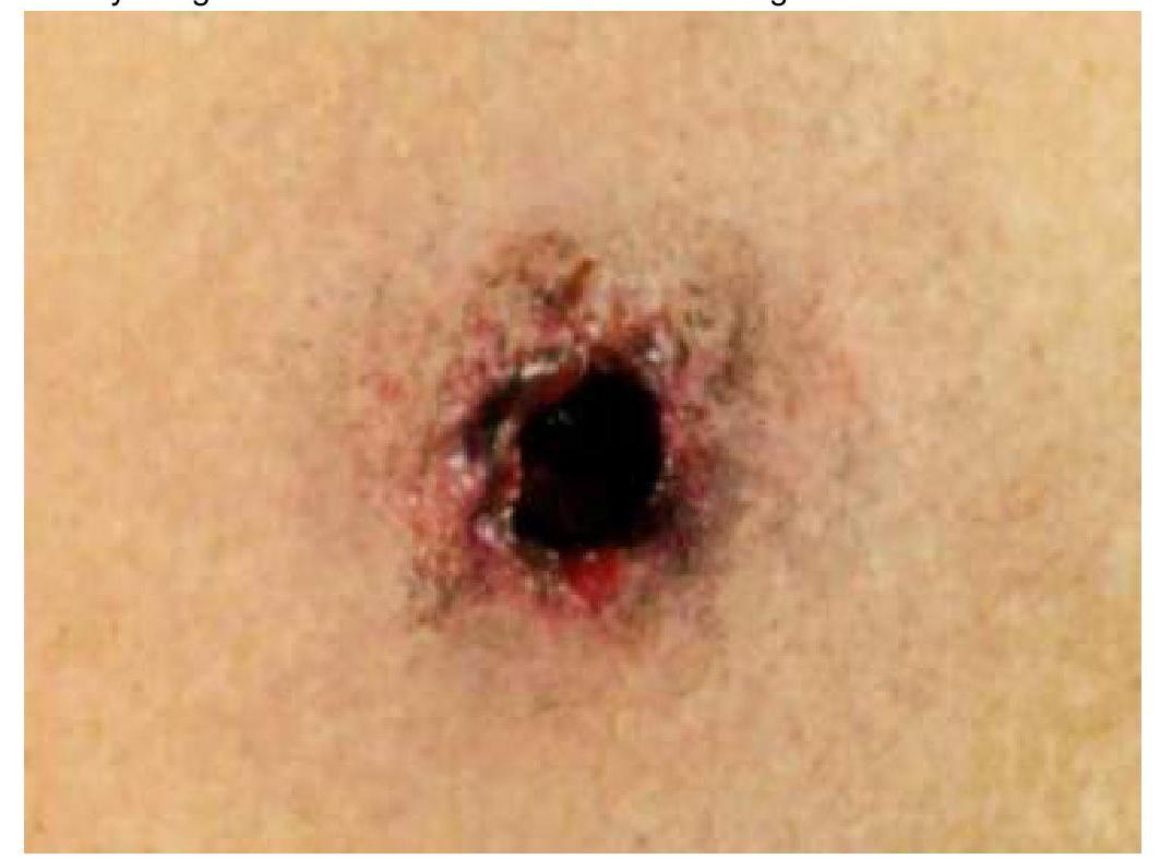
Based on the provided wound characteristics, identify the type of firearm used and the estimated range of the shot.
Q92
A 45-year-old female patient is told about the benefits and complications of a hysterectomy, and she agrees to the procedure. What kind of consent is this?
Q93
A surgeon is called to perform an emergency operation after attending a party. During the operation, the assisting staff notices the surgeon's hands shaking and instruments falling from his grasp. He eventually nicks an artery, leading to the patient's collapse and significant blood loss. Under which legal term is this incident most likely to be classified in Indian medical jurisprudence?