NEET-PG 2022 — Dermatology
4 Previous Year Questions with Answers & Explanations
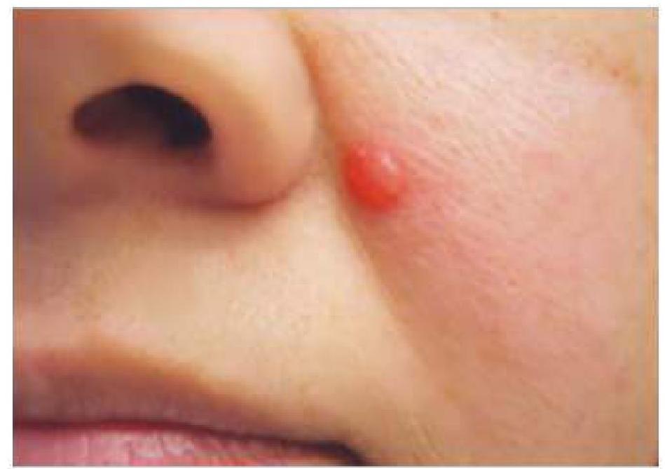
A male patient presented with a 0.3 cm nodule on the left nasolabial fold. A pathological examination revealed a basaloid appearance with peripheral palisading. What is the most likely diagnosis?
A 22-year-old woman presents with diffuse hair loss for 1 month. She had a past history of enteric fever 4 months ago. What is the likely cause?
Pitting of nails is seen in:
A farmer has an ulcer on leg with indurated margin and multiple sinuses with discharging granules. The likely diagnosis is -
NEET-PG 2022 - Dermatology NEET-PG Practice Questions and MCQs
Question 1: A male patient presented with a 0.3 cm nodule on the left nasolabial fold. A pathological examination revealed a basaloid appearance with peripheral palisading. What is the most likely diagnosis?
- A. Basal cell carcinoma (Correct Answer)
- B. Melanoma
- C. Squamous cell carcinoma
- D. Nevus
Explanation: ***Basal cell carcinoma*** - The description of a **basaloid appearance with peripheral palisading** on pathological examination is a classic histological feature of basal cell carcinoma (BCC). - BCC commonly presents as a nodule on sun-exposed areas like the **nasolabial fold** and is the most common skin cancer. *Melanoma* - Melanoma is characterized by the **malignant proliferation of melanocytes** and histologically shows atypical melanocytes with pagetoid spread or nest formation. - While it can appear as a nodule, the described **basaloid appearance with peripheral palisading** is not characteristic of melanoma. *Squamous cell carcinoma* - Squamous cell carcinoma typically shows **atypical keratinocytes** with keratinization, intercellular bridges, and sometimes desmoplasia. - It usually presents as an **erythematous, scaly patch** or nodule, often with ulceration, and the described histology does not match. *Nevus* - A nevus (mole) is a benign proliferation of melanocytes, showing **uniform nests of melanocytes** with maturation as they descend into the dermis. - The term **basaloid appearance** refers to cells resembling basal keratinocytes, which is not typical for a nevus.
Question 2: A 22-year-old woman presents with diffuse hair loss for 1 month. She had a past history of enteric fever 4 months ago. What is the likely cause?
- A. Telogen effluvium (Correct Answer)
- B. Androgenic alopecia
- C. Alopecia areata
- D. Anagen effluvium
Explanation: ***Telogen effluvium*** - **Telogen effluvium** is characterized by diffuse hair shedding, often occurring 2-4 months after a significant physiological or psychological stressor, such as **enteric fever**. - The stress prematurely shifts a large number of hair follicles from the **anagen (growth)** phase into the **telogen (resting)** phase, leading to synchronized shedding. *Androgenic alopecia* - This condition presents as a gradual, patterned hair loss, typically characterized by **receding hairline** and thinning at the crown in men. - In women, it often appears as **diffuse thinning** over the crown, but it's not usually acute or triggered by an infection in the manner described. *Alopecia areata* - **Alopecia areata** is an autoimmune condition causing **sudden, well-demarcated patches of hair loss**, not diffuse shedding. - It is frequently associated with other autoimmune diseases, and the hair loss pattern is distinct from the patient's presentation. *Anagen effluvium* - **Anagen effluvium** causes rapid, diffuse hair loss during the **anagen (growth)** phase, often triggered by chemotherapy or radiation. - The onset is typically much faster (days to weeks) after the trigger, unlike the delayed onset seen in this case.
Question 3: Pitting of nails is seen in:
- A. Psoriasis and Alopecia areata (Correct Answer)
- B. Psoriasis only
- C. Psoriasis and Lichen planus
- D. Alopecia areata and Eczema
Explanation: ***Psoriasis and Alopecia areata*** - **Nail pitting** is a very common and characteristic finding in **psoriasis**, resulting from defective keratinization of the nail matrix. - While less common, nail pitting can also occur in **alopecia areata**, typically due to inflammation affecting the nail matrix. *Psoriasis only* - While **psoriasis** is a primary cause of nail pitting, stating it as "only" is incorrect as other conditions also present with this sign. - This option incorrectly limits the differential diagnosis for nail pitting. *Psoriasis and Lichen planus* - **Psoriasis** does cause nail pitting, but **lichen planus** typically causes **longitudinal ridging**, splitting, subungual hyperkeratosis, and sometimes pterygium formation, rather than classic pitting. - This option includes a condition that usually manifests with different nail changes. *Alopecia areata and Eczema* - **Alopecia areata** can cause nail pitting, but **eczema** of the hands or fingers more commonly leads to **nail plate dystrophy**, discoloration, ridging, or thickening, rather than distinct pitting. - While eczema can affect nails, pitting is not its characteristic nail manifestation.
Question 4: A farmer has an ulcer on leg with indurated margin and multiple sinuses with discharging granules. The likely diagnosis is -
- A. Lupus vulgaris
- B. Actinomycosis
- C. Scrofuloderma
- D. Mycetoma (Correct Answer)
Explanation: ***Mycetoma*** - This is the **correct diagnosis** characterized by the classic triad: **tumefaction** (swelling with indurated margin), multiple **draining sinuses**, and discharge of **granules**. - The **occupational history** (farmer with soil exposure) and **location on the leg** are highly suggestive of mycetoma, particularly common in agricultural workers. - The granules are **colonies of microorganisms** (either fungi [eumycetoma] or bacteria [actinomycetoma]) aggregated and encased in a cement-like matrix, a distinctive feature of this chronic infection. - **Key distinguisher**: Mycetoma has a predilection for the **lower extremities**, especially the foot and leg, in individuals with occupational soil exposure. *Actinomycosis* - Actinomycosis is a bacterial infection caused by *Actinomyces* species, which also forms abscesses and draining sinuses with characteristic **"sulfur granules."** - **Why incorrect**: While actinomycosis shares features of sinuses and granules, it most commonly affects the **cervicofacial (50-60%)**, **thoracic**, or **abdominal** regions. - **Leg involvement is rare** for actinomycosis, making mycetoma the more likely diagnosis in this clinical scenario. - The occupational history and typical location favor mycetoma over actinomycosis. *Lupus vulgaris* - This is a form of **cutaneous tuberculosis** presenting as red-brown plaques or nodules, often with an **"apple-jelly" appearance** on diascopy. - While it can cause ulcers, it typically does **not present with deep-seated sinuses and discharging granules**, which are pathognomonic for mycetoma. *Scrofuloderma* - This is a form of cutaneous tuberculosis that develops from the direct extension of underlying **tuberculous adenitis** or **osteomyelitis** to the skin. - It presents as cold abscesses that eventually rupture, forming irregular ulcers and sinuses, but typically **lacks the distinct discharging granules** of mycetoma. - The clinical presentation with granular discharge clearly differentiates mycetoma from scrofuloderma.