All SubjectsAnatomy (18)Anesthesiology (4)Biochemistry (26)Community Medicine (33)Dental (2)Dermatology (7)ENT (7)Forensic Medicine (13)Internal Medicine (54)Microbiology (21)Obstetrics and Gynecology (28)Ophthalmology (8)Orthopaedics (5)Pathology (25)Pediatrics (18)Pharmacology (27)Physiology (21)Psychiatry (9)Radiology (17)Surgery (23)
Q11
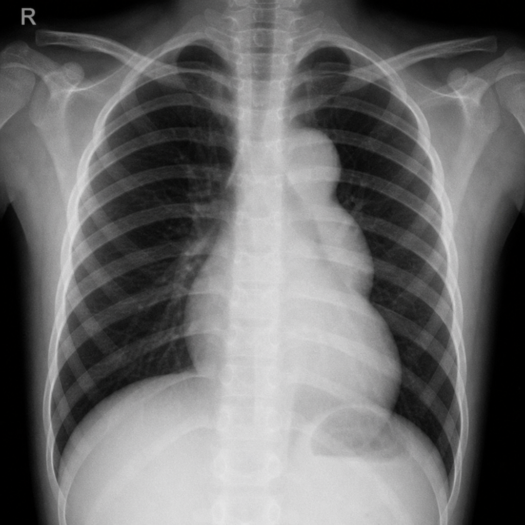
The Figure of 3 sign on chest X-ray is seen in which condition?
Q12
Radiation Dose Monitoring in Occupational Workers is done by
Q13
Which of the following is a FALSE statement regarding radiation?
Q14
Which radiotherapy technique involves the use of remote afterloading to deliver radiation directly to the tumor?
Q15
A 7-week pregnant lady underwent a chest X-ray by mistake. What is to be done?
Q16
A 55 year old woman diagnosed with ca cervix stage IIb is advised for chemoradiation. Which of the following is the true statement regarding radiation use?
Q17
Cancer patient undergoes radiotherapy, pick the true statement for radiosensitivity of tissues?