NEET-PG 2020 — Radiology
16 Previous Year Questions with Answers & Explanations
Identify the condition in the X-ray given below:
The chest radiograph shown below is from a 25-year-old male patient presenting with hypertension. The image demonstrates bilateral inferior rib notching. What is the most likely diagnosis?
Identify the imaging modality and the location of pathology shown in the image.
Steeple sign is seen in which of the following conditions?
What is the angle shown in the image known as?
Which radiological finding is shown in the image?
The Barium Swallow examination shows a filling defect in the esophagus. What is the most probable diagnosis?
Identify the radiological sign of Ischemic colitis from the image provided.
A radiograph is obtained from a child with scoliosis. What is the name of the angle used to measure spinal curvature?
Radiation Dose Monitoring in Occupational Workers is done by
NEET-PG 2020 - Radiology NEET-PG Practice Questions and MCQs
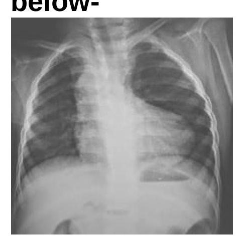
Question 1: Identify the condition in the X-ray given below:
- A. TGA
- B. TAPVC
- C. TOF (Correct Answer)
- D. Ebstein's anomaly
Explanation: ***TOF*** - The chest X-ray shows a **boot-shaped heart (coeur en sabot)**, which is highly characteristic of **Tetralogy of Fallot** due to right ventricular hypertrophy and pulmonary artery hypoplasia. - There is also **reduced pulmonary vascular markings** (oligemia), indicating decreased blood flow to the lungs, a typical finding in TOF. *TGA* - Transposition of the Great Arteries (TGA) typically presents with a **"egg-on-a-string" appearance** on chest X-ray, characterized by a narrow mediastinum and cardiomegaly, which is not seen here. - Pulmonary vascularity can be increased or normal in TGA, unlike the decreased vascularity observed in the image. *TAPVC* - Total Anomalous Pulmonary Venous Connection (TAPVC) usually shows a **"snowman" or "figure-of-8" heart** shadow on chest X-ray, due to enlarged SVC and innominate vein. - This condition is also associated with **increased pulmonary vascular markings** and often cardiomegaly, which are absent in the provided image. *Ebstein's anomaly* - Ebstein's anomaly is characterized by a **massively enlarged heart** on chest X-ray due to right atrial enlargement and tricuspid regurgitation. - It often shows **reduced pulmonary vascular markings** due to functional pulmonary stenosis, but the characteristic "boot shape" is not typically present.
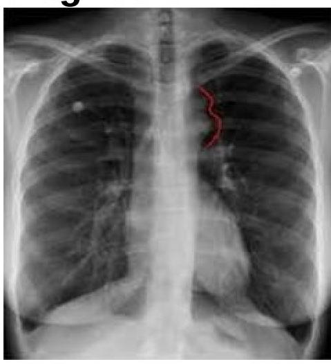
Question 2: The chest radiograph shown below is from a 25-year-old male patient presenting with hypertension. The image demonstrates bilateral inferior rib notching. What is the most likely diagnosis?
- A. Tetralogy of Fallot
- B. Ebstein's Anomaly
- C. TAPVC
- D. Coarctation of Aorta (Correct Answer)
Explanation: ***Coarctation of Aorta*** - The chest radiograph shows findings consistent with **rib notching**, which is a classic sign of coarctation of the aorta due to increased collateral circulation through intercostal arteries. - The history of **hypertension** in a male patient, especially if presenting at a younger age or with differential blood pressures between upper and lower extremities, strongly suggests coarctation of the aorta. *Tetralogy of Fallot* - Characterized by a **boot-shaped heart** due to right ventricular hypertrophy and pulmonary outflow obstruction. - Would typically present with **cyanosis** and decreased pulmonary vascular markings, not rib notching or isolated hypertension. *Ebstein's Anomaly* - Involves apical displacement of the **tricuspid valve**, leading to atrialization of the right ventricle and severe tricuspid regurgitation. - Chest X-rays often show **severe cardiomegaly** (huge heart due to right atrial enlargement) and decreased pulmonary vascularity, which are not depicted here. *TAPVC* - Total anomalous pulmonary venous connection (TAPVC) involves all pulmonary veins draining into the systemic circulation. - The classic chest X-ray finding for supracardiac TAPVC is a **"snowman" or "figure of 8" sign** due to dilated anomalous vessels and superior vena cava, which is absent in this image.
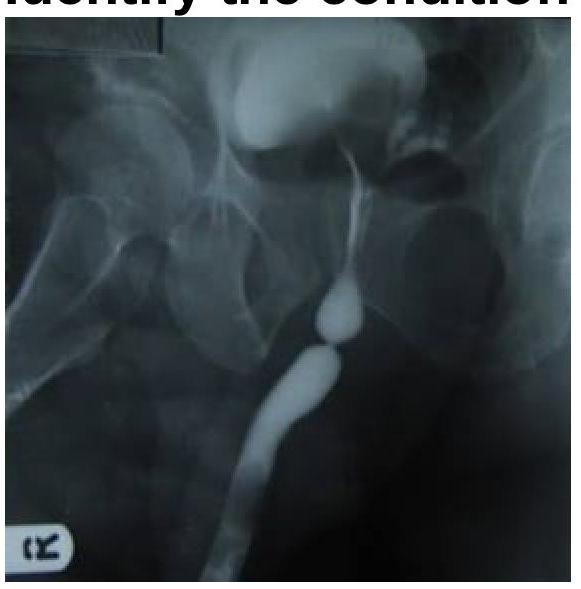
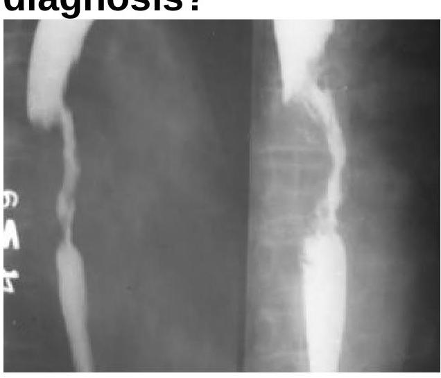
Question 3: Identify the imaging modality and the location of pathology shown in the image.
- A. MCU with Bulbar urethral stricture (Correct Answer)
- B. MCU with penile stricture
- C. RGU with membranous stricture
- D. RGU with prostatic stricture
Explanation: ***MCU with Bulbar urethral stricture*** - The image shown is a **Micturating Cystourethrogram (MCU)** because the bladder is filled with contrast and the urethra is being visualized during urination. - There is a clear **narrowing (stricture)** in the **bulbar portion of the urethra**, appearing as a segment with reduced caliber, consistent with a bulbar urethral stricture. *MCU with penile stricture* - While it is an MCU, the stricture is located in the **bulbar urethra**, which is proximal to the penile (pendulous) urethra. - A penile stricture would be seen further distally in the urethra. *RGU with membranous stricture* - This is an **MCU**, not a Retrograde Urethrogram (RGU), which is performed by inserting contrast from the urethral meatus. In an MCU, the contrast flows antegrade from the bladder. - A **membranous stricture** would be located more proximally, within the deep perineal pouch, between the bulbar urethra and the prostatic urethra. *RGU with prostatic stricture* - As mentioned, this is an **MCU**, not an RGU. - A prostatic stricture would be located within the **prostatic urethra**, which is much more proximal than the stricture seen in this image.
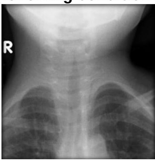
Question 4: Steeple sign is seen in which of the following conditions?
- A. Acute epiglottitis
- B. Acute laryngotracheobronchitis (Correct Answer)
- C. Laryngeal papillomatosis
- D. Bilateral abductor paralysis
Explanation: ***Acute laryngotracheobronchitis*** - The **steeple sign** on an anteroposterior (AP) neck radiograph is a classic finding in acute laryngotracheobronchitis, also known as **croup**. - This sign refers to the **subglottic narrowing** of the trachea, resembling a church steeple, due to edema caused by viral infection. *Acute epiglottitis* - Acute epiglottitis is characterized by the **thumb sign** on a lateral neck radiograph, where the swollen epiglottis appears enlarged. - This condition involves inflammation primarily of the epiglottis, not the subglottic region. *Laryngeal papillomatosis* - Laryngeal papillomatosis is characterized by **wart-like growths** (papillomas) on the vocal cords and larynx, often leading to hoarseness. - Radiographically, it typically appears as irregular soft tissue masses, not the diffuse subglottic narrowing seen in croup. *Bilateral abductor paralysis* - Bilateral abductor paralysis involves the inability of both vocal cords to abduct, leading to a **fixed, narrowed glottic opening**. - This condition presents as a smooth, constant narrowing at the level of the vocal cords rather than the subglottic, conical narrowing of the steeple sign.
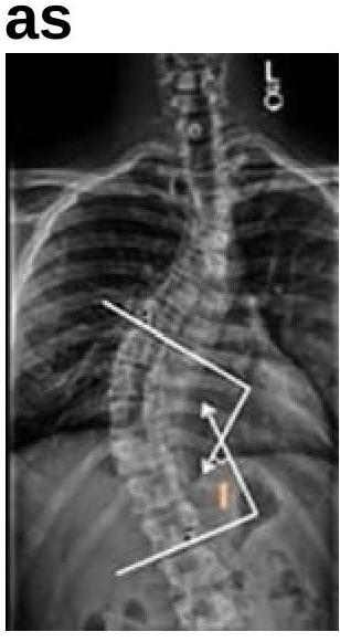
Question 5: What is the angle shown in the image known as?
- A. Cobb angle (Correct Answer)
- B. Bohler angle
- C. Ferguson angle
- D. Baumann angle
Explanation: ***Correct Option: Cobb angle*** - The image displays a method for measuring the angle of a spinal curvature, which is known as the **Cobb angle** - This measurement is routinely used to assess the severity of **scoliosis** by drawing lines parallel to the vertebral endplates at the extreme ends of the curve and then determining the angle between these lines - The Cobb angle is the **gold standard** for quantifying scoliosis and monitoring curve progression *Incorrect Option: Bohler angle* - The **Bohler angle** is a measurement used in the assessment of **calcaneal fractures** - It is formed by two lines drawn on a lateral foot X-ray and is not relevant to spinal deformities *Incorrect Option: Ferguson angle* - The **Ferguson angle**, also known as the lumbosacral angle, measures the angle of the sacral base relative to the horizontal - It describes the degree of **lordosis** and is not used to quantify scoliosis as depicted in the image *Incorrect Option: Baumann angle* - The **Baumann angle** is an important measurement used in pediatric orthopedics to assess the alignment of the **distal humerus** after a supracondylar fracture - It is irrelevant to spinal imaging and curvature assessment
Question 6: Which radiological finding is shown in the image?
- A. Intussusception (Correct Answer)
- B. Colon carcinoma
- C. Sigmoid volvulus
- D. Ileus
Explanation: ***Intussusception*** - The image clearly displays the classic "coiled spring" appearance, which is pathognomonic for **intussusception** on a barium enema study. This pattern is created by barium trapped between the intussusceptum and intussuscipiens. - The arrow specifically points to the leading edge of the intussusception, where the bowel telescopes into an adjacent segment. *Colon carcinoma* - Colon carcinoma typically presents as an **irregular narrowing** or an **apple-core lesion** on barium studies, a sign of luminal stricture due to a mass. - The radiological appearance for carcinoma would not show the distinct layered or coiled pattern seen in the provided image. *Sigmoid volvulus* - Sigmoid volvulus is characterized by a **"coffee bean" sign** on plain radiographs due to the massively dilated, inverted U-shaped loop of colon, or a **"bird's beak" appearance** on contrast studies at the twisted obstruction point. - This contrasts significantly with the concentric rings and linear striations indicative of intussusception. *Ileus* - Ileus, or paralytic ileus, involves generalized **bowel dilation** without a clear point of mechanical obstruction, often with gas present throughout the large and small bowel. - The image shows a very specific, localized abnormality with a characteristic pattern, not generalized bowel distension associated with ileus.
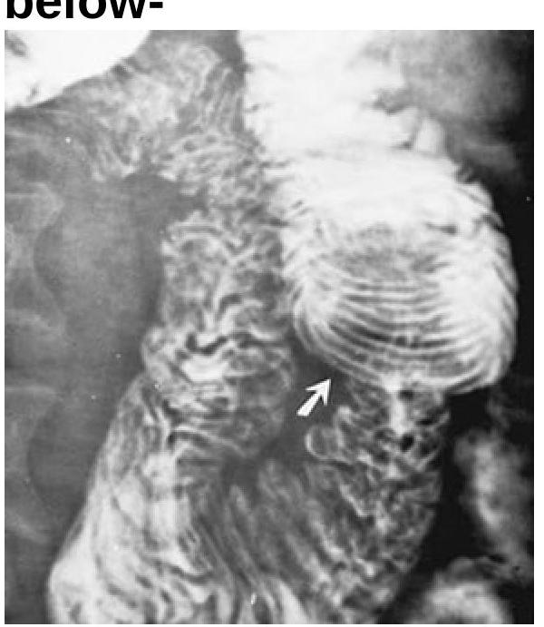
Question 7: The Barium Swallow examination shows a filling defect in the esophagus. What is the most probable diagnosis?
- A. Esophageal Carcinoma (Correct Answer)
- B. Esophageal Ring
- C. Esophageal Tear
- D. Achalasia Cardia
Explanation: ***Esophageal Carcinoma*** - A filling defect on a barium swallow study, especially with irregular borders and luminal narrowing, is highly suggestive of an **esophageal carcinoma**. - The image appears to show an **irregular, obstructing lesion** that displaces the barium column, characteristic of a mass. *Esophageal Ring* - An esophageal ring, such as a **Schatzki ring**, typically presents as a thin, circumferential narrowing of the distal esophagus, forming a smooth, shelf-like indentation, which is not seen here. - Esophageal rings usually cause **intermittent dysphagia** to solids but do not present as a large, irregular filling defect. *Esophageal Tear* - An esophageal tear (e.g., **Mallory-Weiss tear**) is a mucosal laceration that would present with **hematemesis** and would typically appear as a linear defect or streak on a barium swallow if visible, not a filling defect. - A tear is not usually associated with a persistent mass effect or irregular luminal obstruction seen in the image. *Achalasia Cardia* - **Achalasia** is characterized by the failure of the lower esophageal sphincter to relax and **absent peristalsis** in the esophageal body, leading to a classic "bird's beak" or "rat tail" appearance on barium swallow due to distal narrowing and proximal dilation. - While it causes luminal narrowing, it does not typically present as an irregular filling defect within the lumen, but rather as a smooth tapering of the distal esophagus.
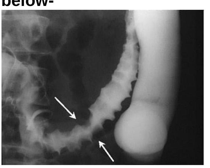
Question 8: Identify the radiological sign of Ischemic colitis from the image provided.
- A. Diverticulitis
- B. Appendicitis
- C. Thumbprinting sign of Ischemic colitis (Correct Answer)
- D. None of the options
Explanation: ***Thumbprinting sign of Ischemic colitis*** - The image displays prominent **indentations (thumbprinting)** along the bowel wall, especially in the descending colon (indicated by arrows). These indentations are caused by **edema** and **hemorrhage** in the submucosal layer due to ischemia. - This characteristic appearance on a barium enema or CT scan is a classic radiological sign highly suggestive of **ischemic colitis**. *Diverticulitis* - Diverticulitis typically presents with **saccular outpouchings** (diverticula) that become inflamed, potentially showing wall thickening or **pericolonic fat stranding**. - This image does not show typical diverticula or signs of severe inflammation associated with diverticulitis, but rather diffuse mucosal changes. *Appendicitis* - Appendicitis is characterized by inflammation of the **vermiform appendix**, typically seen as a **dilated**, non-compressible appendix with surrounding fat stranding in the right lower quadrant. - The radiological findings in the image are of the colon, not the appendix, and are inconsistent with acute appendicitis. *None of the options* - The image presents a clear and characteristic radiological sign that points to a specific diagnosis, making this option incorrect. - The presence of **thumbprinting** is a well-established indicator for ischemic colitis.
Question 9: A radiograph is obtained from a child with scoliosis. What is the name of the angle used to measure spinal curvature?
- A. Bohler's Angle
- B. Ferguson's Angle
- C. Cobb's Angle (Correct Answer)
- D. Pauwels' Angle
Explanation: **Cobb's Angle** - **Cobb's angle** is the primary method for measuring the severity of **scoliosis** on radiographs. - It is measured by drawing lines parallel to the superior endplate of the most tilted superior vertebra and the inferior endplate of the most tilted inferior vertebra of the curve; the angle between these two lines (or their perpendiculars) is the Cobb angle. *Bohler's Angle* - **Bohler's angle** is used in the assessment of **calcaneus fractures** and is measured on a lateral foot radiograph. - A decrease in this angle is indicative of a calcaneal fracture. *Ferguson's Angle* - **Ferguson's angle**, also known as the lumbosacral angle, measures the inclination of the sacrum relative to the horizontal in the standing position. - It is primarily used in the assessment of **spondylolisthesis** and other lumbosacral conditions. *Pauwels' Angle* - **Pauwels' angle** is used to classify **femoral neck fractures** based on the angle of the fracture line relative to the horizontal. - It helps determine the severity and stability of femoral neck fractures, guiding treatment decisions.
Question 10: Radiation Dose Monitoring in Occupational Workers is done by
- A. TLD Badge (Correct Answer)
- B. Collimators
- C. Grid
- D. Linear Accelerator
Explanation: ***TLD Badge (used for monitoring radiation exposure)*** - **Thermoluminescent Dosimeter (TLD) badges** are widely used for monitoring an individual's exposure to ionizing radiation over time. - They work by storing energy from radiation exposure and releasing it as **light when heated**, which is then measured to calculate the accumulated dose. *Collimators (used to shape radiation beams)* - **Collimators** are devices used in radiation therapy and diagnostic imaging to **restrict and shape the radiation beam**, ensuring it only targets the intended area. - They do not measure or monitor the dose received by an individual, but rather **control the spatial distribution** of the radiation. *Grid (used to reduce scatter in imaging)* - An **anti-scatter grid** is placed between the patient and the image receptor in radiography to **absorb scattered radiation**, which degrades image quality. - While essential for image quality, grids do not directly measure or monitor the radiation dose received by an occupational worker. *Linear Accelerator (used for delivering radiation therapy)* - A **linear accelerator (linac)** is a machine used to deliver **external beam radiation treatment** for cancer. - It generates high-energy X-rays or electrons, but it is a **source of radiation** for treatment, not a device for monitoring occupational exposure.