All (366)Anatomy (18)Anesthesiology (4)Biochemistry (26)Community Medicine (33)Dental (2)Dermatology (7)ENT (7)Forensic Medicine (13)Internal Medicine (54)Microbiology (21)Obstetrics and Gynecology (28)Ophthalmology (8)Orthopaedics (5)Pathology (25)Pediatrics (18)Pharmacology (27)Physiology (21)Psychiatry (9)Radiology (17)Surgery (23)
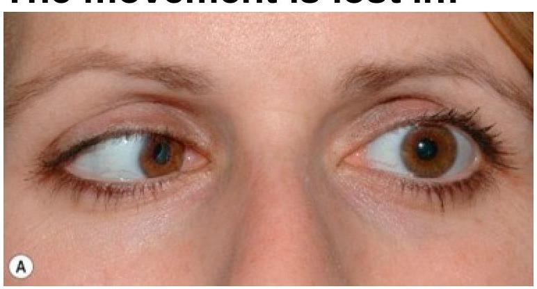
Q41
In trochlear palsy, which specific eye movement is lost?
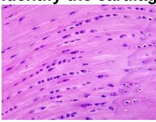
Q42
Identify the type of cartilage shown in the image.
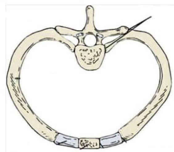
Q43
Identify the type of joint in the image provided.
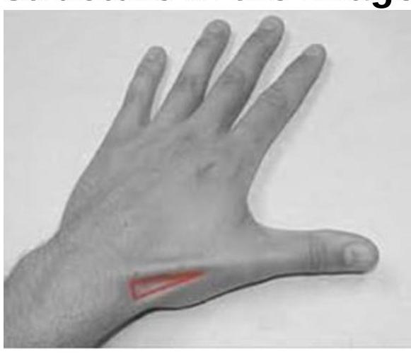
Q44
The image shows a highlighted region on the dorsal aspect of the hand (anatomical snuffbox). Which of the following anatomical structures form the boundaries or floor of this region?
Q45
The patient is presenting with pain around the base of the thumb. Which tendons are likely involved?