NEET-PG 2020 — Dermatology
6 Previous Year Questions with Answers & Explanations
Elderly man with a long-standing mole on his face that is increasing in size and showing an irregular border. Diagnosis:
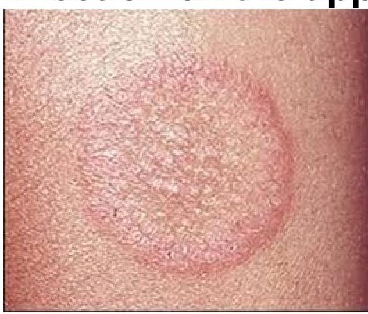
Identify the condition causing this infection on the upper arm
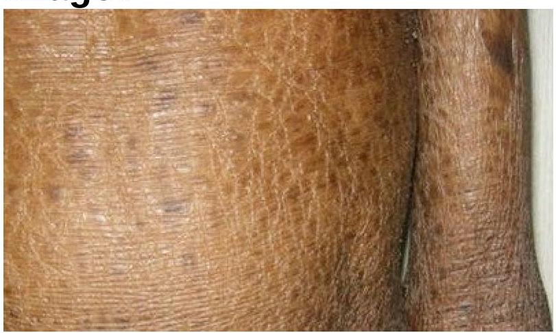
Identify the skin condition depicted in the image.
A farmer presented with a black mole on the cheek. It increased in size, more than 6mm with irregular borders and a central black lesion, what could be the diagnosis?
Dermatological manifestation of which of the following diseases?

Baby born with membrane around him at the time of birth. Which of the following conditions is depicted?
NEET-PG 2020 - Dermatology NEET-PG Practice Questions and MCQs
Question 1: Elderly man with a long-standing mole on his face that is increasing in size and showing an irregular border. Diagnosis:
- A. Superficial spreading melanoma
- B. Nodular melanoma
- C. Acral melanoma
- D. Lentigo maligna (Correct Answer)
Explanation: ***Lentigo maligna*** - This type of melanoma commonly affects **elderly individuals** and presents as a **slowly enlarging, irregularly bordered, flat or slightly raised pigmented lesion** on sun-exposed areas like the face. - It often has a **long radial growth phase** before progressing to invasive lentigo maligna melanoma. *Superficial spreading melanoma* - While common, it typically presents on the **trunk or extremities** and has a faster growth rate compared to lentigo maligna. - It often appears as a **flat, asymmetrical lesion with varied colors and irregular borders**, but the age and location details point away from this. *Nodular melanoma* - This is an **aggressive form** that grows vertically from the start, presenting as a **dark, raised, often ulcerated nodule** and typically has a shorter history of rapid growth. - It lacks the characteristic long-standing, flat growth pattern described in the elderly patient's face. *Acral melanoma* - This rare type occurs on the **palms, soles, or under the nails (subungual)**, not typically on the face. - It often appears as a **pigmented streak or patch** in these acral locations.
Question 2: Identify the condition causing this infection on the upper arm
- A. Tinea capitis (scalp ringworm)
- B. Tinea cruris (jock itch)
- C. Tinea manus (hand ringworm)
- D. Tinea corporis (body ringworm) (Correct Answer)
Explanation: ***Tinea corporis (body ringworm)*** - This lesion, depicted on the upper arm, is characteristic of **tinea corporis** due to its **annular, erythematous, and scaly border with central clearing**. - The term "corporis" refers to the **body surface**, excluding the scalp, hands, feet, groin, and nails. *Tinea capitis (scalp ringworm)* - Tinea capitis specifically affects the **scalp** and can present with scaling, hair loss, and inflammation. - The image clearly shows a lesion on the **upper arm**, not the scalp. *Tinea cruris (jock itch)* - Tinea cruris is a fungal infection found in the **groin area**, often extending to the inner thighs and buttocks. - The location of the lesion in the image, on the **upper arm**, rules out tinea cruris. *Tinea manus (hand ringworm)* - Tinea manus affects the **hands**, typically causing dryness, scaling, and sometimes blister formation on the palms or between the fingers. - The lesion in the image is located on the **upper arm**, not the hand.
Question 3: Identify the skin condition depicted in the image.
- A. Ichthyosis (Correct Answer)
- B. Syndromic ichthyosis
- C. Cutaneous sarcoidosis
- D. Leprosy
Explanation: ***Ichthyosis*** - The image clearly displays widespread **dry, scaling, and thickened skin**, consistent with the characteristic presentation of ichthyosis. - This condition is characterized by a defect in **skin barrier function** leading to excessive dryness and accumulation of scales. *Syndromic ichthyosis* - While syndromic ichthyosis also involves skin scaling, it is associated with **additional systemic symptoms** or **organ involvement**, which cannot be determined from this image alone. - The term "ichthyosis" broadly covers this appearance, and without more clinical information, specifying it as syndromic is not the most direct identification. *Leprosy* - Leprosy typically presents with **hypopigmented, anesthetic skin patches** or **nodules**, which are not seen in the image. - The texture and color changes in the image are not characteristic of the primarily neurological and dermatological manifestations of leprosy. *Cutaneous sarcoidosis* - Cutaneous sarcoidosis manifests as **reddish-brown papules, plaques, or nodules**, often on the face, neck, or extremities. - The widespread, fine scaling and dryness seen in the image do not align with the typical granulomatous lesions of sarcoidosis.
Question 4: A farmer presented with a black mole on the cheek. It increased in size, more than 6mm with irregular borders and a central black lesion, what could be the diagnosis?
- A. Superficial spreading melanoma (Correct Answer)
- B. Acral lentigo melanoma
- C. Lentigo maligna melanoma
- D. Nodular melanoma
Explanation: ***Superficial spreading melanoma*** - This is the most common type of melanoma and often presents as a **mole with irregular borders**, varying colors, and a diameter greater than 6mm, consistent with the description. - The lesion typically grows **radially** across the skin surface before beginning vertical growth, indicated by the increase in size. *Acral lentigo melanoma* - This type of melanoma primarily affects the **palms, soles, and nail beds**, which is inconsistent with a lesion on the cheek. - It often appears as a **dark brown or black patch** that slowly enlarges, but its location is characteristic. *Lentigo maligna melanoma* - This melanoma typically occurs in **chronically sun-damaged skin** of the elderly, often on the head and neck, but usually presents as a **flat, irregularly shaped, tan or brown patch** with varying shades, which may not fit the description of a central black lesion within a larger mole. - It has a dominant **radial growth phase** and progresses slowly over many years before developing a nodular component. *Nodular melanoma* - This type is characterized by its **rapid vertical growth** and appearance as a **raised, dark, often dome-shaped lesion** from the outset. - While it can be black, the description of an "increased in size" mole with irregular borders and a central black lesion points more towards a spreading type rather than a rapidly growing nodule from the beginning.
Question 5: Dermatological manifestation of which of the following diseases?
- A. Photo dermatitis
- B. Pellagra (Correct Answer)
- C. Acrodermatitis enteropathica
- D. Vitamin B deficiency
Explanation: ***Pellagra*** - The image shows a classic "butterfly" rash on the face, specifically a photosensitive dermatitis, which is a hallmark of **pellagra**. - Pellagra is caused by a deficiency of **niacin (vitamin B3)**, characterized by the "3 D's": **dermatitis**, **diarrhea**, and **dementia**. *Photo dermatitis* - While pellagra often presents with photosensitive dermatitis, "photo dermatitis" is a general term for **skin inflammation caused by light exposure** and not a specific disease itself. - It could be caused by various factors, including medication, immune reactions, or other underlying conditions, but the pattern seen here is highly suggestive of pellagra. *Acrodermatitis enteropathica* - This condition is a **hereditary zinc deficiency** that typically presents with a periorificial and acral dermatitis. - The skin lesions are typically **vesicular-pustular or eczematous** and do not usually have the distinct butterfly pattern of photosensitive dermatitis seen in the image. *Vitamin B deficiency* - While pellagra is a vitamin B **(niacin, B3)** deficiency, this option is too broad. - Other vitamin B deficiencies, such as **riboflavin (B2)** or **pyridoxine (B6)** deficiency, have different dermatological manifestations like angular cheilitis, glossitis, or seborrheic dermatitis, but not the characteristic facial rash seen here.
Question 6: Baby born with membrane around him at the time of birth. Which of the following conditions is depicted?
- A. X-linked ichthyosis (steroid sulfatase deficiency)
- B. Generalized hyperkeratosis (thickened skin)
- C. Ichthyosis vulgaris (dry, scaly skin)
- D. Lamellar ichthyosis (collodion membrane at birth) (Correct Answer)
Explanation: ***Lamellar ichthyosis (collodion membrane at birth)*** - This condition is characterized by a "collodion membrane" at birth, which is a **tight, shiny, parchment-like membrane** that covers the entire body. - The membrane typically **sheds within weeks**, revealing underlying scaling and erythema characteristic of lamellar ichthyosis. *X-linked ichthyosis (steroid sulfatase deficiency)* - Marked by **dark brown, adherent scales**, primarily affecting the neck, trunk, and extensor surfaces. - It usually becomes apparent **several weeks or months after birth** and is not typically associated with a collodion membrane. *Generalized hyperkeratosis (thickened skin)* - This is a general term for **thickening of the outermost layer of the epidermis** and is a feature of many ichthyoses, not a specific condition with a "membrane at birth." - It describes a **symptom** rather than a primary diagnosis presenting with a specific birth membrane. *Ichthyosis vulgaris (dry, scaly skin)* - Presents with **fine, white scaling**, most prominent on the extensor surfaces of the limbs, but it **rarely appears at birth**. - It is typically **mild** and often worsens in dry, cold weather, lacking the characteristic "membrane around him" at birth.