All SubjectsAnatomy (167)Anesthesiology (9)Biochemistry (108)Community Medicine (87)Dental (8)Dermatology (19)ENT (30)Forensic Medicine (62)General Medicine (3)Internal Medicine (136)Microbiology (101)Obstetrics and Gynecology (65)Ophthalmology (60)Orthopaedics (33)Pathology (107)Pediatrics (37)Pharmacology (123)Physiology (116)Psychiatry (2)Psychiatry (38)Radiology (25)Surgery (81)
Q71
What is the best management for a human bite?
Q72
A high-riding prostate is indicative of which injury?
Q73
Under what guidelines is treatment started for a patient presenting with appendicular mass on a CT scan?
Q74
Supraomohyoid dissection is a type of?
Q75
Treatment of choice for mucinous carcinoma of the gall bladder in the early stage is -
Q76
A 55 year old woman presented with history of recurrent episodes of right upper abdominal pain for the last one year. She presented to emergency with history of jaundice and fever for 2 days. On examination, the patient appeared toxic and had a blood pressure of 100/60 mmHg. She was started on intravenous antibiotics. Ultrasound of the abdomen showed presence of stones in the common bile duct. What would be the best treatment option for her -
Q77
In which of the following surgeries is monopolar cautery preferred over bipolar cautery?
Q78
Reactionary Hemorrhage occurs due to?
Q79
An Incisional wound heals by
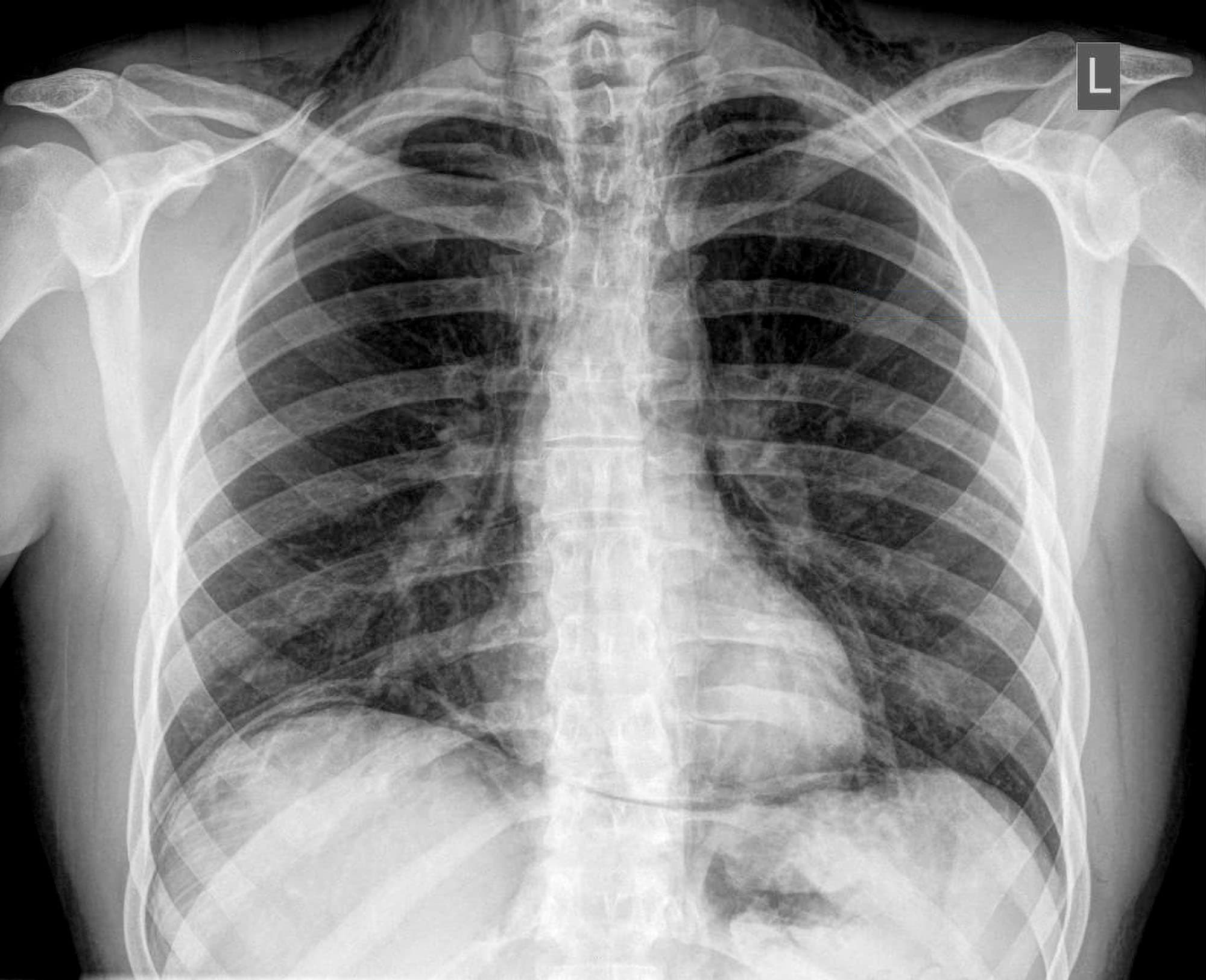
Q80
A patient presents with abdominal pain. On physical examination there was abdominal guarding and tenderness. A plain erect chest X-ray reveals air under diaphragm. Probable diagnosis is