All (1417)Anatomy (167)Anesthesiology (9)Biochemistry (108)Community Medicine (87)Dental (8)Dermatology (19)ENT (30)Forensic Medicine (62)General Medicine (3)Internal Medicine (136)Microbiology (101)Obstetrics and Gynecology (65)Ophthalmology (60)Orthopaedics (33)Pathology (107)Pediatrics (37)Pharmacology (123)Physiology (116)Psychiatry (2)Psychiatry (38)Radiology (25)Surgery (81)
Q1331
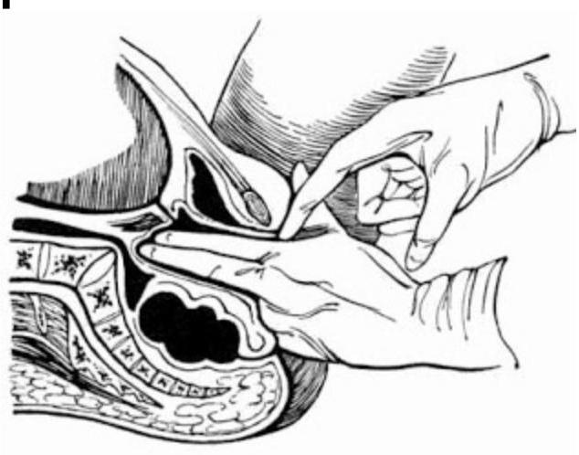
Which of the following describes the points marked in the diagram of pelvic measurements?